
Article Figures & Data
Figures
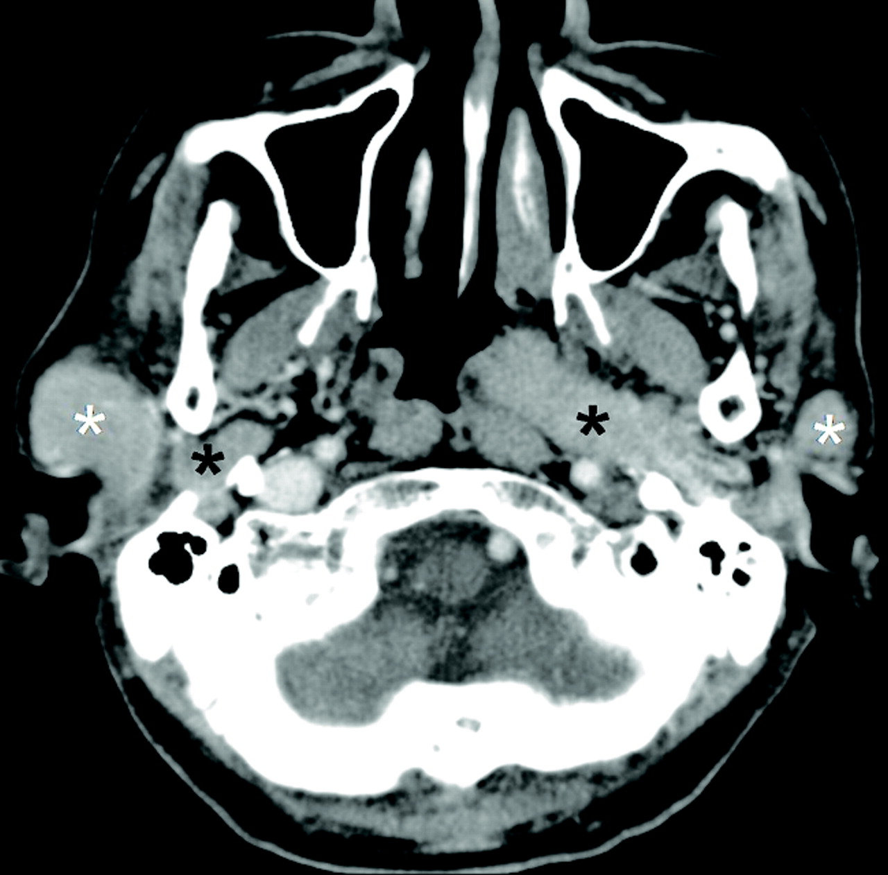
- Fig 1.
A 60-year-old man with bilateral parotid oncocytomas. CT scan shows bilateral multifocal and homogeneously enhancing parotid tumors with well-defined margins involving both the superficial (white asterisks) and deep (black asterisks) lobes of the parotid gland.
- Fig 2.
A 49-year-old woman with a unilateral solitary parotid oncocytoma. A well-defined tumor with a lobulated contour and homogeneous enhancement is identified on the CT scan in the superficial lobe of the left parotid gland (white asterisk).
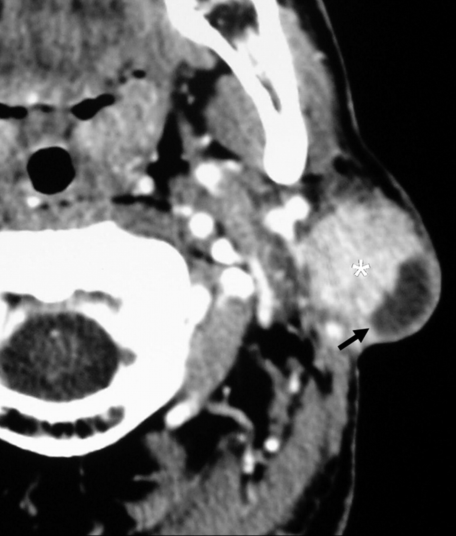
- Fig 3.
A left parotid oncocytoma in a 68-year-old woman. CT scan reveals a large deformable tumor (white asterisk), which extends medially into the parapharyngeal space through the stylomandibular gap. The contour of the tumor is distorted by the styloid process (black arrow) and the left medial pterygoid muscle (black arrowheads).
- Fig 4.
A, A 70-year-old woman with a large right parotid oncocytoma. The large tumor (white asterisk) extends medially through the stylomandibular gap into the parapharyngeal space and demonstrates a nonenhancing curvilinear cleft (black arrow). B, Synchronous bilateral parotid oncocytomas in another 70-year-old woman. CT demonstrates tumors in the right parotid gland (white asterisks), which show homogeneous enhancement, and a large left parotid tumor (black asterisk) with a nonenhancing curvilinear cleft (black arrow).
- Fig 5.
A 49-year-old man with a unilateral solitary parotid oncocytoma. The parotid tumor in the superficial lobe of the left parotid gland (white asterisk) has a lobulated contour and shows a cystic component (black arrow).


Tables
Age and sex distribution including the radiologic features of the 10 cases of parotid gland oncocytomas
No. Sex/Age (yr) Bilaterality/Multifocality Location Size (mm) Margins Contour Enhancement Presence of Nonenhancing Curvilinear Cleft/Cystic Component 1 F/55 Unilateral/Solitary Superficial lobe 11 × 8 Sharp Lobulated Heterogeneous Nonenhancing curvilinear cleft 2 F/70 Bilateral/Multifocal Superficial and deep lobes 32 × 15 Sharp Smooth Heterogeneous Nonenhancing curvilinear cleft 3 F/74 Bilateral/Multifocal Superficial and deep lobes 9 × 6 Sharp Smooth Homogeneous 4 F/49 Unilateral/Solitary Superficial lobe 10 × 8 Sharp Smooth Homogeneous 5 M/49 Unilateral/Solitary Superficial lobe 13 × 12 Sharp Lobulated Heterogeneous Cystic component 6 M/60 Bilateral/Multifocal Superficial and deep lobes 12 × 10 Sharp Lobulated Homogeneous 7 F/49 Unilateral/Solitary Superficial lobe 6 × 6 Sharp Lobulated Heterogeneous Cystic component 8 F/68 Bilateral/Multifocal Superficial and deep lobes 12 × 8 Sharp Smooth Heterogeneous Nonenhancing curvilinear cleft 9 F/70 Bilateral/Multifocal Superficial and deep lobes 66 × 42 Sharp Smooth Heterogeneous Nonenhancing curvilinear cleft 10 M/66 Unilateral/Multifocal Superficial and deep lobes 27 × 18 Sharp Smooth Homogeneous


