
Article Figures & Data
Figures
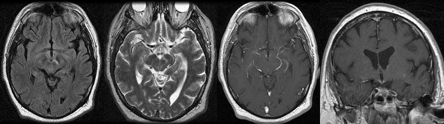
- Fig 1.
Axial noncontrast FLAIR and fast spin-echo T2 MR images as well as postgadolinium axial and coronal T1 images demonstrate a 9-mm focus of enhancing abnormal T2 signal intensity without mass effect within the inferomedial aspect of the hypothalamus bilaterally, extending into the left cerebral peduncle.
- Fig 2.
Axial noncontrast FLAIR MR images demonstrate bilaterally symmetric T2 signal-intensity abnormality without mass effect, involving the corticospinal tracts, brain stem, and brachium pontis.
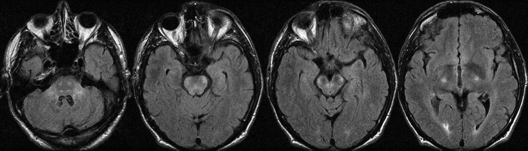
- Fig 3.
Axial noncontrast FLAIR and axial and coronal T1 postgadolinium images demonstrate enhancing abnormally increased T2 signal intensity in the hypothalamus and anteromedial aspect of the right temporal lobe, including the hippocampal head.
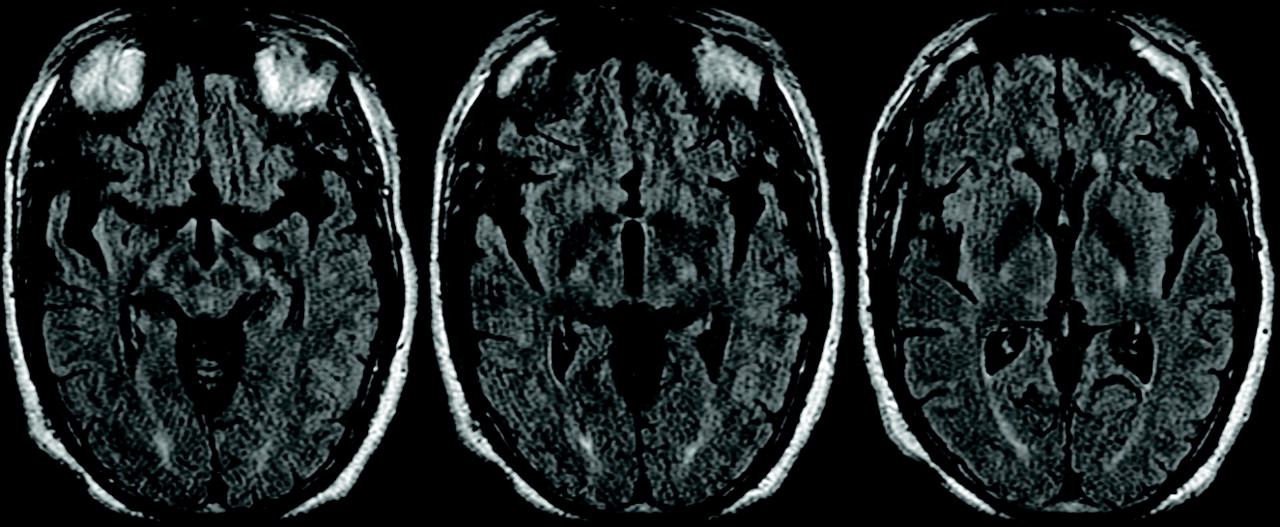
- Fig 4.
Axial noncontrast FLAIR images demonstrate symmetric high T2 signal intensity in the hypothalami and corticospinal tracts.

Tables
Subject No. Age (yr)/Sex Primary MR Imaging PCR CSF PCR Serum PCR GI GI Biopsy (PAS) 1 54/M CNS + + + NP – 2 59/M CNS (asxs GI) + + – + – 3 38/M GI→CNS + + – NP + 4 40/M GI→CNS + – – + + 5 40/M GI→CNS – + NP + – 6 52/M GI→CNS – + – – + 7 63/M GI→CNS – + – – + Note:—+ indicates positive result; –, negative result.
Subject No. Interval Symptoms→Rx Outcome 1 9 months Stabilized on antibiotics but died 15 months later 2 4 months From wheelchair-bound to a cane on antibiotic therapy 3 1 month Complete recovery on antibiotics 4 6 months Lost to follow-up 5 ∼yrs? Severe neurologic impairment 6 ∼1 month Mild attention and concentration deficits 7 ∼3 months Mild symptoms when lost to follow-up


