Advertisement

Main menu

  • Home
  • Content
    • Current Issue
    • Accepted Manuscripts
    • Article Preview
    • Past Issue Archive
    • Video Articles
    • AJNR Case Collection
    • Case of the Week Archive
    • Case of the Month Archive
    • Classic Case Archive
  • Special Collections
    • AJNR Awards
    • ASNR Foundation Special Collection
    • Most Impactful AJNR Articles
    • Photon-Counting CT
    • Spinal CSF Leak Articles (Jan 2020-June 2024)
  • Multimedia
    • AJNR Podcasts
    • AJNR SCANtastic
    • Trainee Corner
    • MRI Safety Corner
    • Imaging Protocols
  • For Authors
    • Submit a Manuscript
    • Submit a Video Article
    • Submit an eLetter to the Editor/Response
    • Manuscript Submission Guidelines
    • Statistical Tips
    • Fast Publishing of Accepted Manuscripts
    • Graphical Abstract Preparation
    • Imaging Protocol Submission
    • Author Policies
  • About Us
    • About AJNR
    • Editorial Board
    • Editorial Board Alumni
  • More
    • Become a Reviewer/Academy of Reviewers
    • Subscribers
    • Permissions
    • Alerts
    • Feedback
    • Advertisers
    • ASNR Home

User menu

  • Alerts
  • Log in

Search

  • Advanced search
American Journal of Neuroradiology

American Journal of Neuroradiology

ASHNR American Society of Functional Neuroradiology ASHNR American Society of Pediatric Neuroradiology ASSR
  • Alerts
  • Log in

Advanced Search

  • Home
  • Content
    • Current Issue
    • Accepted Manuscripts
    • Article Preview
    • Past Issue Archive
    • Video Articles
    • AJNR Case Collection
    • Case of the Week Archive
    • Case of the Month Archive
    • Classic Case Archive
  • Special Collections
    • AJNR Awards
    • ASNR Foundation Special Collection
    • Most Impactful AJNR Articles
    • Photon-Counting CT
    • Spinal CSF Leak Articles (Jan 2020-June 2024)
  • Multimedia
    • AJNR Podcasts
    • AJNR SCANtastic
    • Trainee Corner
    • MRI Safety Corner
    • Imaging Protocols
  • For Authors
    • Submit a Manuscript
    • Submit a Video Article
    • Submit an eLetter to the Editor/Response
    • Manuscript Submission Guidelines
    • Statistical Tips
    • Fast Publishing of Accepted Manuscripts
    • Graphical Abstract Preparation
    • Imaging Protocol Submission
    • Author Policies
  • About Us
    • About AJNR
    • Editorial Board
    • Editorial Board Alumni
  • More
    • Become a Reviewer/Academy of Reviewers
    • Subscribers
    • Permissions
    • Alerts
    • Feedback
    • Advertisers
    • ASNR Home
  • Follow AJNR on Twitter
  • Visit AJNR on Facebook
  • Follow AJNR on Instagram
  • Join AJNR on LinkedIn
  • RSS Feeds

AJNR is seeking candidates for the AJNR Podcast Editor. Read the position description.

Research ArticleInterventional

Curative Embolization with Onyx of Dural Arteriovenous Fistulas with Cortical Venous Drainage

W.J. van Rooij and M. Sluzewski
American Journal of Neuroradiology September 2010, 31 (8) 1516-1520; DOI: https://doi.org/10.3174/ajnr.A2101
W.J. van Rooij
  • Find this author on Google Scholar
  • Find this author on PubMed
  • Search for this author on this site
M. Sluzewski
  • Find this author on Google Scholar
  • Find this author on PubMed
  • Search for this author on this site
  • Article
  • Figures & Data
  • Info & Metrics
  • Responses
  • References
  • PDF
Loading

Abstract

BACKGROUND AND PURPOSE: Dural fistulas with cortical venous drainage often present with hemorrhage or neurologic deficit, and prompt treatment is indicated. Occlusion of the draining vein by endovascular techniques or surgical disconnection is considered curative. We present our first endovascular experience with use of Onyx via the arterial route in these aggressive fistulas.

MATERIALS AND METHODS: Between October 2008 and October 2009, 8 consecutive patients with a dural fistula with exclusive cortical venous drainage were treated with Onyx. Three patients presented with hemorrhage; 3, with hemianopsia; 1 with bruit; and 1 after incomplete coil occlusion of a benign sigmoid sinus dural fistula. Fistula location was the sigmoid sinus segment in 4, the occipital area in 2, the cavernous sinus in 1, and the torcula in 1.

RESULTS: In all 8 patients, it was possible to occlude the proximal venous site of the fistula with Onyx via arterial feeders, resulting in complete cure in all. In 6 patients, a prolonged (17- to 29-minute) single middle meningeal artery injection was sufficient to occlude the venous site of the fistula with retrograde occlusion of other arterial feeders; in the other 2 patients, 2 injections in supplying arteries were needed. Follow-up angiograms after 6–12 weeks confirmed lasting complete occlusion of the fistulas. All patients were clinically cured, there were no complications.

CONCLUSIONS: In this small series, curative embolization of dural fistulas with exclusive cortical venous drainage by using Onyx via the arterial route was possible in all patients.

Abbreviations

AP
anteroposterior
DAVF
dural arteriovenous fistula
IPH
intraparenchymal hemorrhage

Intracranial DAVFs with retrograde cortical venous drainage are aggressive lesions that can present with intracranial hemorrhage, seizures, progressive neurologic deficit, intracranial hypertension, or dementia.1–4 Because the natural history of untreated lesions is associated with a poor prognosis, prompt treatment is indicated.5,6 Dural fistulas are considered multiple-hole fistulas with multiple arterial feeders draining into 1 single vein. Treatment of DAVFs with cortical venous drainage is aimed at occlusion of the venous drainage or occlusion of all arterial supply and can be surgical, endovascular, or a combination.7–13 Endovascular treatment has traditionally included embolization via an arterial or venous route with use of acrylic glue, particles, coils, or a combination. Recently, the use of Onyx (ev3, Irvine, California) via the arterial route has been reported with promising results.14–18 We report our initial experience with arterial injection of Onyx in the endovascular treatment of dural fistulas with drainage exclusively into the cortical veins.

Materials and Methods

Between October 2008 and October 2009, 8 consecutive patients with dural fistulas with exclusive cortical venous drainage were treated with Onyx. Patient characteristics are summarized in the Table. There were 5 men and 3 women with a mean age of 57 years (range, 36–67 years). Three patients presented with hemorrhage; 3, with hemianopsia; and 1, with bruit. One patient was referred after incomplete coil occlusion of a benign sigmoid sinus dural fistula with residual shunt on a trapped sinus segment. Fistula location was the sigmoid sinus segment in 4, the right occipital area in 2, the left cavernous sinus in 1, and the torcula in 1. Diagnosis was confirmed after angiography of all cerebral vessels on a biplane angiographic unit (Integris Allura Neuro; Philips Healthcare, Best, the Netherlands).

View this table:
  • View inline
  • View popup

Patient and treatment characteristics of 8 patients with DAVFs with exclusive cortical venous drainage that were treated with intra-arterial Onyx injection

Endovascular treatment was performed with the patient under general anesthesia. A 4F or 5F catheter was positioned in the external carotid artery, and an Onyx-compatible microcatheter (Rebar 105-5081-153, ev3) was coaxially advanced in a dural feeding artery as close as possible to the fistula site. Then, Onyx 18 was slowly injected. When reflux occurred, the injection was paused for 1–3 minutes. Usually, reflux occurred several times before Onyx would advance into the fistula site. In some cases with middle meningeal artery injection, more than a 5-cm reflux was accepted. Once Onyx advanced, it was intermittently injected until the draining veins were filled and all feeders were filled retrograde, thereby completely occluding the fistula. In 1 patient with a cavernous sinus DAVF supplied by branches of the maxillary artery, the maxillary artery was blocked with a microballoon (HyperForm, ev3) to prevent reflux and promote advancement of Onyx into the cavernous sinus.18 The duration of Onyx injections was calculated from time prints on angiographic images. Follow-up angiography was scheduled after 6–12 weeks in all patients. After the procedure, pain was controlled with medication as needed, including morphine or fentanyl. Illustrative cases are provided in Figs 1⇓–3.

Fig 1.
  • Download figure
  • Open in new tab
  • Download powerpoint
Fig 1.

A 58-year-old man (patient 1) with a good-grade (Hunt and Hess II) subarachnoid hemorrhage from a torcular DAVF that was cured with a single Onyx injection from the middle meningeal artery. A and B, Left external carotid angiograms in arterial (A) and venous (B) phases demonstrate a DAVF located on the torcula supplied by the middle meningeal and occipital arteries with marked congestion of cerebellar and even perimedullary veins. C, Supply to the DAVF by the posterior meningeal artery arising from the right vertebral artery. D, Microcatheter distally in the middle meningeal artery close to the fistula used for Onyx injection. E, AP view of selective angiography of the posterior meningeal artery (arrow) arising from the right vertebral artery. F, Same view as in E shows an Onyx cast in the draining vein and retrograde in the posterior meningeal artery (double arrows). G–I, Lateral views of Onyx cast (G), external carotid (H), and right vertebral (I) angiogams 10 weeks after embolization demonstrate complete occlusion of the DAVF.

Fig 2.
  • Download figure
  • Open in new tab
  • Download powerpoint
Fig 2.

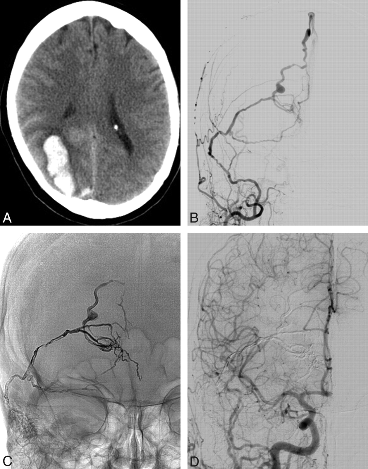
A 36-year-old man (patient 2) with a parenchymal hemorrhage from a DAVF. A, CT scan demonstrates a right parenchymal hematoma. B and C, Early (B) and later (C) phases of a right external carotid angiogram reveal a DAVF located on an isolated open segment of an occluded transverse sinus. The fistula is supplied by the middle meningeal, posterior auricular, and occipital arteries with drainage to the vein of Labbé and a posterior cortical vein (arrows in C). D, Early phase of Onyx injection through a microcatheter, which was positioned close to the fistula in the middle meningeal artery (arrow). Onyx is injected slowly and first fills the dural feeders in the sinus wall. E, Note the Onyx cast at end of injection. The open sinus segment, the proximal draining veins (long arrows), and dural supply from the occipital artery (short arrow) are occluded. F, Complete occlusion of the DAVF is confirmed by postprocedural and follow-up angiography.

Fig 3.
  • Download figure
  • Open in new tab
  • Download powerpoint
Fig 3.

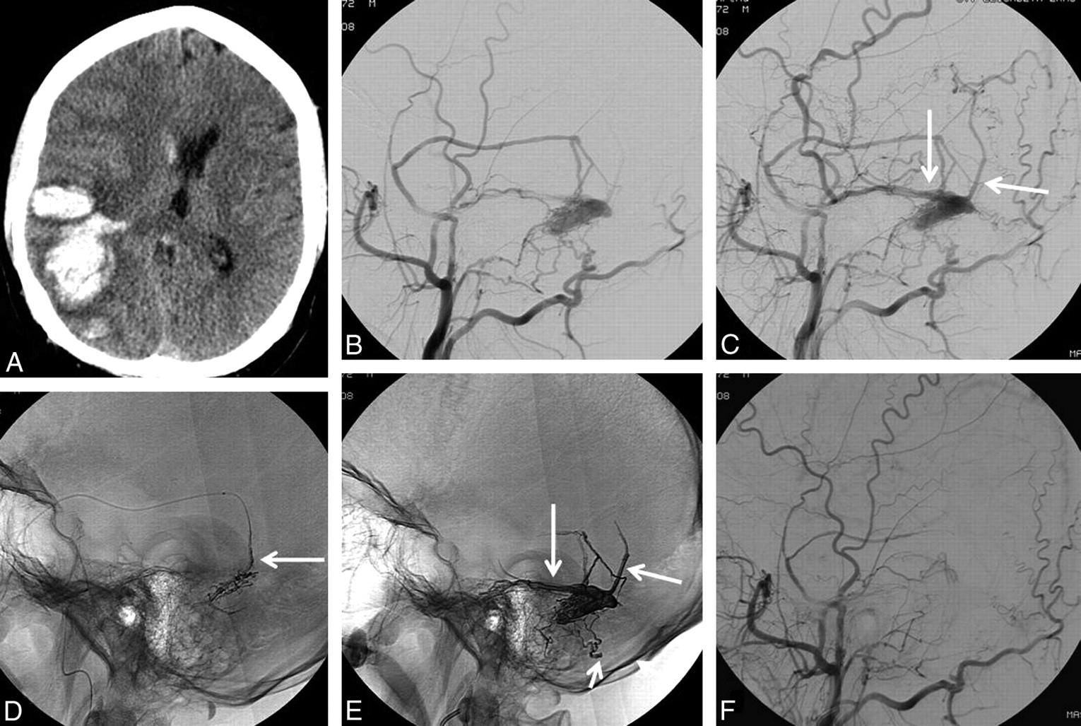
A 59-year-old woman (patient 5) with a right occipital parenchymal hemorrhage from a DAVF. A, CT scan demonstrates a right parenchymal hematoma. B, AP view of a right external carotid angiogram shows a DAVF mainly supplied by the middle meningeal artery. Note the aneurysm on the occipital draining vein. C, Onyx cast after embolization through the middle meningeal artery. All dural supply and the draining veins together with the venous aneurysm are occluded. D, Follow-up carotid angiogram after 12 weeks confirms complete occlusion of the DAVF.

Results

All DAVFs could be cured in a single session, in 6 patients with a single Onyx injection and in 2 patients with 2 injections each. The duration of the Onyx injection in the 6 patients with a single injection was 17–29 minutes (mean, 23 minutes). In all cases, removal of the microcatheter was uneventful, despite sometimes long segments of reflux. Follow-up angiography in all patients confirmed durable obliteration. There were no complications of treatment. Radiation-induced hair loss did not occur. Six patients experienced postprocedural pain, probably because of dural ischemia. Morphine was needed in 2 patients. and fentanyl, in 1 to control the pain. Patients treated electively were discharged home the day following embolization. At the time of follow-up angiography, all patients were pain-free without medication. In 3 patients with hemianopsia as a result of venous congestion in the occipital lobe, visual fields were restored to normal within 12 weeks.

Discussion

Our initial experience in a small patient group by using Onyx for DAVFs with cortical venous drainage is hopeful and confirms the good results reported by others.14–18 Since we adopted Onyx as treatment of choice for these aggressive and rare lesions, all patients could be cured in a single session and mostly with only 1 Onyx injection of less than a half-hour duration. The technique we used to inject Onyx in DAVFs is comparable with the technique for the treatment of brain arteriovenous malformations19: Once a distal position of the tip of the microcatheter in a dural feeder close to the fistula is reached, reflux is slowly built up with a 1- to 3-minute pause between injection attempts until the feeding artery is plugged and Onyx starts to advance into the fistula and the draining veins. Sometimes 5–15 attempts are needed before Onyx advances; thus, patience is required. When in a long feeding vessel (mostly the anterior division of the middle meningeal artery), a distal position is obtained, reflux of 5 cm or even more can be accepted, and many injection attempts are possible. Even after injections of almost half an hour with a long segment of reflux, the Rebar microcatheter could be retrieved without difficulty. Thus, the length of reflux in these dural arteries is not directly related to the possibility of removal of the microcatheter. Onyx does not react with or stick to the vessel wall; it merely fills the vessel lumen.20 Therefore, it is important to completely fill all vessels involved in the fistulous complex; deposition of Onyx in the proximal draining vein only may not be sufficient to completely occlude the fistula or may give rise to recurrence at follow-up.

We suspend the injection for a short period of time when Onyx starts to fill the veins. With repeated injection, additional Onyx often initially is deposited more proximally, including retrograde filling of other dural feeders, before further distal advancement in the veins. This technique can be repeated several times, thereby slowly achieving complete occlusion of the fistula, all dural feeders, and the proximal draining veins with complete luminal filling. In selected cases, a microballoon proximal to the tip of the Onyx microcatheter can help in this injection process by preventing reflux and stimulating advancement of the Onyx.18 In our experience, confirmed by others,15,17 the middle meningeal artery is the most suitable for Onyx injection; it is often involved in dural fistulas and can be readily catheterized to a distal position, and long segments of reflux can be accepted because microcatheter removal proved to be easy in such cases. Whenever this artery contributes to the fistula, we try to use it for embolization.

Other vessels such as the occipital artery are mostly more tortuous and less easy to catheterize. When the tentorial artery is involved and has to be used as access for embolization, we probably still prefer injection of acrylic glue under protection of a microballoon across the origin in the internal carotid artery.21 Although we had no complications of treatment, postprocedural pain was experienced by most patients, and adequate pain medication was needed in some patients. Pain is probably related to ischemia of the dura as a result of occlusion of dural arteries. Pain usually gradually decreased in the following days to weeks, and at the time of follow-up angiography after 6–12 weeks, no patients used analgesics.

Another matter of concern is the radiation dose. Often many angiographic runs are necessary to document the dural fistula. In addition, prolonged Onyx injection during fluoroscopy adds to this dose. For dose reduction, we avoid simultaneous biplane fluoroscopy, and we approximate the flat panels as near as possible to the patient's head.

As with other embolic agents, complications with Onyx may occur. Ischemic damage to the trigeminal and facial nerve may be associated with too proximal reflux of Onyx in the middle meningeal artery beyond the foramen spinosum when occlusion of cavernous and petrosal branches may occur. Reflexive bradycardia may be associated with the treatment of tentorial DAVFs. When anastomoses of external carotid artery branches to the vertebrobasilar system remain unrecognized, migration of Onyx may cause posterior circulation infarctions.22–24 However, in our opinion, with proper technique and sufficient anatomic knowledge, these complications can mostly be avoided.

Conclusions

In conclusion, our limited experience with the use of Onyx for arterial embolization of DAVFs with cortical venous drainage is encouraging, with complete cure in all 8 patients without complications.

References

  1. 1.↵
    1. Cognard C,
    2. Gobin YP,
    3. Pierot L,
    4. et al
    . Cerebral dural arteriovenous fistulas: clinical and angiographic correlation with a revised classification of venous drainage. Radiology 1995;194:671–80
    PubMed
  2. 2.↵
    1. Mullan S
    . Reflections upon the nature and management of intracranial and intraspinal vascular malformations and fistulae. J Neurosurg 1994;80:606–16
    PubMed
  3. 3.↵
    1. Cognard C,
    2. Casasco A,
    3. Toevi M,
    4. et al
    . Dural arteriovenous fistulas as a cause of intracranial hypertension due to impairment of cranial venous outflow. J Neurol Neurosurg Psychiatry 1998;65:308–16
    Abstract/FREE Full Text
  4. 4.↵
    1. Hurst RW,
    2. Bagley LJ,
    3. Galetta S,
    4. et al
    . Dementia resulting from dural arteriovenous fistulas: the pathologic findings of venous hypertensive encephalopathy. AJNR Am J Neuroradiol 1998;19:1267–73
    Abstract
  5. 5.↵
    1. van Dijk JM,
    2. terBrugge KG,
    3. Willinsky RA,
    4. et al
    . Clinical course of cranial dural arteriovenous fistulas with long-term persistent cortical venous reflux. Stroke 2002;33:1233–36
    Abstract/FREE Full Text
  6. 6.↵
    1. Duffau H,
    2. Lopes M,
    3. Janosevic V,
    4. et al
    . Early rebleeding from intracranial dural arteriovenous fistulas: report of 20 cases and review of the literature. J Neurosurg 1999;90:78–84
    PubMed
  7. 7.↵
    1. Hoh BL,
    2. Choudhri TF,
    3. Connolly ES Jr.,
    4. et al
    . Surgical management of high-grade intracranial dural arteriovenous fistulas: leptomeningeal venous disruption without nidus excision. Neurosurgery 1998;42:796–804
    PubMed
  8. 8.↵
    1. van Dijk JM,
    2. TerBrugge KG,
    3. Willinsky RA,
    4. et al
    . Selective disconnection of cortical venous reflux as treatment for cranial dural arteriovenous fistulas. J Neurosurg 2004;101:31–35
    PubMed
  9. 9.
    1. Nelson PK,
    2. Russell SM,
    3. Woo HH,
    4. et al
    . Use of a wedged microcatheter for curative transarterial embolization of complex intracranial dural arteriovenous fistulas: indications, endovascular technique, and outcome in 21 patients. J Neurosurg 2003;98:498–506
    CrossRefPubMed
  10. 10.↵
    1. Defreyne L,
    2. Vanlangenhove P,
    3. Vandekerckhove T,
    4. et al
    . Transvenous embolization of a dural arteriovenous fistula of the anterior cranial fossa: preliminary results. AJNR Am J Neuroradiol 2000;21:761–65
    Abstract/FREE Full Text
  11. 11.↵
    1. Kallmes DF,
    2. Jensen ME,
    3. Cloft HJ,
    4. et al
    . Percutaneous transvenous coil embolization of a Djindjian type 4 tentorial dural arteriovenous malformation. AJNR Am J Neuroradiol 1997;18:673–76
    Abstract/FREE Full Text
  12. 12.↵
    1. Layton KF,
    2. Nelson MD,
    3. Kallmes DF
    . Transarterial coil embolization of the venous component of aggressive type 4 dural arteriovenous fistulas. AJNR Am J Neuroradiol 2006;27:750–52
    Abstract/FREE Full Text
  13. 13.↵
    1. van Rooij WJ,
    2. Sluzewski M,
    3. Beute GN
    . Dural arteriovenous fistulas with cortical venous drainage: incidence, clinical presentation, and treatment. AJNR Am J Neuroradiol 2007;28:651–55
    Abstract/FREE Full Text
  14. 14.↵
    1. Stiefel MF,
    2. Albuquerque FC,
    3. Park MS,
    4. et al
    . Endovascular treatment of intracranial dural arteriovenous fistulae using Onyx: a case series. Neurosurgery 2009;65(6 suppl):132–39
    CrossRefPubMed
  15. 15.↵
    1. Macdonald JH,
    2. Millar JS,
    3. Barker CS
    . Endovascular treatment of cranial dural arteriovenous fistulae: a single-centre, 14-year experience and the impact of Onyx on local practise. Neuroradiology 2010;52:387–95
    CrossRefPubMed
  16. 16.↵
    1. Huang Q,
    2. Xu Y,
    3. Hong B,
    4. et al
    . Use of Onyx in the management of tentorial dural arteriovenous fistulae. Neurosurgery 2009;65:287–92
    CrossRefPubMed
  17. 17.↵
    1. Chew J,
    2. Weill A,
    3. Guilbert F,
    4. et al
    . Arterial Onyx embolisation of intracranial DAVFs with cortical venous drainage. Can J Neurol Sci 2009;36:168–75
    PubMed
  18. 18.↵
    1. Shi ZS,
    2. Loh Y,
    3. Duckwiler GR,
    4. et al
    . Balloon-assisted transarterial embolization of intracranial dural arteriovenous fistulas. J Neurosurg 2009;110:921–28
    CrossRefPubMed
  19. 19.↵
    1. van Rooij WJ,
    2. Sluzewski M,
    3. Beute GN
    . Brain AVM embolization with Onyx. AJNR Am J Neuroradiol 2007;28:172–77
    Abstract/FREE Full Text
  20. 20.↵
    1. Van Loock K,
    2. Menovsky T,
    3. Voormolen MH,
    4. et al
    . Microsurgical removal of Onyx HD-500 from an aneurysm for relief of brainstem compression. J Neurosurg 2009 Nov 6. [Epub ahead of print]
  21. 21.↵
    1. van Rooij WJ,
    2. Sluzewski M,
    3. Beute GN
    . Tentorial artery embolization in tentorial dural arteriovenous fistulas. Neuroradiology 2006;48:737–43
    CrossRefPubMed
  22. 22.↵
    1. Lv X,
    2. Jiang C,
    3. Zhang J,
    4. et al
    . Complications related to percutaneous transarterial embolization of intracranial dural arteriovenous fistulas in 40 patients. AJNR Am J Neuroradiol 2009;30:462–68
    Abstract/FREE Full Text
  23. 23.↵
    1. Schaller B,
    2. Cornelius JF,
    3. Prabhakar H,
    4. et al
    . The trigemino-cardiac reflex: an update of the current knowledge. J Neurosurg Anesthesiol 2009;21:187–95
    CrossRefPubMed
  24. 24.↵
    1. Geibprasert S,
    2. Pongpech S,
    3. Armstrong D,
    4. et al
    . Dangerous extracranial-intracranial anastomoses and supply to the cranial nerves: vessels the neurointerventionalist needs to know. AJNR Am J Neuroradiol 2009;30:1459–68
    Abstract/FREE Full Text
  • Received December 21, 2009.
  • Accepted after revision February 20, 2010.
  • Copyright © American Society of Neuroradiology
View Abstract
PreviousNext
Back to top

In this issue

American Journal of Neuroradiology: 31 (8)
American Journal of Neuroradiology
Vol. 31, Issue 8
1 Sep 2010
  • Table of Contents
  • Index by author
Advertisement
Print
Download PDF
Email Article

Thank you for your interest in spreading the word on American Journal of Neuroradiology.

NOTE: We only request your email address so that the person you are recommending the page to knows that you wanted them to see it, and that it is not junk mail. We do not capture any email address.

Enter multiple addresses on separate lines or separate them with commas.
Curative Embolization with Onyx of Dural Arteriovenous Fistulas with Cortical Venous Drainage
(Your Name) has sent you a message from American Journal of Neuroradiology
(Your Name) thought you would like to see the American Journal of Neuroradiology web site.
CAPTCHA
This question is for testing whether or not you are a human visitor and to prevent automated spam submissions.
Cite this article
W.J. van Rooij, M. Sluzewski
Curative Embolization with Onyx of Dural Arteriovenous Fistulas with Cortical Venous Drainage
American Journal of Neuroradiology Sep 2010, 31 (8) 1516-1520; DOI: 10.3174/ajnr.A2101

Citation Manager Formats

  • BibTeX
  • Bookends
  • EasyBib
  • EndNote (tagged)
  • EndNote 8 (xml)
  • Medlars
  • Mendeley
  • Papers
  • RefWorks Tagged
  • Ref Manager
  • RIS
  • Zotero
0 Responses
Respond to this article
Share
Bookmark this article
Curative Embolization with Onyx of Dural Arteriovenous Fistulas with Cortical Venous Drainage
W.J. van Rooij, M. Sluzewski
American Journal of Neuroradiology Sep 2010, 31 (8) 1516-1520; DOI: 10.3174/ajnr.A2101
del.icio.us logo Twitter logo Facebook logo Mendeley logo
  • Tweet Widget
  • Facebook Like
  • Google Plus One
Purchase

Jump to section

  • Article
    • Abstract
    • Abbreviations
    • Materials and Methods
    • Results
    • Discussion
    • Conclusions
    • References
  • Figures & Data
  • Info & Metrics
  • Responses
  • References
  • PDF

Related Articles

  • No related articles found.
  • PubMed
  • Google Scholar

Cited By...

  • Quantifying the Cerebral Hemodynamics of Dural Arteriovenous Fistula in Transverse Sigmoid Sinus Complicated by Sinus Stenosis: A Retrospective Cohort Study
  • Intracranial Dural Arteriovenous Fistulae: Clinical Presentation and Management Strategies
  • Onyx embolization of dural arteriovenous fistulas of the cavernous sinus through the superior pharyngeal branch of the ascending pharyngeal artery
  • Onyx embolization of dural arteriovenous fistulas of the cavernous sinus through the superior pharyngeal branch of the ascending pharyngeal artery
  • Angioarchitecture of Transverse-Sigmoid Sinus Dural Arteriovenous Fistulas: Evaluation of Shunted Pouches by Multiplanar Reformatted Images of Rotational Angiography
  • Coiling of a carotid cavernous sinus fistula via microsurgical venotomy: recommendation of a combined neurosurgical and endovascular approach
  • Middle Cranial Fossa Sphenoidal Region Dural Arteriovenous Fistulas: Anatomic and Treatment Considerations
  • Endovascular Treatment of Ruptured Brain AVMs in the Acute Phase of Hemorrhage
  • Crossref (54)
  • Google Scholar

This article has been cited by the following articles in journals that are participating in Crossref Cited-by Linking.

  • Intracranial Dural Arteriovenous Fistulae
    Timothy R. Miller, Dheeraj Gandhi
    Stroke 2015 46 7
  • Evolution of treatment and a detailed analysis of occlusion, recurrence, and clinical outcomes in an endovascular library of 260 dural arteriovenous fistulas
    Bradley A. Gross, Felipe C. Albuquerque, Karam Moon, Cameron G. McDougall
    Journal of Neurosurgery 2016 126 6
  • Stereotactic radiosurgery for intracranial dural arteriovenous fistulas: a systematic review
    Ching-Jen Chen, Cheng-Chia Lee, Dale Ding, Robert M. Starke, Srinivas Chivukula, Chun-Po Yen, Shayan Moosa, Zhiyuan Xu, David Hung-Chi Pan, Jason P. Sheehan
    Journal of Neurosurgery 2015 122 2
  • Natural history, current concepts, classification, factors impacting endovascular therapy, and pathophysiology of cerebral and spinal dural arteriovenous fistulas
    Lotfi Hacein-Bey, Angelos Aristeidis Konstas, John Pile-Spellman
    Clinical Neurology and Neurosurgery 2014 121
  • Intracranial Dural Arteriovenous Shunts: Transarterial Glue Embolization—Experience in 115 Consecutive Patients
    Dong Joon Kim, Robert A. Willinsky, Timo Krings, Ronit Agid, Karel terBrugge
    Radiology 2011 258 2
  • Recurrence of “cured” dural arteriovenous fistulas after Onyx embolization
    Peter Adamczyk, Arun Paul Amar, William J. Mack, Donald W. Larsen
    Neurosurgical Focus 2012 32 5
  • Endovascular Treatment of Ruptured Brain AVMs in the Acute Phase of Hemorrhage
    W.J. van Rooij, S. Jacobs, M. Sluzewski, G.N. Beute, B. van der Pol
    American Journal of Neuroradiology 2012 33 6
  • Middle Cranial Fossa Sphenoidal Region Dural Arteriovenous Fistulas: Anatomic and Treatment Considerations
    Z.-S. Shi, J. Ziegler, L. Feng, N.R. Gonzalez, S. Tateshima, R. Jahan, N.A. Martin, F. Viñuela, G.R. Duckwiler
    American Journal of Neuroradiology 2013 34 2
  • Endovascular Management of Intracranial Dural Arteriovenous Fistulae
    Stylianos Rammos, Carlo Bortolotti, Giuseppe Lanzino
    Neurosurgery Clinics of North America 2014 25 3
  • Endovascular management of intracranial dural arteriovenous fistulas: A review
    Alejandro Santillan, Michael Nanaszko, Jan-Karl Burkhardt, Athos Patsalides, Y. Pierre Gobin, Howard A. Riina
    Clinical Neurology and Neurosurgery 2013 115 3

More in this TOC Section

  • SAVE vs. Solumbra Techniques for Thrombectomy
  • Contrast-Induced Encephalopathy after NeuroIR
  • CT Perfusion&Reperfusion in Acute Ischemic Stroke
Show more Interventional

Similar Articles

Advertisement

Indexed Content

  • Current Issue
  • Accepted Manuscripts
  • Article Preview
  • Past Issues
  • Editorials
  • Editor's Choice
  • Fellows' Journal Club
  • Letters to the Editor
  • Video Articles

Cases

  • Case Collection
  • Archive - Case of the Week
  • Archive - Case of the Month
  • Archive - Classic Case

Special Collections

  • AJNR Awards
  • ASNR Foundation Special Collection
  • Most Impactful AJNR Articles
  • Photon-Counting CT
  • Spinal CSF Leak Articles (Jan 2020-June 2024)

More from AJNR

  • Trainee Corner
  • Imaging Protocols
  • MRI Safety Corner

Multimedia

  • AJNR Podcasts
  • AJNR Scantastics

Resources

  • Turnaround Time
  • Submit a Manuscript
  • Submit a Video Article
  • Submit an eLetter to the Editor/Response
  • Manuscript Submission Guidelines
  • Statistical Tips
  • Fast Publishing of Accepted Manuscripts
  • Graphical Abstract Preparation
  • Imaging Protocol Submission
  • Evidence-Based Medicine Level Guide
  • Publishing Checklists
  • Author Policies
  • Become a Reviewer/Academy of Reviewers
  • News and Updates

About Us

  • About AJNR
  • Editorial Board
  • Editorial Board Alumni
  • Alerts
  • Permissions
  • Not an AJNR Subscriber? Join Now
  • Advertise with Us
  • Librarian Resources
  • Feedback
  • Terms and Conditions
  • AJNR Editorial Board Alumni

American Society of Neuroradiology

  • Not an ASNR Member? Join Now

© 2025 by the American Society of Neuroradiology All rights, including for text and data mining, AI training, and similar technologies, are reserved.
Print ISSN: 0195-6108 Online ISSN: 1936-959X

Powered by HighWire