Advertisement

Main menu

  • Home
  • Content
    • Current Issue
    • Publication Preview--Ahead of Print
    • Past Issue Archive
    • Case of the Week Archive
    • Classic Case Archive
    • Case of the Month Archive
    • COVID-19 Content and Resources
  • For Authors
  • About Us
    • About AJNR
    • Editors
    • American Society of Neuroradiology
  • Submit a Manuscript
  • Podcasts
    • Subscribe on iTunes
    • Subscribe on Stitcher
  • More
    • Subscribers
    • Permissions
    • Advertisers
    • Alerts
    • Feedback
  • Other Publications
    • ajnr

User menu

  • Subscribe
  • Alerts
  • Log in

Search

  • Advanced search
American Journal of Neuroradiology

American Journal of Neuroradiology

  • Subscribe
  • Alerts
  • Log in

Advanced Search

  • Home
  • Content
    • Current Issue
    • Publication Preview--Ahead of Print
    • Past Issue Archive
    • Case of the Week Archive
    • Classic Case Archive
    • Case of the Month Archive
    • COVID-19 Content and Resources
  • For Authors
  • About Us
    • About AJNR
    • Editors
    • American Society of Neuroradiology
  • Submit a Manuscript
  • Podcasts
    • Subscribe on iTunes
    • Subscribe on Stitcher
  • More
    • Subscribers
    • Permissions
    • Advertisers
    • Alerts
    • Feedback
  • Follow AJNR on Twitter
  • Visit AJNR on Facebook
  • Follow AJNR on Instagram
  • Join AJNR on LinkedIn
  • RSS Feeds
Research ArticleInterventional

Testing Flow Diverters in Giant Fusiform Aneurysms: A New Experimental Model Can Show Leaks Responsible for Failures

T.E. Darsaut, F. Bing, I. Salazkin, G. Gevry and J. Raymond
American Journal of Neuroradiology December 2011, 32 (11) 2175-2179; DOI: https://doi.org/10.3174/ajnr.A2657
T.E. Darsaut
aFrom Centre hospitalier de l'Université de Montréal (CHUM), Notre-Dame Hospital, Department of Radiology (T.E.D., F.B., J.R.)
bCHUM Research Centre, Interventional Neuroradiology Research Laboratory (T.E.D., I.S., G.G., J.R.), Montreal, Quebec, Canada
cUniversity of Alberta, Mackenzie Health Science Centre, Division of Neurosurgery (T.E.D.), Edmonton, Alberta, Canada.
  • Find this author on Google Scholar
  • Find this author on PubMed
  • Search for this author on this site
F. Bing
aFrom Centre hospitalier de l'Université de Montréal (CHUM), Notre-Dame Hospital, Department of Radiology (T.E.D., F.B., J.R.)
  • Find this author on Google Scholar
  • Find this author on PubMed
  • Search for this author on this site
I. Salazkin
bCHUM Research Centre, Interventional Neuroradiology Research Laboratory (T.E.D., I.S., G.G., J.R.), Montreal, Quebec, Canada
  • Find this author on Google Scholar
  • Find this author on PubMed
  • Search for this author on this site
G. Gevry
bCHUM Research Centre, Interventional Neuroradiology Research Laboratory (T.E.D., I.S., G.G., J.R.), Montreal, Quebec, Canada
  • Find this author on Google Scholar
  • Find this author on PubMed
  • Search for this author on this site
J. Raymond
aFrom Centre hospitalier de l'Université de Montréal (CHUM), Notre-Dame Hospital, Department of Radiology (T.E.D., F.B., J.R.)
bCHUM Research Centre, Interventional Neuroradiology Research Laboratory (T.E.D., I.S., G.G., J.R.), Montreal, Quebec, Canada
  • Find this author on Google Scholar
  • Find this author on PubMed
  • Search for this author on this site
  • Article
  • Figures & Data
  • Info & Metrics
  • References
  • PDF
Loading

Article Figures & Data

Figures

  • Tables
  • Fig 1.
    • Download figure
    • Open in new tab
    • Download powerpoint
    Fig 1.

    Surgical construction of the model. Schematic depicting surgical construction of lingual fusiform model with or without branches, with venous patch graft remodeling to form large or giant aneurysms.

  • Fig 2.
    • Download figure
    • Open in new tab
    • Download powerpoint
    Fig 2.

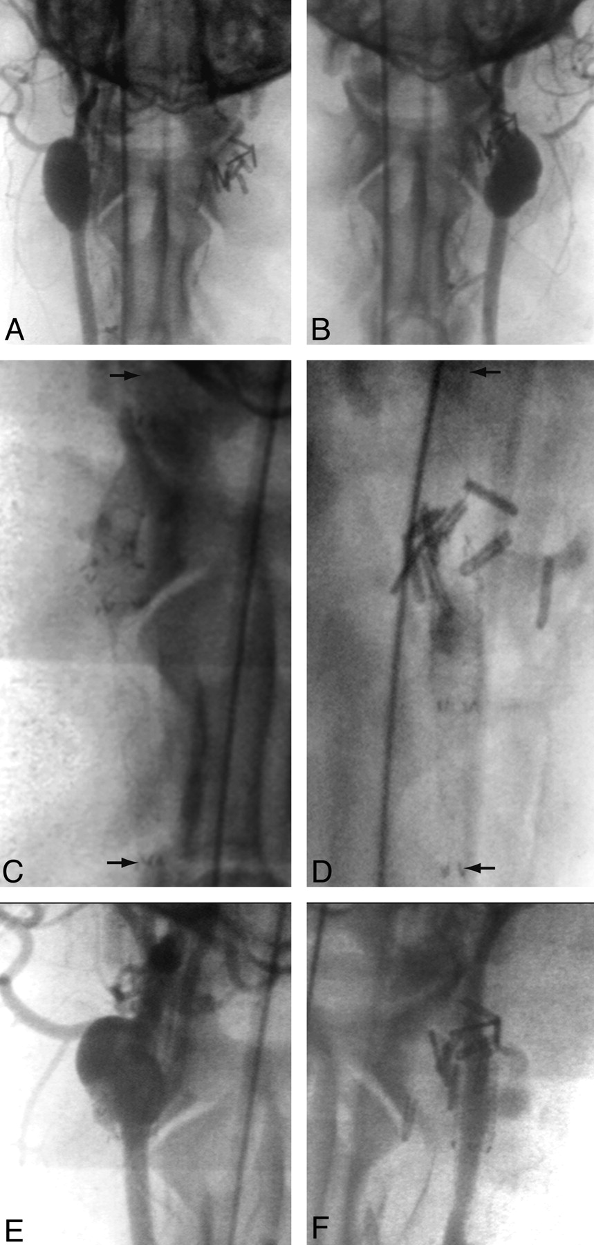
    Angiographic results of treatments. Angiographic demonstration of giant fusiform aneurysm with (A) or without (B) arterial branches, 6 weeks after construction, immediately before treatment. Both have been treated with multiple FD constructs to span the aneurysmal dilation (arrows in C and D). Stent placement in the aneurysm with arterial branches led to treatment failure (E), whereas, in the aneurysm with surgically occluded branches led to almost complete aneurysm thrombosis after FD (F).

  • Fig 3.
    • Download figure
    • Open in new tab
    • Download powerpoint
    Fig 3.

    Residual aneurysm, neointima formation, and leaks. Note patent, open aneurysms (A; corresponding to Fig 2E), when the aneurysm serves as a reservoir for feeding branches. When branches are absent (B) the aneurysm is almost completely occluded, with pouches containing thrombus in various stages or organization (*). A small crescentic remnant, fed by a small leak (curved arrows) is present (**; aneurysm corresponding to Fig 2F). The FDs are covered with neointima both inside and outside (C), and leaks penetrate both neointimal layers covering the stents (D and E). There is good neointimal coverage of the stent struts apposed to the parent artery (E). Note patent arterial branch ostium but partially covered with neointimal tissue and organizing clot (F).

Tables

  • Figures
  • Angiographic and pathologic outcomes after flow diversion in experimental fusiform aneurysms

    AneurysmPatent BranchesAneurysm Size (Length × Width × Height) (mm)No. of FD StentsLength of Deployed Stents (mm in 3.5 mm vessel)Angiographic Outcome% Occlusion at Pathology
    Immediate2 wk12 wk
    1Yes25 × 12.5 × 12.5252 + 52 (104)3330
    2Yes17.5 × 10 × 10152 (52)3330
    3Yes25 × 12.5 × 12.5346 + 30 + 26 (102)3330
    4Yes25 × 12 × 12446 + 40 + 26 + 26 (138)3330
    5No18 × 10 × 12246 + 17 (83)32180
    6No19 × 10 × 12337 + 37 + 26 (100)32180
PreviousNext
Back to top

In this issue

American Journal of Neuroradiology: 32 (11)
American Journal of Neuroradiology
Vol. 32, Issue 11
1 Dec 2011
  • Table of Contents
  • Index by author
Advertisement
Print
Download PDF
Email Article

Thank you for your interest in spreading the word on American Journal of Neuroradiology.

NOTE: We only request your email address so that the person you are recommending the page to knows that you wanted them to see it, and that it is not junk mail. We do not capture any email address.

Enter multiple addresses on separate lines or separate them with commas.
Testing Flow Diverters in Giant Fusiform Aneurysms: A New Experimental Model Can Show Leaks Responsible for Failures
(Your Name) has sent you a message from American Journal of Neuroradiology
(Your Name) thought you would like to see the American Journal of Neuroradiology web site.
CAPTCHA
This question is for testing whether or not you are a human visitor and to prevent automated spam submissions.
Citation Tools
Testing Flow Diverters in Giant Fusiform Aneurysms: A New Experimental Model Can Show Leaks Responsible for Failures
T.E. Darsaut, F. Bing, I. Salazkin, G. Gevry, J. Raymond
American Journal of Neuroradiology Dec 2011, 32 (11) 2175-2179; DOI: 10.3174/ajnr.A2657

Citation Manager Formats

  • BibTeX
  • Bookends
  • EasyBib
  • EndNote (tagged)
  • EndNote 8 (xml)
  • Medlars
  • Mendeley
  • Papers
  • RefWorks Tagged
  • Ref Manager
  • RIS
  • Zotero
Share
Testing Flow Diverters in Giant Fusiform Aneurysms: A New Experimental Model Can Show Leaks Responsible for Failures
T.E. Darsaut, F. Bing, I. Salazkin, G. Gevry, J. Raymond
American Journal of Neuroradiology Dec 2011, 32 (11) 2175-2179; DOI: 10.3174/ajnr.A2657
Reddit logo Twitter logo Facebook logo Mendeley logo
  • Tweet Widget
  • Facebook Like
  • Google Plus One
Purchase

Jump to section

  • Article
    • Abstract
    • ABBREVIATIONS
    • Materials and Methods
    • Results
    • Discussion
    • Conclusions
    • Footnotes
    • References
  • Figures & Data
  • Info & Metrics
  • References
  • PDF

Related Articles

  • No related articles found.
  • PubMed
  • Google Scholar

Cited By...

  • Surgical technique for venous patch aneurysms with no neck in a rabbit model
  • Flow diversion of bifurcation aneurysms is more effective when the jailed branch is occluded: an experimental study in a novel canine model
  • Testing Stenting and Flow Diversion Using a Surgical Elastase-Induced Complex Fusiform Aneurysm Model
  • Compaction of flow diverters improves occlusion of experimental wide-necked aneurysms
  • The Success of Flow Diversion in Large and Giant Sidewall Aneurysms May Depend on the Size of the Defect in the Parent Artery
  • Variable Porosity of the Pipeline Embolization Device in Straight and Curved Vessels: A Guide for Optimal Deployment Strategy
  • Flow diversion to treat aneurysms: the free segment of stent
  • The Varying Porosity of Braided Self-Expanding Stents and Flow Diverters: An Experimental Study
  • Thrombosis Heralding Aneurysmal Rupture: An Exploration of Potential Mechanisms in a Novel Giant Swine Aneurysm Model
  • Flow Diverters Can Occlude Aneurysms and Preserve Arterial Branches: A New Experimental Model
  • Crossref
  • Google Scholar

This article has not yet been cited by articles in journals that are participating in Crossref Cited-by Linking.

More in this TOC Section

  • Diagnostic Performance of High-Resolution Vessel Wall MR Imaging Combined with TOF-MRA in the Follow-up of Intracranial Vertebrobasilar Dissecting Aneurysms after Reconstructive Endovascular Treatment
  • Outcomes with Endovascular Treatment of Patients with M2 Segment MCA Occlusion in the Late Time Window
  • Direct Aspiration versus Combined Technique for Distal Medium-Vessel Occlusions: Comparison on a Human Placenta Model
Show more INTERVENTIONAL

Similar Articles

Advertisement

News and Updates

  • Lucien Levy Best Research Article Award
  • Thanks to our 2022 Distinguished Reviewers
  • Press Releases

Resources

  • Evidence-Based Medicine Level Guide
  • How to Participate in a Tweet Chat
  • AJNR Podcast Archive
  • Ideas for Publicizing Your Research
  • Librarian Resources
  • Terms and Conditions

Opportunities

  • Share Your Art in Perspectives
  • Get Peer Review Credit from Publons
  • Moderate a Tweet Chat

American Society of Neuroradiology

  • Neurographics
  • ASNR Annual Meeting
  • Fellowship Portal
  • Position Statements

© 2023 by the American Society of Neuroradiology | Print ISSN: 0195-6108 Online ISSN: 1936-959X

Powered by HighWire