
Article Figures & Data
Figures
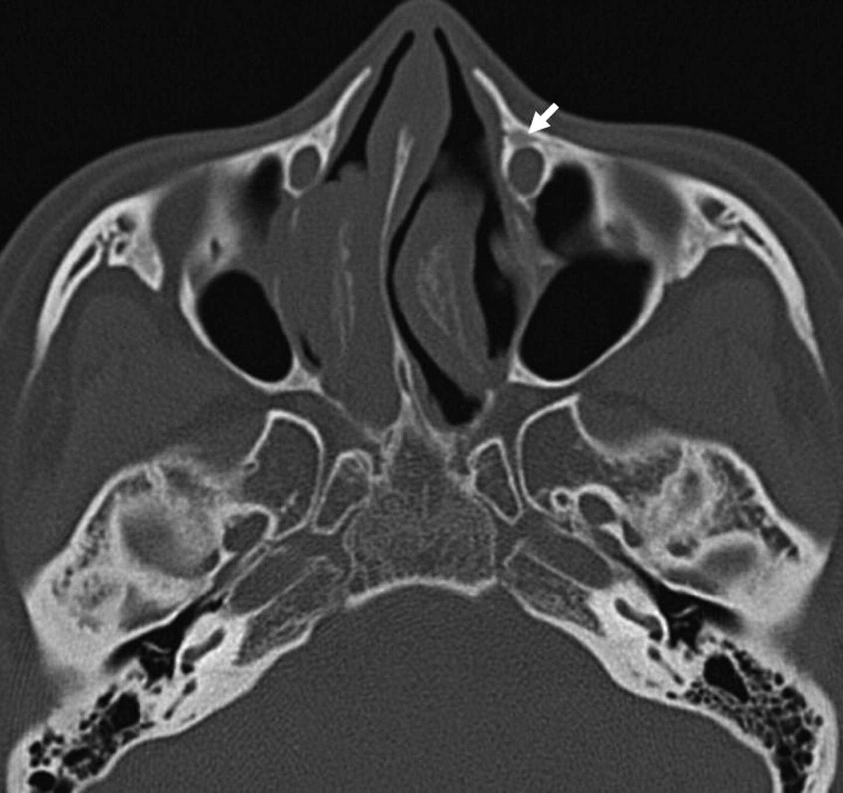
- Fig 1.
Axial CT image shows the typical pagetoid appearance (type I) (arrow) in the left frontal process of the maxilla.
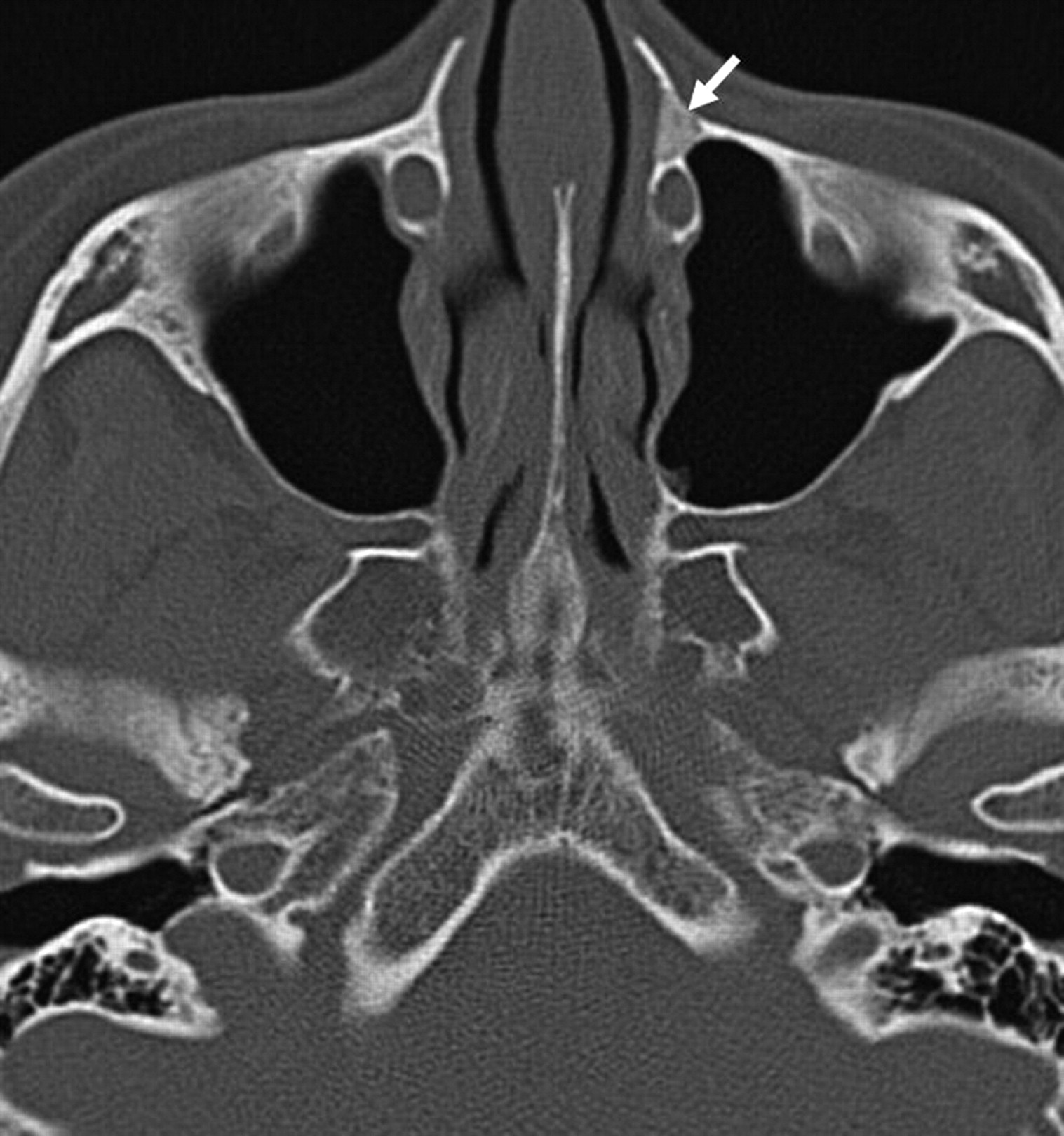
- Fig 2.
Axial CT image shows a ground-glass appearance (type II) (arrow) in the left frontal process of maxilla.
- Fig 3.
Axial CT image shows the cyst-like appearance (type III) (arrow) in the right frontal process of the maxilla.


In this issue


Jump to section
Related Articles
Cited By...
- No citing articles found.