Advertisement

Main menu

  • Home
  • Content
    • Current Issue
    • Publication Preview--Ahead of Print
    • Past Issue Archive
    • Case of the Week Archive
    • Classic Case Archive
    • Case of the Month Archive
    • COVID-19 Content and Resources
  • For Authors
  • About Us
    • About AJNR
    • Editors
    • American Society of Neuroradiology
  • Submit a Manuscript
  • Podcasts
    • Subscribe on iTunes
    • Subscribe on Stitcher
  • More
    • Subscribers
    • Permissions
    • Advertisers
    • Alerts
    • Feedback
  • Other Publications
    • ajnr

User menu

  • Subscribe
  • Alerts
  • Log in

Search

  • Advanced search
American Journal of Neuroradiology

American Journal of Neuroradiology

  • Subscribe
  • Alerts
  • Log in

Advanced Search

  • Home
  • Content
    • Current Issue
    • Publication Preview--Ahead of Print
    • Past Issue Archive
    • Case of the Week Archive
    • Classic Case Archive
    • Case of the Month Archive
    • COVID-19 Content and Resources
  • For Authors
  • About Us
    • About AJNR
    • Editors
    • American Society of Neuroradiology
  • Submit a Manuscript
  • Podcasts
    • Subscribe on iTunes
    • Subscribe on Stitcher
  • More
    • Subscribers
    • Permissions
    • Advertisers
    • Alerts
    • Feedback
  • Follow AJNR on Twitter
  • Visit AJNR on Facebook
  • Follow AJNR on Instagram
  • Join AJNR on LinkedIn
  • RSS Feeds
LetterLetter

Third Cerebellar Hemisphere: An Unusual New Cerebellar Anomaly

S. Agarwal and G. Gathwala
American Journal of Neuroradiology April 2011, 32 (4) E73; DOI: https://doi.org/10.3174/ajnr.A2473
S. Agarwal
  • Find this author on Google Scholar
  • Find this author on PubMed
  • Search for this author on this site
G. Gathwala
  • Find this author on Google Scholar
  • Find this author on PubMed
  • Search for this author on this site

Fig 2.

Fig 2.
  • Download figure
  • Open in new tab
  • Download powerpoint
Fig 2.

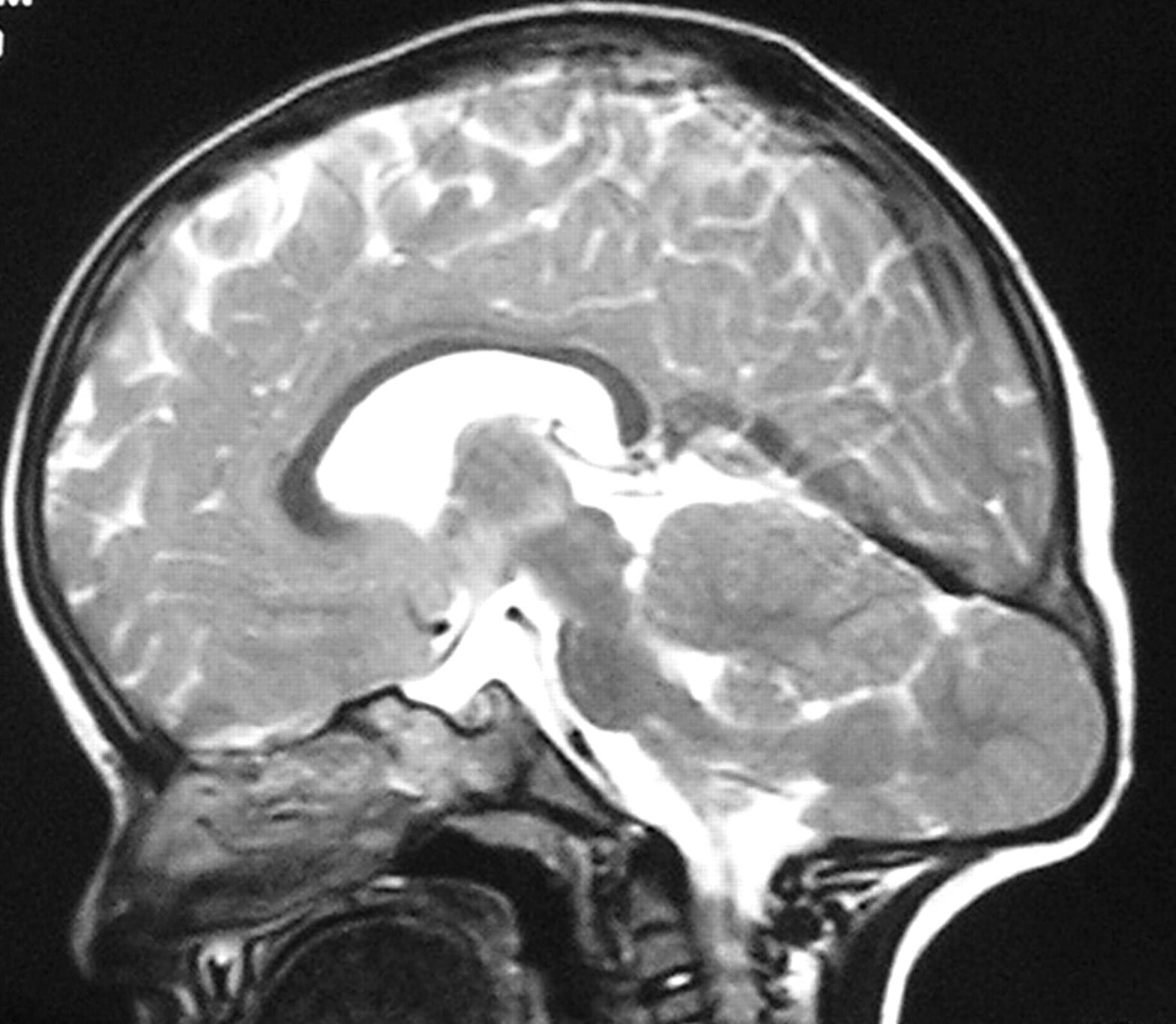
Sagittal T2-weighted MR image reveals a mildly dysplastic third cerebellar hemisphere located within a bony outpouching connected to the medulla, with a thin band of soft tissue of signal intensity similar to that of the cortex. In addition, it also reveals a low-lying torcular herophilli and mild basilar invagination. The visualized supratentorial parenchyma appears normal.

  View article

In this issue

American Journal of Neuroradiology: 44 (3)
American Journal of Neuroradiology
Vol. 44, Issue 3
1 Mar 2023
  • Table of Contents
  • Index by author
  • Complete Issue (PDF)
Print
Download PDF
Email Article

Thank you for your interest in spreading the word on American Journal of Neuroradiology.

NOTE: We only request your email address so that the person you are recommending the page to knows that you wanted them to see it, and that it is not junk mail. We do not capture any email address.

Enter multiple addresses on separate lines or separate them with commas.
Fig 2.
(Your Name) has sent you a message from American Journal of Neuroradiology
(Your Name) thought you would like to see the American Journal of Neuroradiology web site.
CAPTCHA
This question is for testing whether or not you are a human visitor and to prevent automated spam submissions.
Citation Tools
Third Cerebellar Hemisphere: An Unusual New Cerebellar Anomaly
S. Agarwal, G. Gathwala
American Journal of Neuroradiology Apr 2011, 32 (4) E73; DOI: 10.3174/ajnr.A2473

Citation Manager Formats

  • BibTeX
  • Bookends
  • EasyBib
  • EndNote (tagged)
  • EndNote 8 (xml)
  • Medlars
  • Mendeley
  • Papers
  • RefWorks Tagged
  • Ref Manager
  • RIS
  • Zotero
Share
Third Cerebellar Hemisphere: An Unusual New Cerebellar Anomaly
S. Agarwal, G. Gathwala
American Journal of Neuroradiology Apr 2011, 32 (4) E73; DOI: 10.3174/ajnr.A2473
Reddit logo Twitter logo Facebook logo Mendeley logo
  • Tweet Widget
  • Facebook Like
  • Google Plus One

Related Articles

  • No related articles found.
  • PubMed
  • Google Scholar

Cited By...

  • No citing articles found.
  • Crossref
  • Google Scholar

This article has not yet been cited by articles in journals that are participating in Crossref Cited-by Linking.

More in this TOC Section

  • Fair Performance of CT in Diagnosing Unilateral Vocal Fold Paralysis
  • Reply:
  • Regarding “Altered Blood Flow in the Ophthalmic and Internal Carotid Arteries in Patients with Age-Related Macular Degeneration Measured Using Noncontrast MR Angiography at 7T”
Show more LETTERS

Similar Articles

Advertisement

News and Updates

  • Lucien Levy Best Research Article Award
  • Thanks to our 2022 Distinguished Reviewers
  • Press Releases

Resources

  • Evidence-Based Medicine Level Guide
  • How to Participate in a Tweet Chat
  • AJNR Podcast Archive
  • Ideas for Publicizing Your Research
  • Librarian Resources
  • Terms and Conditions

Opportunities

  • Share Your Art in Perspectives
  • Get Peer Review Credit from Publons
  • Moderate a Tweet Chat

American Society of Neuroradiology

  • Neurographics
  • ASNR Annual Meeting
  • Fellowship Portal
  • Position Statements

© 2023 by the American Society of Neuroradiology | Print ISSN: 0195-6108 Online ISSN: 1936-959X

Powered by HighWire