Advertisement

Main menu

  • Home
  • Content
    • Current Issue
    • Accepted Manuscripts
    • Article Preview
    • Past Issue Archive
    • Video Articles
    • AJNR Case Collection
    • Case of the Week Archive
    • Case of the Month Archive
    • Classic Case Archive
  • Special Collections
    • AJNR Awards
    • ASNR Foundation Special Collection
    • Most Impactful AJNR Articles
    • Photon-Counting CT
    • Spinal CSF Leak Articles (Jan 2020-June 2024)
  • Multimedia
    • AJNR Podcasts
    • AJNR SCANtastic
    • Trainee Corner
    • MRI Safety Corner
    • Imaging Protocols
  • For Authors
    • Submit a Manuscript
    • Submit a Video Article
    • Submit an eLetter to the Editor/Response
    • Manuscript Submission Guidelines
    • Statistical Tips
    • Fast Publishing of Accepted Manuscripts
    • Graphical Abstract Preparation
    • Imaging Protocol Submission
    • Author Policies
  • About Us
    • About AJNR
    • Editorial Board
    • Editorial Board Alumni
  • More
    • Become a Reviewer/Academy of Reviewers
    • Subscribers
    • Permissions
    • Alerts
    • Feedback
    • Advertisers
    • ASNR Home

User menu

  • Alerts
  • Log in

Search

  • Advanced search
American Journal of Neuroradiology

American Journal of Neuroradiology

ASHNR American Society of Functional Neuroradiology ASHNR American Society of Pediatric Neuroradiology ASSR
  • Alerts
  • Log in

Advanced Search

  • Home
  • Content
    • Current Issue
    • Accepted Manuscripts
    • Article Preview
    • Past Issue Archive
    • Video Articles
    • AJNR Case Collection
    • Case of the Week Archive
    • Case of the Month Archive
    • Classic Case Archive
  • Special Collections
    • AJNR Awards
    • ASNR Foundation Special Collection
    • Most Impactful AJNR Articles
    • Photon-Counting CT
    • Spinal CSF Leak Articles (Jan 2020-June 2024)
  • Multimedia
    • AJNR Podcasts
    • AJNR SCANtastic
    • Trainee Corner
    • MRI Safety Corner
    • Imaging Protocols
  • For Authors
    • Submit a Manuscript
    • Submit a Video Article
    • Submit an eLetter to the Editor/Response
    • Manuscript Submission Guidelines
    • Statistical Tips
    • Fast Publishing of Accepted Manuscripts
    • Graphical Abstract Preparation
    • Imaging Protocol Submission
    • Author Policies
  • About Us
    • About AJNR
    • Editorial Board
    • Editorial Board Alumni
  • More
    • Become a Reviewer/Academy of Reviewers
    • Subscribers
    • Permissions
    • Alerts
    • Feedback
    • Advertisers
    • ASNR Home
  • Follow AJNR on Twitter
  • Visit AJNR on Facebook
  • Follow AJNR on Instagram
  • Join AJNR on LinkedIn
  • RSS Feeds

AJNR is seeking candidates for the AJNR Podcast Editor. Read the position description.

Case ReportCase Report

Extensive Intracranial Juvenile Xanthogranulomas

P. Lalitha, M.Ch.B. Reddy and K.J. Reddy
American Journal of Neuroradiology August 2011, 32 (7) E132-E133; DOI: https://doi.org/10.3174/ajnr.A2209
P. Lalitha
  • Find this author on Google Scholar
  • Find this author on PubMed
  • Search for this author on this site
M.Ch.B. Reddy
  • Find this author on Google Scholar
  • Find this author on PubMed
  • Search for this author on this site
K.J. Reddy
  • Find this author on Google Scholar
  • Find this author on PubMed
  • Search for this author on this site
  • Article
  • Figures & Data
  • Info & Metrics
  • Responses
  • References
  • PDF
Loading

Abstract

SUMMARY: JXG is a disorder classified under histiocytosis. It usually affects children and commonly presents with skin lesions. Intracranial lesions are uncommon and usually solitary. We present the case of a child who had extensive intracranial involvement with multiple enhancing solid lesions in the cerebellum, brain stem, thalami, and bilateral cerebral hemispheres on MR imaging.

Abbreviations

JXG
juvenile xanthogranuloma

JXG is a part of the spectrum of histiocytosis. Unlike the more commonly seen cutaneous lesions, intracranial lesions do not respond to medical management and require surgery.

Case Report

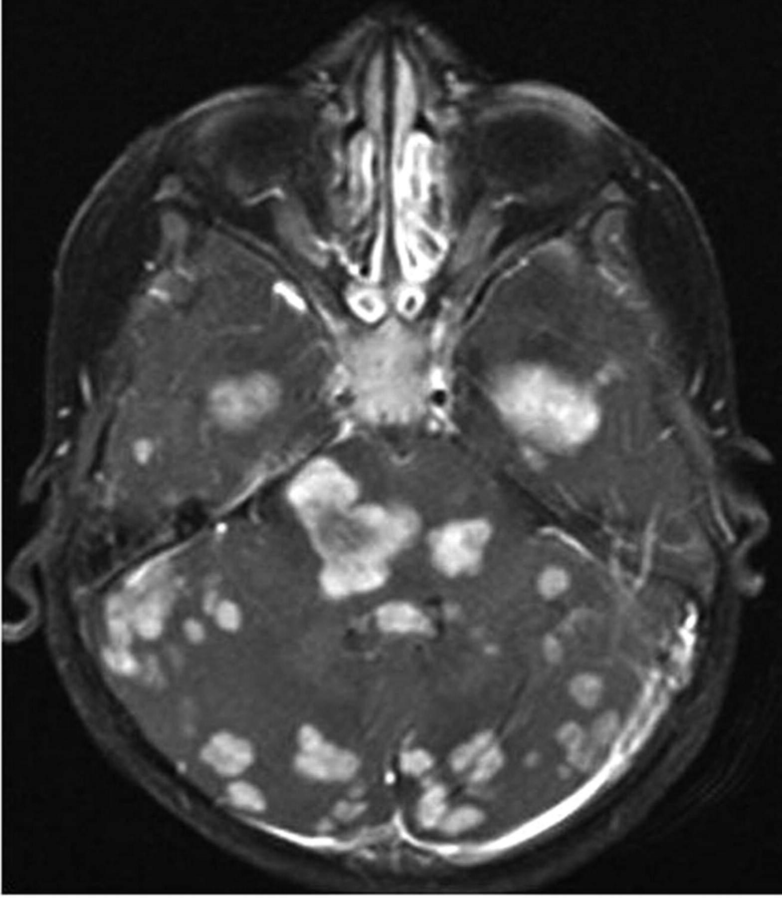
A 3-year-old child was admitted with generalized seizures, headache, dysarthria, and gait disturbance with weakness of the limbs since 1 year of age. He had a history of extensive macula-papular skin lesions from 8 months of age. Biopsy of the skin lesions revealed JXG. At that time, findings of sonography of the abdomen and a CT scan of the brain were normal. A contrast-enhanced MR imaging of the brain was requested in view of presenting symptoms. It revealed extensive intracranial involvement, with multiple lesions in the cerebellum, brain stem, thalami, and cerebral hemispheres. The lesions were of varying sizes. They were isointense on T1-weighted images and mildly hyperintense on T2-weighted images, with a mild degree of perilesional edema (Fig 1). The larger lesions were located in both hippocampi. All the lesions showed homogeneous contrast enhancement (Figs 2–5). Mild supratentorial hydrocephalous was seen secondary to compression and distortion of the aqueduct by the brain stem lesions. The results of a biopsy of 1 of the lesions revealed xanthogranulomas.

Fig 1.
  • Download figure
  • Open in new tab
  • Download powerpoint
Fig 1.

Axial T2-weighted MR image showing isohyperintense lesions in both thalami and frontoparietal lobes with mild perilesional edema.

Fig 2.
  • Download figure
  • Open in new tab
  • Download powerpoint
Fig 2.

Axial contrast-enhanced T1-weighted image showing multiple enhancing cerebellar, brain stem, and parenchymal lesions.

Fig 3.
  • Download figure
  • Open in new tab
  • Download powerpoint
Fig 3.

Axial contrast-enhanced T1-weighted image showing enhancing solid lesions in both hippocampi, brain stem, and temporal lobes.

Fig 4.
  • Download figure
  • Open in new tab
  • Download powerpoint
Fig 4.

Axial contrast-enhanced T1-weighted image showing enhancing solid lesions in both frontoparietal lobes and the subependymal region.

Fig 5.
  • Download figure
  • Open in new tab
  • Download powerpoint
Fig 5.

Sagittal contrast-enhanced T1-weighted image showing enhancing solid lesions in the brain stem, cerebellum, and cerebral parenchyma.

Discussion

JXG is disorder of histiocytes, which is classified under histiocytosis syndromes in children. Histiocytosis refers to a group of disorders characterized by abundance of histiocytes (tissue macrophages) within the affected tissues. It is usually diagnosed in children younger than 10 years of age.

The World Health Organization has proposed a classification for histiocytosis syndromes in children, with 3 main classes1: Class I includes Langerhans cell histiocytosis. Class II includes histiocytosis of mononuclear phagocytes other than Langerhans cells, familial and reactive hemophagocytic lymphohistiocytosis, sinus histiocytosis with massive lymphadenopathy, Rosai-Dorfman disease, JXG, and reticulohistiocytoma. Class III includes malignant histiocytic disorders, acute monocytic leukemia, malignant histiocytosis, and true histiocytic lymphoma. According to the Histiocyte Society classification of histiocytosis syndromes, JXG is classified under dendritic cell−related disorders.1

JXG is a disorder that commonly affects the skin. Other organs like the liver, spleen, kidneys, lungs, and musculoskeletal system can also be involved. Intracranial involvement is uncommon and has been reported previously.2,3 Solitary intracranial lesions are more commonly seen than multiple lesions. However, such an extensive dural and parenchymal involvement as seen in our case is extremely uncommon. Intracranial JXG is usually seen secondary to skin or other organ involvement that has been there for a considerable time.2 The lesions may arise from mesenchymal stem cells, dura, blood vessel walls, or from the perivascular soft tissues. Intracranial lesions may be seen in the cerebral parenchyma, meninges, choroid plexus, or cranial nerves. Clinically, the affected children may be asymptomatic or may present with seizures, ataxia, weakness, or growth retardation. Intracranial involvement in JXG is associated with increased mortality and morbidity. The intracranial lesions have a tendency to grow slowly and must be followed up.4 Treatment can be surgical or nonsurgical. Isolated symptomatic lesions can undergo surgery. Conservative management with steroid therapy provides symptomatic relief, and radiotherapy has also been tried in multiple lesions.

References

  1. 1.↵
    1. Tebbi CK,
    2. Arceci RJ,
    3. Loew TW
    . Histiocytosis. http://emedicine.medscape.com/article/958026-overview. Accessed July 30, 2009
  2. 2.↵
    1. Sun LP,
    2. Jin HM,
    3. Yang B,
    4. et al
    . Intracranial solitary juvenile xanthogranuloma in an infant. World J Pediatr 2009;5:71–73. Epub 2009 Jan 27
    CrossRefPubMed
  3. 3.↵
    1. Chidambaram B,
    2. Santosh V
    . Giant orbital and intracranial xanthogranuloma: a short report. Neurol India 2001;49:208–10
    PubMed
  4. 4.↵
    1. Kasliwal MK,
    2. Suri A,
    3. Rishi A,
    4. et al
    . Symptomatic bilateral cerebellar mass lesions: an unusual presentation of intracranial xanthogranuloma. J Clin Neurosci 2008;15:1401–04. Epub 2008 Oct 25
    CrossRefPubMed
  • Received May 13, 2010.
  • Accepted after revision May 18, 2010.
  • Copyright © American Society of Neuroradiology
View Abstract
PreviousNext
Back to top

In this issue

American Journal of Neuroradiology: 32 (7)
American Journal of Neuroradiology
Vol. 32, Issue 7
1 Aug 2011
  • Table of Contents
  • Index by author
Advertisement
Print
Download PDF
Email Article

Thank you for your interest in spreading the word on American Journal of Neuroradiology.

NOTE: We only request your email address so that the person you are recommending the page to knows that you wanted them to see it, and that it is not junk mail. We do not capture any email address.

Enter multiple addresses on separate lines or separate them with commas.
Extensive Intracranial Juvenile Xanthogranulomas
(Your Name) has sent you a message from American Journal of Neuroradiology
(Your Name) thought you would like to see the American Journal of Neuroradiology web site.
CAPTCHA
This question is for testing whether or not you are a human visitor and to prevent automated spam submissions.
Cite this article
P. Lalitha, M.Ch.B. Reddy, K.J. Reddy
Extensive Intracranial Juvenile Xanthogranulomas
American Journal of Neuroradiology Aug 2011, 32 (7) E132-E133; DOI: 10.3174/ajnr.A2209

Citation Manager Formats

  • BibTeX
  • Bookends
  • EasyBib
  • EndNote (tagged)
  • EndNote 8 (xml)
  • Medlars
  • Mendeley
  • Papers
  • RefWorks Tagged
  • Ref Manager
  • RIS
  • Zotero
0 Responses
Respond to this article
Share
Bookmark this article
Extensive Intracranial Juvenile Xanthogranulomas
P. Lalitha, M.Ch.B. Reddy, K.J. Reddy
American Journal of Neuroradiology Aug 2011, 32 (7) E132-E133; DOI: 10.3174/ajnr.A2209
del.icio.us logo Twitter logo Facebook logo Mendeley logo
  • Tweet Widget
  • Facebook Like
  • Google Plus One
Purchase

Jump to section

  • Article
    • Abstract
    • Abbreviations
    • Case Report
    • Discussion
    • References
  • Figures & Data
  • Info & Metrics
  • Responses
  • References
  • PDF

Related Articles

  • No related articles found.
  • PubMed
  • Google Scholar

Cited By...

  • Neuroimaging in Pediatric Patients with Juvenile Xanthogranuloma of the CNS
  • Imaging Features of Juvenile Xanthogranuloma of the Pediatric Head and Neck
  • Consensus guidelines for the diagnosis and clinical management of Erdheim-Chester disease
  • Crossref (15)
  • Google Scholar

This article has been cited by the following articles in journals that are participating in Crossref Cited-by Linking.

  • Consensus guidelines for the diagnosis and clinical management of Erdheim-Chester disease
    Eli L. Diamond, Lorenzo Dagna, David M. Hyman, Giulio Cavalli, Filip Janku, Juvianee Estrada-Veras, Marina Ferrarini, Omar Abdel-Wahab, Mark L. Heaney, Paul J. Scheel, Nancy K. Feeley, Elisabetta Ferrero, Kenneth L. McClain, Augusto Vaglio, Thomas Colby, Laurent Arnaud, Julien Haroche
    Blood 2014 124 4
  • The clinical diagnosis and management options for intracranial juvenile xanthogranuloma in children: based on four cases and another 39 patients in the literature
    Baocheng Wang, Huiming Jin, Yang Zhao, Jie Ma
    Acta Neurochirurgica 2016 158 7
  • Imaging Features of Juvenile Xanthogranuloma of the Pediatric Head and Neck
    D. T. Ginat, S. O. Vargas, V. M. Silvera, M. S. Volk, B. A. Degar, C. D. Robson
    American Journal of Neuroradiology 2016 37 5
  • Fatal juvenile xanthogranuloma presenting as a sellar lesion: case report and literature review
    Sherise D. Ferguson, Steven G. Waguespack, Lauren A. Langford, Joann L. Ater, Ian E. McCutcheon
    Child's Nervous System 2015 31 5
  • Diagnostic and management difficulties in a case of multiple intracranial juvenile xanthogranuloma
    Kentaro Chiba, Yasuo Aihara, Seiichiro Eguchi, Masahiko Tanaka, Takashi Komori, Yoichi Nakazato, Yoshikazu Okada
    Child's Nervous System 2013 29 6
  • Cerebral and Cutaneous Involvements of Xanthoma Disseminatum Successfully Treated with an Interleukin-1 Receptor Antagonist: A Case Report and Minireview
    Mario Campero, Sebastián Campero, Julia Guerrero, Achille Aouba, Alex Castro
    Dermatology 2016 232 2
  • Elevated taurine and glutamate in cerebral juvenile xanthogranuloma on MR spectroscopy
    Ken Matsubara, Harushi Mori, Nozomi Hirai, Kumi Yasukawa, Takafumi Honda, Jun-ichi Takanashi
    Brain and Development 2016 38 10
  • Juvenile xanthogranuloma of central nervous system: Imaging of two cases report and literature review
    Wangqiang Chen, Yue Cheng, Shengfa Zhou, Yu Chen, Xuming Chen, Shuang Xia
    Radiology of Infectious Diseases 2017 4 3
  • Histiocytic Disorders
    Eli L. Diamond, Benjamin H. Durham
    2018
  • Intradural Juvenile Xanthogranuloma with Involvement of Multiple Nerve Roots: A Case Report and Review of the Literature
    Cody Wolfe, Tarek Y. El Ahmadieh, Salah G. Aoun, Awais Z. Vance, Kimmo J. Hatanpaa, Bryan Wohlfeld
    World Neurosurgery 2018 119

More in this TOC Section

  • Atypical Diffusion-Restricted Lesion in 5-Fluorouracil Encephalopathy
  • Dural Infantile Hemangioma Masquerading as a Skull Vault Lesion
  • Multimodal Imaging of Spike Propagation: A Technical Case Report
Show more Case Reports

Similar Articles

Advertisement

Indexed Content

  • Current Issue
  • Accepted Manuscripts
  • Article Preview
  • Past Issues
  • Editorials
  • Editor's Choice
  • Fellows' Journal Club
  • Letters to the Editor
  • Video Articles

Cases

  • Case Collection
  • Archive - Case of the Week
  • Archive - Case of the Month
  • Archive - Classic Case

Special Collections

  • AJNR Awards
  • ASNR Foundation Special Collection
  • Most Impactful AJNR Articles
  • Photon-Counting CT
  • Spinal CSF Leak Articles (Jan 2020-June 2024)

More from AJNR

  • Trainee Corner
  • Imaging Protocols
  • MRI Safety Corner

Multimedia

  • AJNR Podcasts
  • AJNR Scantastics

Resources

  • Turnaround Time
  • Submit a Manuscript
  • Submit a Video Article
  • Submit an eLetter to the Editor/Response
  • Manuscript Submission Guidelines
  • Statistical Tips
  • Fast Publishing of Accepted Manuscripts
  • Graphical Abstract Preparation
  • Imaging Protocol Submission
  • Evidence-Based Medicine Level Guide
  • Publishing Checklists
  • Author Policies
  • Become a Reviewer/Academy of Reviewers
  • News and Updates

About Us

  • About AJNR
  • Editorial Board
  • Editorial Board Alumni
  • Alerts
  • Permissions
  • Not an AJNR Subscriber? Join Now
  • Advertise with Us
  • Librarian Resources
  • Feedback
  • Terms and Conditions
  • AJNR Editorial Board Alumni

American Society of Neuroradiology

  • Not an ASNR Member? Join Now

© 2025 by the American Society of Neuroradiology All rights, including for text and data mining, AI training, and similar technologies, are reserved.
Print ISSN: 0195-6108 Online ISSN: 1936-959X

Powered by HighWire