
Article Figures & Data
Figures
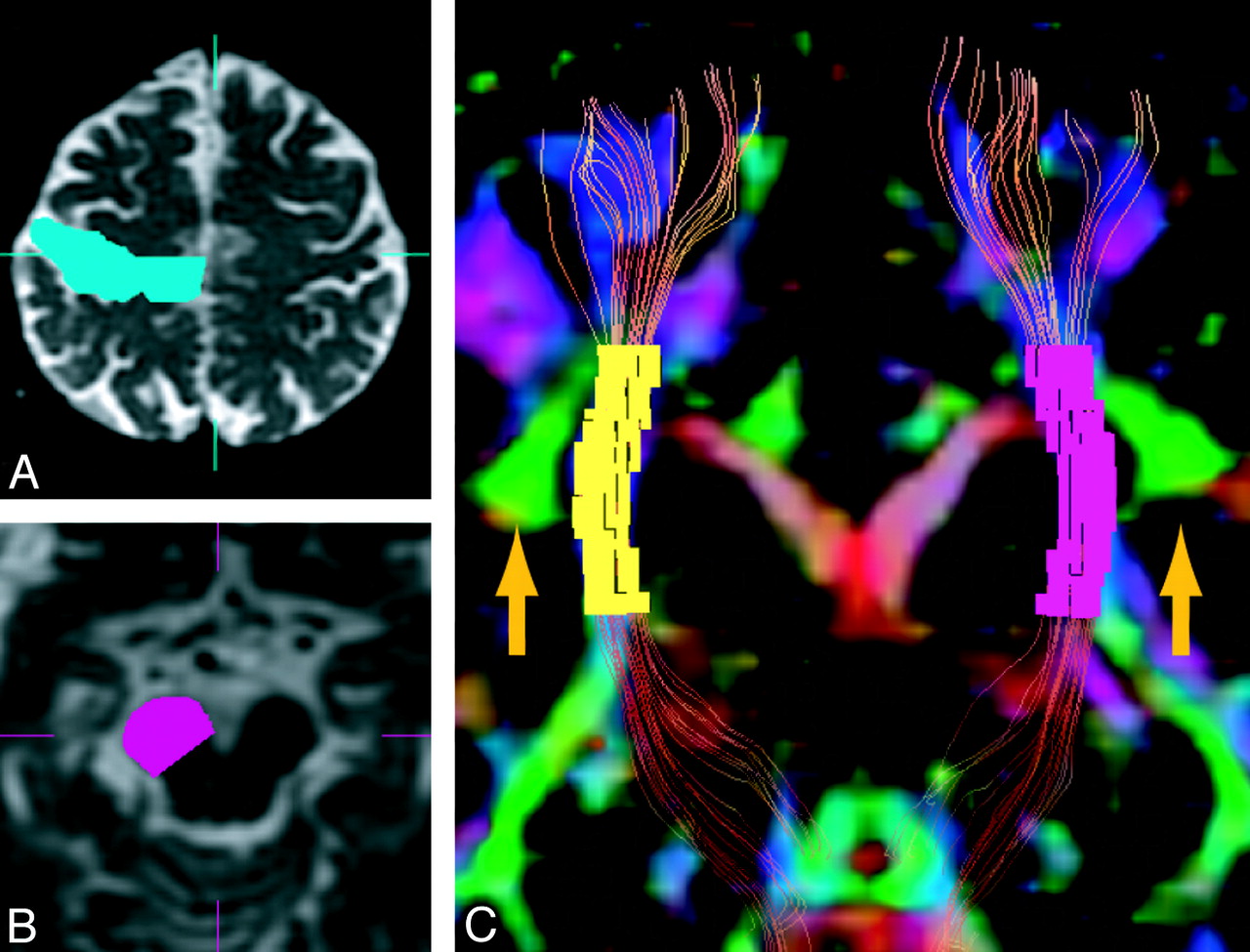
- Fig 1.
Voxelization of part of the CST by tract-specific analysis. Tractographic images of the CST were generated by using the 2-ROI method of seed ROI in the motor cortex (A) and target ROI in the cerebral peduncle (B). C, Tractography of the CST is shown with a color-coded FA map (coronal section). Voxelization along the CST was performed between the uppermost part of the SLF (green area, arrow) and the posterior limb of the internal capsule by using dTV software.
- Fig 2.
Visual evaluation of anisotropy color-coding tractography of the CST. Anisotropy color-coding tractography of the CST in a control subject (A) and a patient with iNPH (B) is shown, with b = 0 images. The areas defined by the yellow squares in A and B were used for visual evaluation in C and D. C, Type A (low anisotropy and rough). D, Type B (high anisotropy and straight).
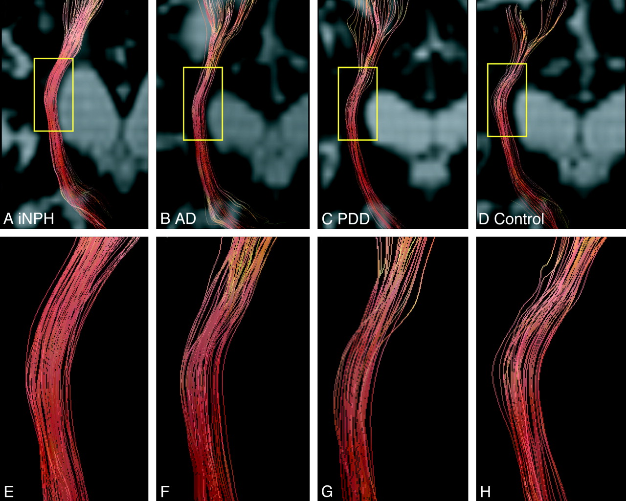
- Fig 3.
Examples of anisotropy color-coding tractography of the CST. Anisotropy color-coding tractography of the CST in patients with iNPH (Evans index: 0.31; A), AD (0.33; B), PDD (0.33; C), and a control subject (0.31; D) are shown, with b = 0 images. The areas defined by the yellow squares in A, B, C, and D are shown in E, F, G, and H, respectively.


Tables
- Table 1:
Demographic and clinical data of patients with iNPH, AD, or PDD and control subjects
Characteristic A, iNPH (n = 18) B, AD (n = 11) C, PDD (n = 11) D, Control (n = 19) P Post Hoc Significance Age (yr) 77.3 ± 4.7 72.1 ± 5.5 75.6 ± 5.5 75.6 ± 5.5 .222 Sex (M:F) 7:11 5:6 6:5 7:12 .805 MMSE score 22.1 ± 3.7 18.3 ± 6.7 22.4 ± 2.7 28.6 ± 0.0 <.001 A, B, C < Da Evans index 0.37 ± 0.04 0.29 ± 0.03 0.29 ± 0.04 0.28 ± 0.03 <.001 A > B, C, Da Vascular risk factor Hypertension (%) 27.8 36.4 18.2 42.1 .561 Diabetes mellitus (%) 16.7 18.2 0.0 0 .157 Dyslipidemia (%) 22.2 27.3 18.2 0 .168 -
Note:—Data are presented as means ± SD.
a P < .001.
-
A, iNPH (n = 18) B, AD (n = 11) C, PDD (n = 11) D, Control (n = 19) P Post Hoc Significance Extent of leukoaraiosis (Fazekas classification) 2.06 ± 0.87 1.64 ± 0.92 1.27 ± 0.90 0.89 ± 0.81 .003 A > C, Da Tract-specific analysis FA value 0.62 ± 0.03 0.54 ± 0.04 0.55 ± 0.04 0.55 ± 0.04 <.001 A > B, C, Db Axial eigenvalue 1.65 ± 0.10 1.49 ± 0.07 1.42 ± 0.08 1.43 ± 0.07 <.001 A > B, C, Db Radial eigenvalue 0.53 ± 0.08 0.56 ± 0.05 0.54 ± 0.05 0.54 ± 0.04 .422 Visual evaluation of tractography Pattern B, n (%) 17 (94.4) 3 (27.2) 1 (9.1) 1 (5.3) <.001 A > B, C, Db -
Note:—Data are presented as means ± SD.
a P < .05.
b P < .001.
-
Variable Sensitivity Specificity Positive Predictive Value Negative Predictive Value Cutoff Value of Each Measure Area Under Curve FA value Compared with AD 94 82 89 90 0.59 0.92 Compared with PDD 94 72 85 89 0.59 0.89 Compared with control 94 84 85 94 0.59 0.91 Compared with AD, PDD, and control 94 80 68 97 0.59 0.91 Axial eigenvalue Compared with AD 94 82 89 90 1.52 0.95 Compared with PDD 100 82 90 100 1.48 0.96 Compared with control 100 84 86 100 1.50 0.98 Compared with AD, PDD, and control 94 83 71 97 1.52 0.97 -
Note:—Data (except for the cutoffs of FA values and axial eigenvalues) are shown as percentages. Receiver operating characteristic analysis was used to determine the optimum cutoff values for evaluating the usefulness of FA values and axial eigenvalues in differentiating iNPH from AD, PDD, and controls.
-
In this issue


Jump to section
Related Articles
Cited By...
- Differential Diagnosis of Normal Pressure Hydrocephalus by MRI Mean Diffusivity Histogram Analysis
- Diffusion tensor imaging in parkinsonian syndromes: A systematic review and meta-analysis
- Different Patterns of Fornix Damage in Idiopathic Normal Pressure Hydrocephalus and Alzheimer Disease
- White Matter Alteration in Idiopathic Normal Pressure Hydrocephalus: Tract-Based Spatial Statistics Study