Advertisement

Main menu

  • Home
  • Content
    • Current Issue
    • Accepted Manuscripts
    • Article Preview
    • Past Issue Archive
    • Video Articles
    • AJNR Case Collection
    • Case of the Week Archive
    • Case of the Month Archive
    • Classic Case Archive
  • Special Collections
    • AJNR Awards
    • ASNR Foundation Special Collection
    • Most Impactful AJNR Articles
    • Photon-Counting CT
    • Spinal CSF Leak Articles (Jan 2020-June 2024)
  • Multimedia
    • AJNR Podcasts
    • AJNR SCANtastic
    • Trainee Corner
    • MRI Safety Corner
    • Imaging Protocols
  • For Authors
    • Submit a Manuscript
    • Submit a Video Article
    • Submit an eLetter to the Editor/Response
    • Manuscript Submission Guidelines
    • Statistical Tips
    • Fast Publishing of Accepted Manuscripts
    • Graphical Abstract Preparation
    • Imaging Protocol Submission
    • Author Policies
  • About Us
    • About AJNR
    • Editorial Board
    • Editorial Board Alumni
  • More
    • Become a Reviewer/Academy of Reviewers
    • Subscribers
    • Permissions
    • Alerts
    • Feedback
    • Advertisers
    • ASNR Home

User menu

  • Alerts
  • Log in

Search

  • Advanced search
American Journal of Neuroradiology

American Journal of Neuroradiology

ASHNR American Society of Functional Neuroradiology ASHNR American Society of Pediatric Neuroradiology ASSR
  • Alerts
  • Log in

Advanced Search

  • Home
  • Content
    • Current Issue
    • Accepted Manuscripts
    • Article Preview
    • Past Issue Archive
    • Video Articles
    • AJNR Case Collection
    • Case of the Week Archive
    • Case of the Month Archive
    • Classic Case Archive
  • Special Collections
    • AJNR Awards
    • ASNR Foundation Special Collection
    • Most Impactful AJNR Articles
    • Photon-Counting CT
    • Spinal CSF Leak Articles (Jan 2020-June 2024)
  • Multimedia
    • AJNR Podcasts
    • AJNR SCANtastic
    • Trainee Corner
    • MRI Safety Corner
    • Imaging Protocols
  • For Authors
    • Submit a Manuscript
    • Submit a Video Article
    • Submit an eLetter to the Editor/Response
    • Manuscript Submission Guidelines
    • Statistical Tips
    • Fast Publishing of Accepted Manuscripts
    • Graphical Abstract Preparation
    • Imaging Protocol Submission
    • Author Policies
  • About Us
    • About AJNR
    • Editorial Board
    • Editorial Board Alumni
  • More
    • Become a Reviewer/Academy of Reviewers
    • Subscribers
    • Permissions
    • Alerts
    • Feedback
    • Advertisers
    • ASNR Home
  • Follow AJNR on Twitter
  • Visit AJNR on Facebook
  • Follow AJNR on Instagram
  • Join AJNR on LinkedIn
  • RSS Feeds

AJNR is seeking candidates for the AJNR Podcast Editor. Read the position description.

Research ArticleResearch Perspectives
Open Access

MR Imaging Features of High-Grade Gliomas in Murine Models: How They Compare with Human Disease, Reflect Tumor Biology, and Play a Role in Preclinical Trials

A.R. Borges, P. Lopez-Larrubia, J.B. Marques and S.G. Cerdan
American Journal of Neuroradiology January 2012, 33 (1) 24-36; DOI: https://doi.org/10.3174/ajnr.A2959
A.R. Borges
  • Find this author on Google Scholar
  • Find this author on PubMed
  • Search for this author on this site
P. Lopez-Larrubia
  • Find this author on Google Scholar
  • Find this author on PubMed
  • Search for this author on this site
J.B. Marques
  • Find this author on Google Scholar
  • Find this author on PubMed
  • Search for this author on this site
S.G. Cerdan
  • Find this author on Google Scholar
  • Find this author on PubMed
  • Search for this author on this site
  • Article
  • Figures & Data
  • Info & Metrics
  • Responses
  • References
  • PDF
Loading

Article Figures & Data

Figures

  • Tables
  • Fig 1.
    • Download figure
    • Open in new tab
    • Download powerpoint
    Fig 1.

    Dedicated MR imaging magnet for small animals and scanning devices. A, 7.4T Bruker Pharmascan (Bruker BioSpin, Rheinstetten, Germany) with a 160-mm horizontal bore (small arrows). A dedicated fixation device for mice brain studies is seen in place, equipped with continuous isofluorane anesthesia (long arrow), respiratory monitoring, and a thermostatic water mat set for 37°C (curved arrows). B, Dedicated mouse head coil (22-mm diameter). C, In-house built poly-methyl-methacrylate bed and fixation device with a Swiss mouse in place.
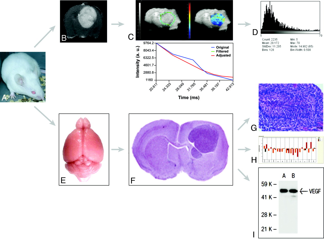
  • Fig 2.
    • Download figure
    • Open in new tab
    • Download powerpoint
    Fig 2.

    Flow chart for integrated MR imaging, histology, and genomic and proteomic approaches in models of HGG. A, Representative Swiss mouse. B, Gadolinium-enhanced axial T1-weighted image shows an implanted Gl261 HGG. C, Parametric T2*map of the tumor in B. D, Parametric histogram of T2* values of C. E, Isolated normal brain from a representative Swiss mouse. F, Representative hematoxylin-eosin–stained section across a formalin-fixed paraffin-embedded mouse brain carrying an implanted HGG. Reproduced from Lal et al17 with permission from the Journal of Neurosurgery Publishing Group. G, Microscopic view (×40) of a fixed and stained section across an HGG (small black bar on the lower right corner represents 50 μm) shows pseudopalisading necrosis characteristic of HGG. Reproduced from Collier et al18 with permission from the American Association for Cancer Research. H, Gene-expression profiling (RT qPCR results) from a representative Gl261 HGG. I, Representative VEGF-A protein expression as detected by Western blot analysis.
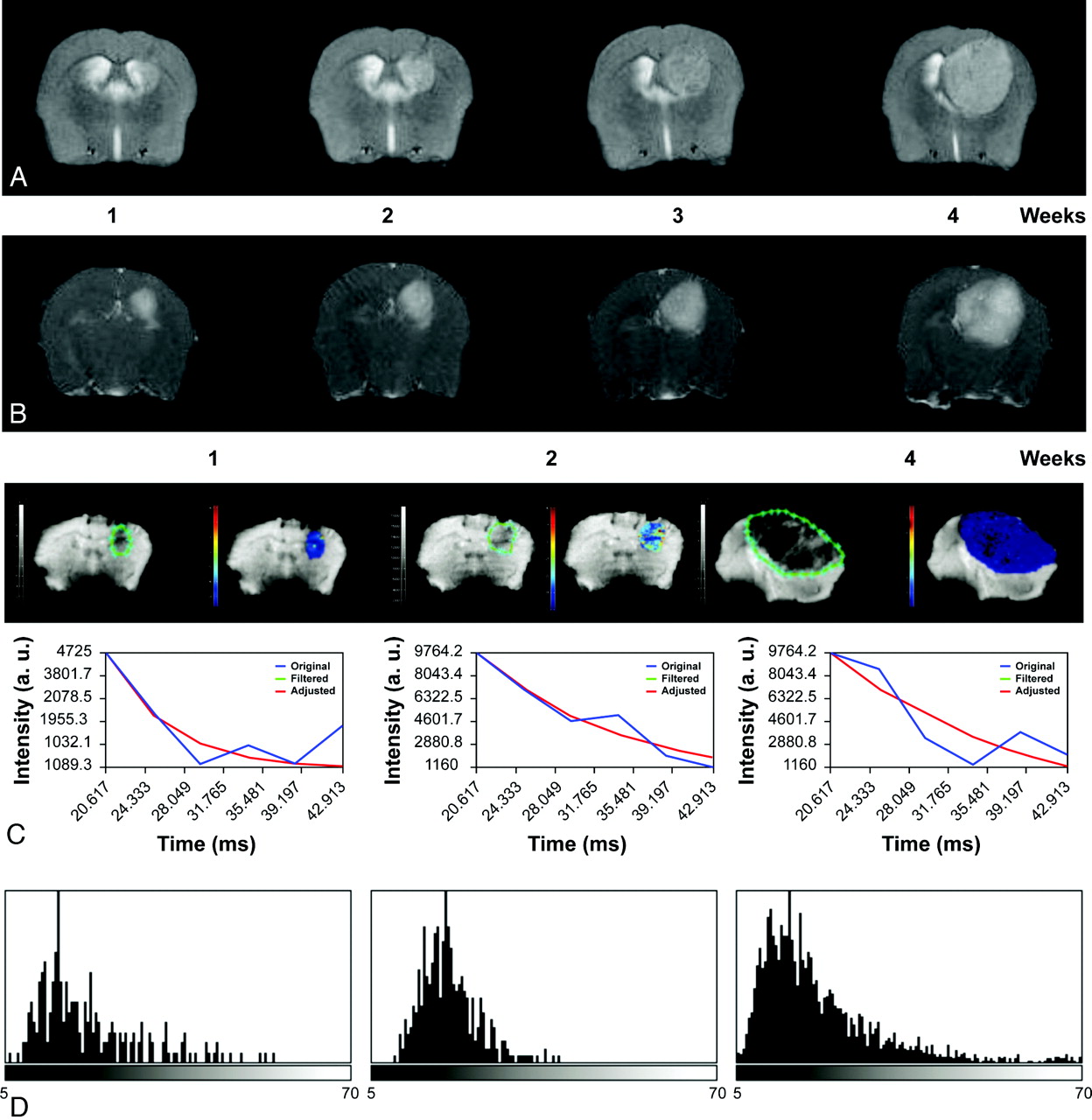
  • Fig 3.
    • Download figure
    • Open in new tab
    • Download powerpoint
    Fig 3.

    Serial axial T2-weighted (A) and contrast-enhanced T1-weighted (B) images of a Gl261 C57/Bl6 mouse model of HGG, showing tumor growth and increased enhancement over a period of 4 weeks. C, ROIs delineating the tumor (green limit) 1, 2, and 4 weeks after implantation; and nonlinear fitting (red) of signal intensity versus TE (blue) in representative pixel and parametric T2* maps depicting T2* values in the selected ROI. D, T2* histograms nicely depict the temporal variation of T2* values with tumor progression modified by small differences in oxygen blood saturation reflected by the paramagnetic effect of deoxyhemoglobin.
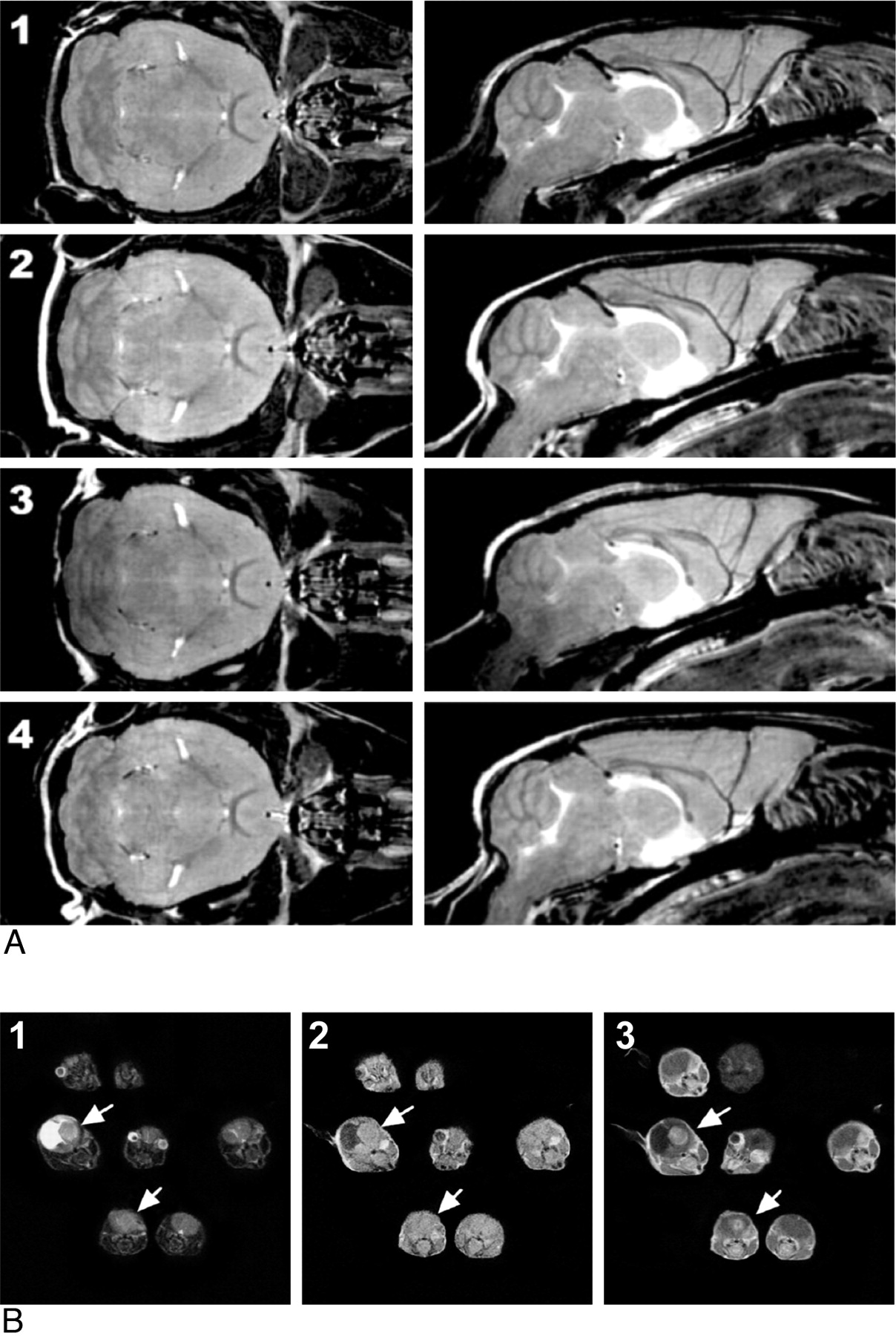
  • Fig 4.
    • Download figure
    • Open in new tab
    • Download powerpoint
    Fig 4.

    A, Multiple mouse parallel imaging. Coronal (left) and sagittal (right) sections from FSE multiple mouse MRI images of 4 mice (numbered 1–4). Each 3D image has a 100-μm isotropic resolution. Scan duration was 2 hours 50 minutes. Reproduced from Nieman et al16 with permission from John Wiley and Sons. B, Multiple mouse MRI of GEMMs of HGG T2-weighted (1), T1-weighted (2), and gadolinium-enhanced T1-weighted (3) images, obtained on a 1.5T Signa clinical scanner (GE Healthcare, Milwaukee, Wisconsin) by using a common receiver coil, clearly demonstrates which mice developed brain tumors (white arrows) and differentiates brain tumors from hydrocephalus. Reproduced from Koutcher et al with the authors' permission.26
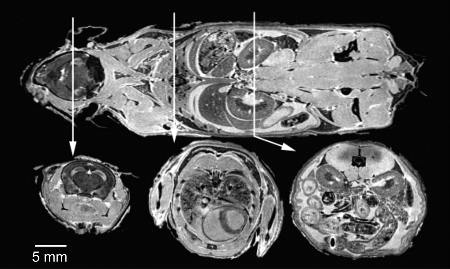
  • Fig 5.
    • Download figure
    • Open in new tab
    • Download powerpoint
    Fig 5.

    The visible mouse project (3D MR imaging of a whole fixed C57Bl/6J mouse). Images acquired with an isotropic array with a 256 × 256 × 1024 matrix and 110 × 110 × 110 μm in-plane resolution. Coronal (top) and representative axial T1-weighted sections from the brain (left), thorax (center), and abdomen (right). Reproduced from Johnson et al21 with permission from the Radiologic Society of North America.
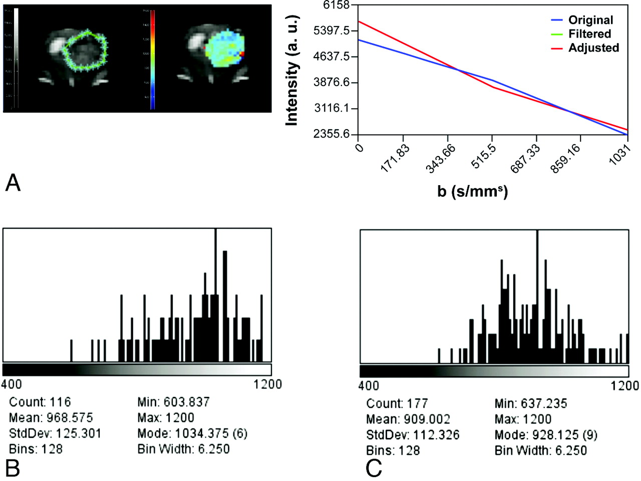
  • Fig 6.
    • Download figure
    • Open in new tab
    • Download powerpoint
    Fig 6.

    Multiple mouse MRI loading system described by Dazai et al.22 Mice loading array equipped with anesthesia and a mouse hive, which holds up to 19 receiver coils. Reproduced from Dazai et al22 with permission from John Wiley and Sons.

  • Fig 7.
    • Download figure
    • Open in new tab
    • Download powerpoint
    Fig 7.

    ADC imaging. A, Delineation of the ROI (left, green limits), intensity versus b value graph of a representative pixel (right), and a parametric color-coded map (center). Serial histograms over time on the second week (B) and on the third week (C) after tumor implantation depict a progressive leftward shift reflecting a decrease in ADC values secondary to increased tumor growth and cellularity.

Tables

  • Figures
    • View popup
    Table 1:

    Distinctive features of the 2 main groups of HGG mouse models

    Features
    Endogenous models (GEMMs and nontargeted mutagenesis/carcinogenesis)
        Unpredictable latency periods, growth curves, tumor location, and morphology; implies regular screening for tumor detection and longitudinal monitoring for progression; more physiologic and best representing human disease
    Exogenous models or xenografts
        Predictable and reproducible, high tumor take; best for high throughput preclinical trials; less physiologic and less representative of the actual human condition
    • View popup
    Table 2:

    Main features of currently available mouse models of HHG

    Animal Model/SubtypeImmunogenicityReproducible Tumor-Host InteractionTumor Take and Growth RateReproducibility of Human HGG Genetics and BiologyStromal DisruptionHigh-Throughput Preclinical Trials
    Exogenous models or xenografts
        SyngeneicNone/lowYes
        HeterogeneicHigh/requires nude miceYes
        OrthotopicYesYes
        HeterotopicNoYes
        Cell suspensionHighYes
        SpheroidsModerateYes
        Tumor explants:LowNo
            From primary cell linesHighDepends on the cell line
            From patient tumorsLowHigh
    Endogenous models
        Carcinogenically induced (RT, QT, viral mutagenesis)No
        GEMMsNo
        Transplantable GEMMsYes
    • Note:—RT indicates radiation therapy; QT, chemotherapy.

PreviousNext
Back to top

In this issue

American Journal of Neuroradiology: 33 (1)
American Journal of Neuroradiology
Vol. 33, Issue 1
1 Jan 2012
  • Table of Contents
  • Index by author
Advertisement
Print
Download PDF
Email Article

Thank you for your interest in spreading the word on American Journal of Neuroradiology.

NOTE: We only request your email address so that the person you are recommending the page to knows that you wanted them to see it, and that it is not junk mail. We do not capture any email address.

Enter multiple addresses on separate lines or separate them with commas.
MR Imaging Features of High-Grade Gliomas in Murine Models: How They Compare with Human Disease, Reflect Tumor Biology, and Play a Role in Preclinical Trials
(Your Name) has sent you a message from American Journal of Neuroradiology
(Your Name) thought you would like to see the American Journal of Neuroradiology web site.
CAPTCHA
This question is for testing whether or not you are a human visitor and to prevent automated spam submissions.
Cite this article
A.R. Borges, P. Lopez-Larrubia, J.B. Marques, S.G. Cerdan
MR Imaging Features of High-Grade Gliomas in Murine Models: How They Compare with Human Disease, Reflect Tumor Biology, and Play a Role in Preclinical Trials
American Journal of Neuroradiology Jan 2012, 33 (1) 24-36; DOI: 10.3174/ajnr.A2959

Citation Manager Formats

  • BibTeX
  • Bookends
  • EasyBib
  • EndNote (tagged)
  • EndNote 8 (xml)
  • Medlars
  • Mendeley
  • Papers
  • RefWorks Tagged
  • Ref Manager
  • RIS
  • Zotero
0 Responses
Respond to this article
Share
Bookmark this article
MR Imaging Features of High-Grade Gliomas in Murine Models: How They Compare with Human Disease, Reflect Tumor Biology, and Play a Role in Preclinical Trials
A.R. Borges, P. Lopez-Larrubia, J.B. Marques, S.G. Cerdan
American Journal of Neuroradiology Jan 2012, 33 (1) 24-36; DOI: 10.3174/ajnr.A2959
del.icio.us logo Twitter logo Facebook logo Mendeley logo
  • Tweet Widget
  • Facebook Like
  • Google Plus One
Purchase

Jump to section

  • Article
    • Abstract
    • ABBREVIATIONS:
    • Outlook
    • Footnotes
    • References
  • Figures & Data
  • Info & Metrics
  • Responses
  • References
  • PDF

Related Articles

  • No related articles found.
  • PubMed
  • Google Scholar

Cited By...

  • No citing articles found.
  • Crossref (14)
  • Google Scholar

This article has been cited by the following articles in journals that are participating in Crossref Cited-by Linking.

  • Trojan Horse Monocyte-Mediated Delivery of Conjugated Polymer Nanoparticles for Improved Photodynamic Therapy of Glioblastoma
    Luis E Ibarra, Lucía Beaugé, Nuria Arias-Ramos, Viviana A Rivarola, Carlos A Chesta, Pilar López-Larrubia, Rodrigo E Palacios
    Nanomedicine 2020 15 17
  • Iron Oxide Incorporated Conjugated Polymer Nanoparticles for Simultaneous Use in Magnetic Resonance and Fluorescent Imaging of Brain Tumors
    Nuria Arias-Ramos, Luis Exequiel Ibarra, María Serrano-Torres, Balbino Yagüe, Matías Daniel Caverzán, Carlos Alberto Chesta, Rodrigo Emiliano Palacios, Pilar López-Larrubia
    Pharmaceutics 2021 13 8
  • Folate Receptor-Targeted Multimodality Imaging of Ovarian Cancer in a Novel Syngeneic Mouse Model
    Meltem Ocak, Andrea G. Gillman, Jamee Bresee, Lixin Zhang, Anda M. Vlad, Cristina Müller, Roger Schibli, W. Barry Edwards, Carolyn J. Anderson, H. Michael Gach
    Molecular Pharmaceutics 2015 12 2
  • Spatial oxygenation profiles in tumors during normo- and hyperbaric hyperoxia
    Oliver Thews, Peter Vaupel
    Strahlentherapie und Onkologie 2015 191 11
  • Process of the Functional Reorganization of the Cortical Centers for Movement in GBM Patients: fMRI Study
    A. Majos, B. Bryszewski, K. N. Kośla, L. Pfaifer, D. Jaskólski, L. Stefańczyk
    Clinical Neuroradiology 2017 27 1
  • Multiparametric magnetic resonance in the assessment of the gender differences in a high-grade glioma rat model
    Rocío Pérez-Carro, Omar Cauli, Pilar López-Larrubia
    EJNMMI Research 2014 4 1
  • Deep learning of MRI contrast enhancement for mapping cerebral blood volume from single-modal non-contrast scans of aging and Alzheimer's disease brains
    Chen Liu, Nanyan Zhu, Haoran Sun, Junhao Zhang, Xinyang Feng, Sabrina Gjerswold-Selleck, Dipika Sikka, Xuemin Zhu, Xueqing Liu, Tal Nuriel, Hong-Jian Wei, Cheng-Chia Wu, J. Thomas Vaughan, Andrew F. Laine, Frank A. Provenzano, Scott A. Small, Jia Guo
    Frontiers in Aging Neuroscience 2022 14
  • Development of a transplantable glioma tumour model from genetically engineered mice: MRI/MRS/MRSI characterisation
    Magdalena Ciezka, Milena Acosta, Cristina Herranz, Josep M. Canals, Martí Pumarola, Ana Paula Candiota, Carles Arús
    Journal of Neuro-Oncology 2016 129 1
  • Preclinical MRI
    Pilar López-Larrubia
    2018 1718
  • Advanced Imaging Techniques in Clinical Pathology
    Francesca Iacobellis, Roberto Grassi, Daniela Berritto, Luigi Mansi
    2016

More in this TOC Section

  • Hot Topics in Research: Preventive Neuroradiology in Brain Aging and Cognitive Decline
  • Evidence Levels for Neuroradiology Articles: Low Agreement among Raters
  • Imaging Biomarkers in Ischemic Stroke Clinical Trials: An Opportunity for Rigor
Show more RESEARCH PERSPECTIVES

Similar Articles

Advertisement

Indexed Content

  • Current Issue
  • Accepted Manuscripts
  • Article Preview
  • Past Issues
  • Editorials
  • Editor's Choice
  • Fellows' Journal Club
  • Letters to the Editor
  • Video Articles

Cases

  • Case Collection
  • Archive - Case of the Week
  • Archive - Case of the Month
  • Archive - Classic Case

Special Collections

  • AJNR Awards
  • ASNR Foundation Special Collection
  • Most Impactful AJNR Articles
  • Photon-Counting CT
  • Spinal CSF Leak Articles (Jan 2020-June 2024)

More from AJNR

  • Trainee Corner
  • Imaging Protocols
  • MRI Safety Corner

Multimedia

  • AJNR Podcasts
  • AJNR Scantastics

Resources

  • Turnaround Time
  • Submit a Manuscript
  • Submit a Video Article
  • Submit an eLetter to the Editor/Response
  • Manuscript Submission Guidelines
  • Statistical Tips
  • Fast Publishing of Accepted Manuscripts
  • Graphical Abstract Preparation
  • Imaging Protocol Submission
  • Evidence-Based Medicine Level Guide
  • Publishing Checklists
  • Author Policies
  • Become a Reviewer/Academy of Reviewers
  • News and Updates

About Us

  • About AJNR
  • Editorial Board
  • Editorial Board Alumni
  • Alerts
  • Permissions
  • Not an AJNR Subscriber? Join Now
  • Advertise with Us
  • Librarian Resources
  • Feedback
  • Terms and Conditions
  • AJNR Editorial Board Alumni

American Society of Neuroradiology

  • Not an ASNR Member? Join Now

© 2025 by the American Society of Neuroradiology All rights, including for text and data mining, AI training, and similar technologies, are reserved.
Print ISSN: 0195-6108 Online ISSN: 1936-959X

Powered by HighWire