Advertisement

Main menu

  • Home
  • Content
    • Current Issue
    • Accepted Manuscripts
    • Article Preview
    • Past Issue Archive
    • Video Articles
    • AJNR Case Collection
    • Case of the Week Archive
    • Case of the Month Archive
    • Classic Case Archive
  • Special Collections
    • AJNR Awards
    • ASNR Foundation Special Collection
    • Most Impactful AJNR Articles
    • Photon-Counting CT
    • Spinal CSF Leak Articles (Jan 2020-June 2024)
  • Multimedia
    • AJNR Podcasts
    • AJNR SCANtastic
    • Trainee Corner
    • MRI Safety Corner
    • Imaging Protocols
  • For Authors
    • Submit a Manuscript
    • Submit a Video Article
    • Submit an eLetter to the Editor/Response
    • Manuscript Submission Guidelines
    • Statistical Tips
    • Fast Publishing of Accepted Manuscripts
    • Graphical Abstract Preparation
    • Imaging Protocol Submission
    • Author Policies
  • About Us
    • About AJNR
    • Editorial Board
    • Editorial Board Alumni
  • More
    • Become a Reviewer/Academy of Reviewers
    • Subscribers
    • Permissions
    • Alerts
    • Feedback
    • Advertisers
    • ASNR Home

User menu

  • Alerts
  • Log in

Search

  • Advanced search
American Journal of Neuroradiology

American Journal of Neuroradiology

ASHNR American Society of Functional Neuroradiology ASHNR American Society of Pediatric Neuroradiology ASSR
  • Alerts
  • Log in

Advanced Search

  • Home
  • Content
    • Current Issue
    • Accepted Manuscripts
    • Article Preview
    • Past Issue Archive
    • Video Articles
    • AJNR Case Collection
    • Case of the Week Archive
    • Case of the Month Archive
    • Classic Case Archive
  • Special Collections
    • AJNR Awards
    • ASNR Foundation Special Collection
    • Most Impactful AJNR Articles
    • Photon-Counting CT
    • Spinal CSF Leak Articles (Jan 2020-June 2024)
  • Multimedia
    • AJNR Podcasts
    • AJNR SCANtastic
    • Trainee Corner
    • MRI Safety Corner
    • Imaging Protocols
  • For Authors
    • Submit a Manuscript
    • Submit a Video Article
    • Submit an eLetter to the Editor/Response
    • Manuscript Submission Guidelines
    • Statistical Tips
    • Fast Publishing of Accepted Manuscripts
    • Graphical Abstract Preparation
    • Imaging Protocol Submission
    • Author Policies
  • About Us
    • About AJNR
    • Editorial Board
    • Editorial Board Alumni
  • More
    • Become a Reviewer/Academy of Reviewers
    • Subscribers
    • Permissions
    • Alerts
    • Feedback
    • Advertisers
    • ASNR Home
  • Follow AJNR on Twitter
  • Visit AJNR on Facebook
  • Follow AJNR on Instagram
  • Join AJNR on LinkedIn
  • RSS Feeds

AJNR is seeking candidates for the AJNR Podcast Editor. Read the position description.

Research ArticleInterventional

A Large and Giant Bifurcation Aneurysm Model in Canines: Proof of Feasibility

R. Ysuda, C.M. Strother, B. Aagaard-Kienitz, K. Pulfer and D. Consigny
American Journal of Neuroradiology March 2012, 33 (3) 507-512; DOI: https://doi.org/10.3174/ajnr.A2789
R. Ysuda
  • Find this author on Google Scholar
  • Find this author on PubMed
  • Search for this author on this site
C.M. Strother
  • Find this author on Google Scholar
  • Find this author on PubMed
  • Search for this author on this site
B. Aagaard-Kienitz
  • Find this author on Google Scholar
  • Find this author on PubMed
  • Search for this author on this site
K. Pulfer
  • Find this author on Google Scholar
  • Find this author on PubMed
  • Search for this author on this site
D. Consigny
  • Find this author on Google Scholar
  • Find this author on PubMed
  • Search for this author on this site
  • Article
  • Figures & Data
  • Info & Metrics
  • Responses
  • References
  • PDF
Loading

Article Figures & Data

Figures

  • Tables
  • Fig 1.
    • Download figure
    • Open in new tab
    • Download powerpoint
    Fig 1.

    Arterial structures of (A) bifurcation and (B) terminal. Adapted from Strother, Graves, and Rappe.28
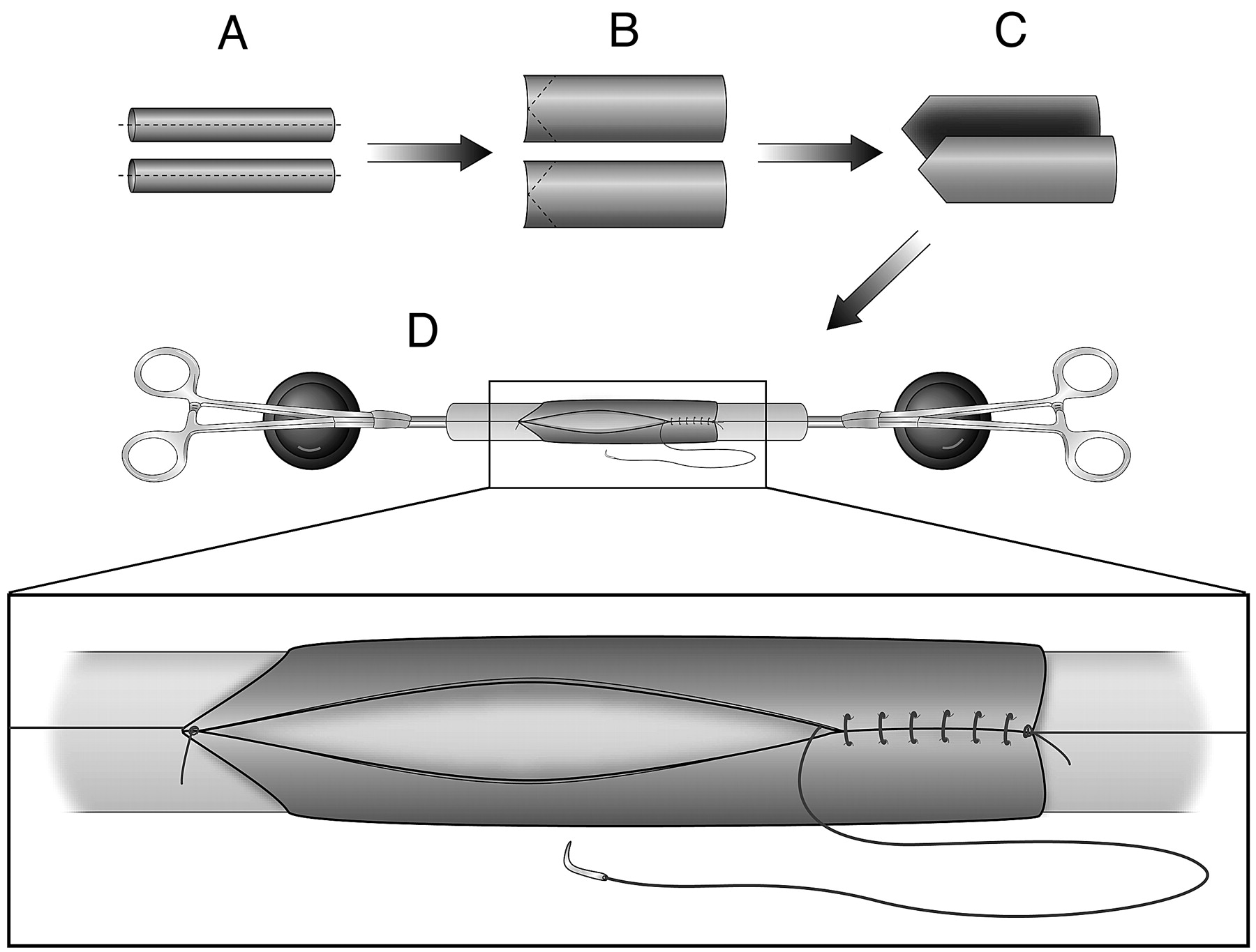
  • Fig 2.
    • Download figure
    • Open in new tab
    • Download powerpoint
    Fig 2.

    Bilateral external jugular veins are harvested. A, The vein grafts are split to make 2 venous sheets. B, These sheets are trimmed into a shape that would fit into the bifurcation arterial structure. The sheets are then mounted on a device, shown in D, in a way such that the original adventitial side is on the exterior surface. C, A handmade device to facilitate venous pouch preparation. Note that, by using 2 clamps, tension is made on the suture line to facilitate the procedure.

  • Fig 3.
    • Download figure
    • Open in new tab
    • Download powerpoint
    Fig 3.

    Incorporation of the venous pouch into the arterial structure. A, At this point, the venous pouch still remains on the device shown in Fig 2D, so as to keep the venous pouch cylindrical and thus facilitate suturing. B, The top of the venous pouch is also sutured. C, Completion of “aneurysm” creation after achieving hemostasis.

  • Fig 4.
    • Download figure
    • Open in new tab
    • Download powerpoint
    Fig 4.

    Images acquired at the second follow-up angiogram. A, B, and C represent images of aneurysms from dogs 1, 2, and 3, respectively. Except in 1 case (F, dog 6), all the aneurysms remain patent. In cases H and I (dogs 8 and 9), PTFE was used along with vein grafts.

Tables

  • Figures
    • View popup
    Table 1:

    Case summary

    Dog #Artery“Aneurysm” Sac1st f/u2nd f/uComment
    1BifurcationVein graftIV (2)IA (4)
    2BifurcationVein graftIV (1)IA (3)
    3BifurcationVein graftIV (1)IA (3)
    4BifurcationVein graftIV (3)IA (6)
    5BifurcationVein graftIV (3)IA (6)
    6BifurcationVein graftIV (3)IA (12)An aneurysm was thrombosed
    7BifurcationVein graftIV (3)IA (6)
    8BifurcationVein graft + PTFEIA (2)*Dog was sacrificed after the 1st f/u due to sickness
    9TerminalVein graft + PTFEIV (3)IA (7)
    • Note:—IV and IA represent a modality employed for the follow-up study. A number in parentheses represents the interval in weeks between aneurysm creation and each follow-up study. Animals were sacrificed on the day following the 2nd follow-up. f/u indicates follow-up study; IV, intravenous digital subtraction angiogram; IA, intra-arterial digital subtraction angiogram.

    • View popup
    Table 2:

    Aneurysm dimensions

    Dog #Height (mm)Dog #Neck Width (mm)
    1st f/u2nd f/uDif.1st f/u2nd f/uDif.
    119.020.01.018.99.90.9
    225.225.1−0.229.610.61.0
    321.724.12.438.69.61.1
    430.131.51.4411.013.22.3
    522.321.7−0.658.48.3−0.1
    6***6***
    722.923.20.378.89.00.2
    821.6**810.4**
    920.920.1−0.898.57.7−0.8
    Mean23.023.7Mean9.39.8
    SD3.44.0SD1.01.8
    Dog #Dome Width (mm)CasesVolume (mm3)/Ostium Area (mm2)
    1st f/u2nd f/uDif.VolumeOstium AreaVOR
    110.111.41.411552.054.828.3
    210.511.81.322576.590.428.5
    310.512.41.932319.995.824.2
    413.414.41.044595.0172.326.7
    510.912.21.352287.568.533.4
    6***6***
    712.112.20.172601.289.229.2
    818.6**82418.164.137.7
    920.722.82.193350.373.445.7
    Mean13.313.9Mean2712.688.631.7
    SD4.14.0SD906.336.77.0
    • Note:—Volume and the area of the ostium were measured only for the second follow-up study.

    • ↵* For dog 6, no measurements were performed because of spontaneous thrombosis. f/u indicates follow-up; Dif., difference.

PreviousNext
Back to top

In this issue

American Journal of Neuroradiology: 33 (3)
American Journal of Neuroradiology
Vol. 33, Issue 3
1 Mar 2012
  • Table of Contents
  • Index by author
Advertisement
Print
Download PDF
Email Article

Thank you for your interest in spreading the word on American Journal of Neuroradiology.

NOTE: We only request your email address so that the person you are recommending the page to knows that you wanted them to see it, and that it is not junk mail. We do not capture any email address.

Enter multiple addresses on separate lines or separate them with commas.
A Large and Giant Bifurcation Aneurysm Model in Canines: Proof of Feasibility
(Your Name) has sent you a message from American Journal of Neuroradiology
(Your Name) thought you would like to see the American Journal of Neuroradiology web site.
CAPTCHA
This question is for testing whether or not you are a human visitor and to prevent automated spam submissions.
Cite this article
R. Ysuda, C.M. Strother, B. Aagaard-Kienitz, K. Pulfer, D. Consigny
A Large and Giant Bifurcation Aneurysm Model in Canines: Proof of Feasibility
American Journal of Neuroradiology Mar 2012, 33 (3) 507-512; DOI: 10.3174/ajnr.A2789

Citation Manager Formats

  • BibTeX
  • Bookends
  • EasyBib
  • EndNote (tagged)
  • EndNote 8 (xml)
  • Medlars
  • Mendeley
  • Papers
  • RefWorks Tagged
  • Ref Manager
  • RIS
  • Zotero
0 Responses
Respond to this article
Share
Bookmark this article
A Large and Giant Bifurcation Aneurysm Model in Canines: Proof of Feasibility
R. Ysuda, C.M. Strother, B. Aagaard-Kienitz, K. Pulfer, D. Consigny
American Journal of Neuroradiology Mar 2012, 33 (3) 507-512; DOI: 10.3174/ajnr.A2789
del.icio.us logo Twitter logo Facebook logo Mendeley logo
  • Tweet Widget
  • Facebook Like
  • Google Plus One
Purchase

Jump to section

  • Article
    • Abstract
    • ABBREVIATIONS:
    • Materials and Methods
    • Results
    • Discussion
    • Footnotes
    • References
  • Figures & Data
  • Info & Metrics
  • Responses
  • References
  • PDF

Related Articles

  • No related articles found.
  • PubMed
  • Google Scholar

Cited By...

  • Surgical technique for venous patch aneurysms with no neck in a rabbit model
  • Crossref (6)
  • Google Scholar

This article has been cited by the following articles in journals that are participating in Crossref Cited-by Linking.

  • In vivo cerebral aneurysm models
    John W. Thompson, Omar Elwardany, David J. McCarthy, Dallas L. Sheinberg, Carlos M. Alvarez, Ahmed Nada, Brian M. Snelling, Stephanie H. Chen, Samir Sur, Robert M. Starke
    Neurosurgical Focus 2019 47 1
  • Preclinical extracranial aneurysm models for the study and treatment of brain aneurysms: A systematic review
    Serge Marbacher, Fabio Strange, Juhana Frösén, Javier Fandino
    Journal of Cerebral Blood Flow & Metabolism 2020 40 5
  • Surgical technique for venous patch aneurysms with no neck in a rabbit model
    Kilian Greim-Kuczewski, Alejandro Berenstein, Sebastian Kis, Anna Hauser, Monika Killer-Oberpfalzer
    Journal of NeuroInterventional Surgery 2018 10 2
  • Very large and giant microsurgical bifurcation aneurysms in rabbits: Proof of feasibility and comparability using computational fluid dynamics and biomechanical testing
    Camillo Sherif, Erwin Herbich, Roberto Plasenzotti, Helga Bergmeister, Ursula Windberger, Georg Mach, Gerhard Sommer, Gerhard A. Holzapfel, Thomas Haider, Martin Krssak, Guenther Kleinpeter
    Journal of Neuroscience Methods 2016 268
  • Introducing a Realistic, Low-Cost Simulation Model for Clipping of Brain Aneurysms
    Gallardo Federico Carlos, Feldman Santiago Enrrique, Targa García Aylen Andrea, Morales Nadia Sabrina, Martinez Silvina, Martin Clara, Bottan Juan Santiago, Orellana Cristian Marcelo, Almerares Leandro Noel, Rubino Pablo Augusto
    World Neurosurgery 2022 158
  • Imaging of intracranial aneurysms in animals: a systematic review of modalities
    Anne F. Cayron, Sandrine Morel, Eric Allémann, Philippe Bijlenga, Brenda R. Kwak
    Neurosurgical Review 2023 46 1

More in this TOC Section

  • SAVE vs. Solumbra Techniques for Thrombectomy
  • Contrast-Induced Encephalopathy after NeuroIR
  • CT Perfusion&Reperfusion in Acute Ischemic Stroke
Show more Interventional

Similar Articles

Advertisement

Indexed Content

  • Current Issue
  • Accepted Manuscripts
  • Article Preview
  • Past Issues
  • Editorials
  • Editor's Choice
  • Fellows' Journal Club
  • Letters to the Editor
  • Video Articles

Cases

  • Case Collection
  • Archive - Case of the Week
  • Archive - Case of the Month
  • Archive - Classic Case

Special Collections

  • AJNR Awards
  • ASNR Foundation Special Collection
  • Most Impactful AJNR Articles
  • Photon-Counting CT
  • Spinal CSF Leak Articles (Jan 2020-June 2024)

More from AJNR

  • Trainee Corner
  • Imaging Protocols
  • MRI Safety Corner

Multimedia

  • AJNR Podcasts
  • AJNR Scantastics

Resources

  • Turnaround Time
  • Submit a Manuscript
  • Submit a Video Article
  • Submit an eLetter to the Editor/Response
  • Manuscript Submission Guidelines
  • Statistical Tips
  • Fast Publishing of Accepted Manuscripts
  • Graphical Abstract Preparation
  • Imaging Protocol Submission
  • Evidence-Based Medicine Level Guide
  • Publishing Checklists
  • Author Policies
  • Become a Reviewer/Academy of Reviewers
  • News and Updates

About Us

  • About AJNR
  • Editorial Board
  • Editorial Board Alumni
  • Alerts
  • Permissions
  • Not an AJNR Subscriber? Join Now
  • Advertise with Us
  • Librarian Resources
  • Feedback
  • Terms and Conditions
  • AJNR Editorial Board Alumni

American Society of Neuroradiology

  • Not an ASNR Member? Join Now

© 2025 by the American Society of Neuroradiology All rights, including for text and data mining, AI training, and similar technologies, are reserved.
Print ISSN: 0195-6108 Online ISSN: 1936-959X

Powered by HighWire