
Article Figures & Data
Figures
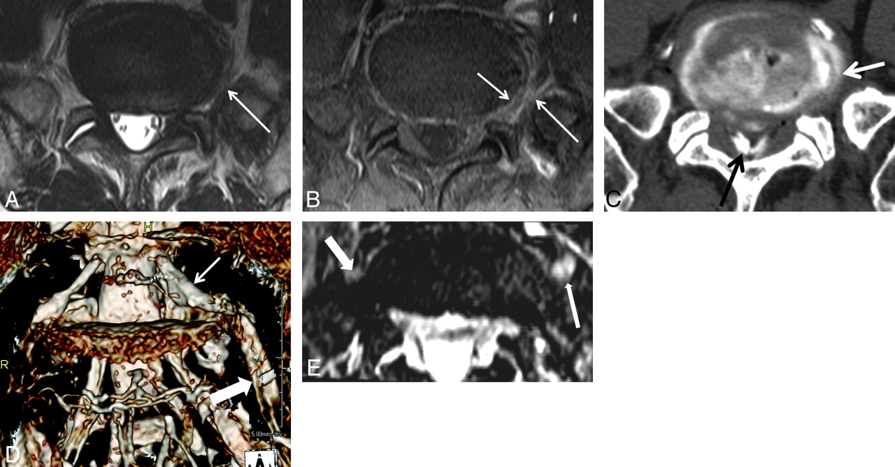
- Fig 1.
A 69-year-old man with left leg pain. A, There is intermediate signal intensity (arrow) between the outer anulus and left L5 nerve root on the T2-weighted image. B, Contrast-enhanced T1-weighted image with fat suppression shows abnormal enhancement at the left perianular extraforaminal zone (large arrow) and outer anulus (small arrow) in L5-S1. Pain reproduction at this level during diskography shows concordant pain. C, CT diskography shows an anular tear and left posterolateral extraforaminal leak (arrow) of the contrast media from an anular tear. There is contrast media (black arrow) at the extradural spinal canal. D, Diffuse swelling of the entire left L5 nerve root (thick arrow) including DRG (thin arrow) is demonstrated on 3D MR radiculography. E, Axial Proset MPR image demonstrates that the left L5 exit nerve root (long arrow) along the ventral surface of the sacral ala is larger and higher in signal intensity compared with the right L5 exit nerve root (thick arrow).
- Fig 2.
A 51-year-old man with left buttock and calf pain. A and B, T1- (A) and T2-weighted (B) images show an anular tear (arrow) at the left lateral margin of the L5-S1 disk. There is intermediate signal intensity due to the fibrovascular and granulation tissue between the outer anulus and left L5 nerve root (short arrow). There is no apparent disk herniation. C, There is abnormal enhancement at the perianular extraforaminal zone (arrow) in L5-S1 on the contrast-enhanced T1-weighted image with fat suppression. D, Diffuse swelling of the left L5 dorsal root ganglion (arrow) and exit nerve root (thick arrow) is demonstrated on 3D MR radiculography. E, Axial Proset MPR image shows the left L5 exit nerve root (long arrow) along the ventral surface of the sacral ala to be larger and higher in signal intensity compared with the contralateral nerve root (short arrow). Star indicates the right S1 nerve root. F, Pain reproduction at this level (arrow) during a selective nerve root block (prone position) shows concordant pain. At 1 month after selective nerve root block at the left L5 nerve root, clinical symptoms are completely improved. G, However, a 3-month follow-up 3D MR radiculography reveals decreased swelling at the left L5 nerve root.


Tables
- Table 1:
Clinical symptoms and MR imaging findings of the patients with chemical radiculitis
No. Age (yr)/Sex Symptoms NR Lesion Segment of Swelling Location of Perianular Enhancement ODI, Before VAS, After Provocation 1 51/M Lt buttock, calf pain Lt L5 DRG, exit For and extra 18.5 0.0 SNI 2 53/M Lt leg pain Lt L5 Entire For and extra 33.4 0.0 SNI 3 62/M Lt leg pain Lt L5 Entire – 44.6 25, 3 SNI 4 49/F Rt buttock, lat thigh pain Rt L5 DRG, exit For and extra 25.4 13.2 SNI 5 69/M Lt leg pain, numbness Lt L5 Entire For and extra 22.3 16.1 Diskography 6 53/M Lt leg pain Lt L5 Exit For and extra 42.5 21.1 SNI 7 62/M Rt leg pain Rt L5 Exit Extra 62.7 33.2 SNI 8 35/M Rt leg pain, toe weakness Rt L5 Exit For and extra 22.3 11.1 SNI 9 76/F Lt buttock, leg pain Lt L5 Entire For and extra 54.7 22.2 Diskography 10 32/M Rt leg pain Rt L5 Exit Extra 33.6 31.3 SNI 11 51/F Lt buttock, leg pain Lt L5 Exit – 18.4 0.0 SNI 12 56/F Lt buttock, leg pain Lt L5 Exit Extra 44.5 22.1 SNI 13 69/F Rt buttock, leg pain Rt L5 Entire For and extra 54.6 No SNI 14 44/M Lt leg pain Lt L4 DRG, exit For and extra No 80% SNI 15 88/M Rt leg pain Rt L4 Entire For and extra 51.5 11.2 SNI 16 54/F Rt anterior, lat leg pain Rt L4 Trans – No 90% Diskography 17 59/F Lt leg pain Lt L2 Entire For and extra 56.6 0.0 Diskography -
Note:—NR indicates nerve root; ODI, Oswestry Disability Index; VAS, visual analogue scale; Rt, right; Lt, left; exit, exit nerve root; Trans, transverse nerve root; Entire, entire segments of nerve root; For, foraminal zone; extra, extraforaminal zone; lat, lateral; SNI, selective nerve root injection; No, no response of symptoms; Before, before treatment; After, after treatment; %, degree of symptom improvement; –, no contrast study.
-
- Table 2:
Comparison between quantitative measurements of the symptomatic nerve roots and those of the contralateral asymptomatic nerve roots on Proset images
Side Mean Value of Nerve Root Diameter (mm) Mean Value of SIRa Transverse DRG Exit Transverse DRG Exit Ipsilateral 4.2 ± 0.9 6.33 ± 0.12 7.2 ± 1.9 2.66 ± 1.48 2.51 ± 1.67 1.77 ± 0.75 Contralateral 3.3 ± 0.77 5.15 ± 0.98 4.72 ± 1.1 2.57 ± 1.41 2.14 ± 1.53 1.27 ± 0.54 -
Note:—Transverse indicates transverse nerve root; Exit, exit nerve root.
↵a Width of the nerve roots with chemical radiculitis is larger than that of the contralateral asymptomatic nerve roots (paired t test, P < .005). SIR of the DRG and exit nerve roots with chemical radiculitis is higher than that of the contralateral asymptomatic nerve roots (paired t test, P < .005).
-


