
Article Figures & Data
Figures
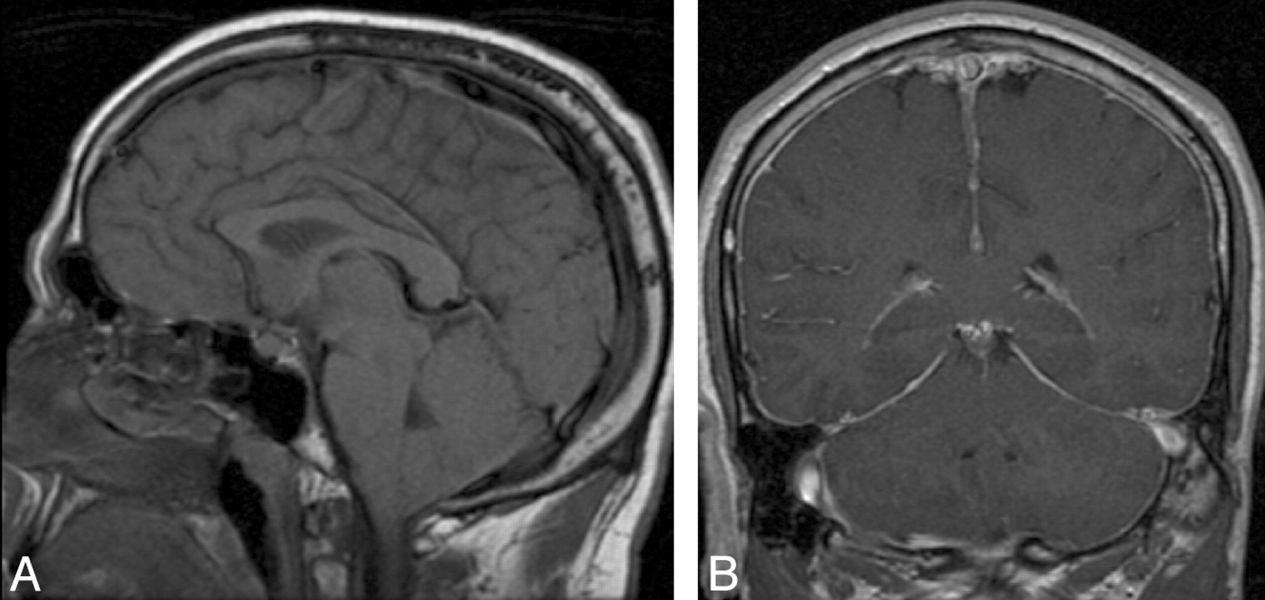
- Fig 1.
Patient with “classic” MR imaging findings of SIH on brain MR imaging. A, Brain sag on precontrast sagittal T1 imaging, with effacement of the suprasellar and prepontine cisterns, descent of the optic chiasm, draping of the floor of the third ventricle over the dorsum sella, descent of the midbrain, and extension of the tonsils through the foramen magnum. B, Diffuse pachymeningeal enhancement on postcontrast coronal T1 imaging.
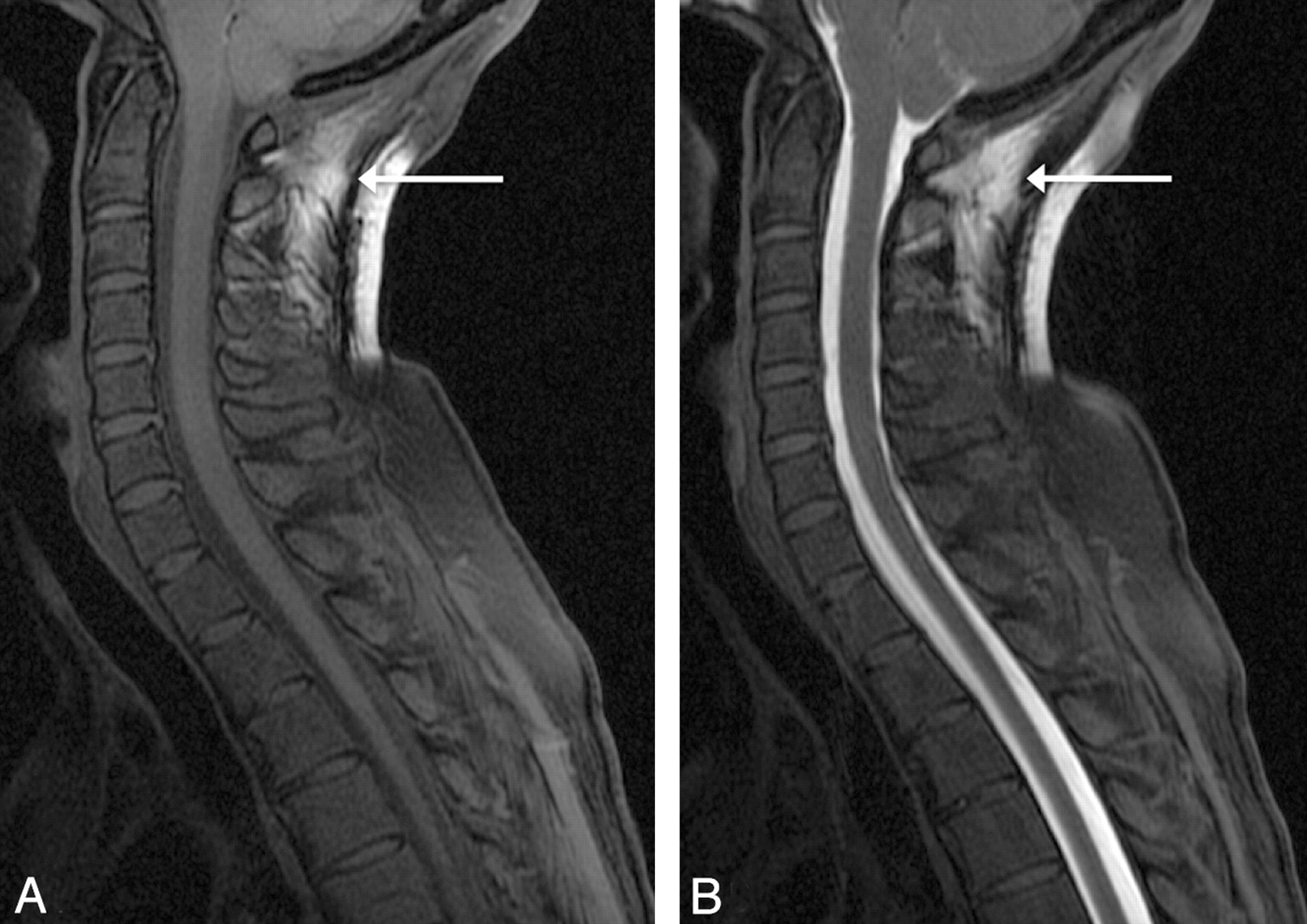
- Fig 2.
A, Pre- and postintrathecal gadolinium fat-suppressed T1 images demonstrate typical artifacts, which may simulate a leak at C1-C2 seen in 6 of 41 of our patients. Note the inhomogeneous fat saturation on this precontrast sagittal T1 image at C1-2 (arrow). B, Recognizing this artifact on precontrast imaging is important because with the addition of intrathecal gadolinium, this inhomogeneous fat saturation can potentially mimic a leak (arrow).
- Fig 3.
Patient with CSF leaks at the left T3–4 and T4–5 interspaces, which are only visible on delayed GdM. A, Immediate left parasagittal fat-suppressed T1 imaging with normal findings. B−D, Three-hour delayed left parasagittal fat-suppressed T1 imaging demonstrates the CSF leaks (arrows), a finding which is also documented in the coronal plane (C and D) with evidence of contrast within the T3 and T4 foramina (arrows).

Tables
Categorization and distribution of patients with CSF leak identified on GdM
No. of Patients CSF Leak on GdM (% of Patients in Group) Group 1: No CSF leak on CTM with at least 1 imaging study with positive findings Group 1a: Classic brain MR imaging with diffuse dural enhancement and brain sag 14a 2 (17%) Group 1b: Extradural fluid collection on MR spine imaging 2a 1 (50%) Group 2: No CSF leak on CTM with equivocal brain imaging (dural enhancement without brain sag or brain sag without dural enhancement) and negative findings on spine imaging 3 1 (33%) Group 3: No CSF leak on CTM and negative findings on MR brain and spine imaging 6 1 (14%) Group 4: Known CSF leak seen on CTM 17 12 (71%) Total number of patients in groups 1–4 41 17 (41%) ↵a One patient without a leak had both classic brain MR imaging and extradural fluid and is included in both groups 1a and 1b.
In this issue


Jump to section
Related Articles
Cited By...
- Myelographic Techniques for the Localization of CSF-Venous Fistulas: Updates in 2024
- Prospective Safety Study of Intrathecal Gadobutrol in Different Doses
- Prospective Safety Study of Intrathecal Gadobutrol in Different Doses
- American Society of Regional Anesthesia and Pain Medicine contrast shortage position statement
- Brain Sagging Dementia--Diagnosis, Treatment, and Outcome: A Review
- Diskogenic Dural Defect Is the Reason for the Ventral Location of the Epidural Spinal Fluid Collection Seen in Superficial Siderosis
- Safety of Consecutive Bilateral Decubitus Digital Subtraction Myelography in Patients with Spontaneous Intracranial Hypotension and Occult CSF Leak
- Spine MRI in Spontaneous Intracranial Hypotension for CSF Leak Detection: Nonsuperiority of Intrathecal Gadolinium to Heavily T2-Weighted Fat-Saturated Sequences
- MR Myelography for the Detection of CSF-Venous Fistulas
- Intrathecal Use of Gadobutrol for Glymphatic MR Imaging: Prospective Safety Study of 100 Patients
- Fatal gadolinium-induced encephalopathy following accidental intrathecal administration: a case report and a comprehensive evidence-based review
- A classification system of spontaneous spinal CSF leaks
- Spinal CSF venous fistula: A treatable etiology for CSF leaks in craniospinal hypovolemia
- MR Myelography for Identification of Spinal CSF Leak in Spontaneous Intracranial Hypotension
- Sensitivity of MRI of the spine compared with CT myelography in orthostatic headache with CSF leak
- Spontaneous intracranial hypotension and venous sinus thrombosis