Advertisement

Main menu

  • Home
  • Content
    • Current Issue
    • Accepted Manuscripts
    • Article Preview
    • Past Issue Archive
    • Video Articles
    • AJNR Case Collection
    • Case of the Week Archive
    • Case of the Month Archive
    • Classic Case Archive
  • Special Collections
    • AJNR Awards
    • ASNR Foundation Special Collection
    • Most Impactful AJNR Articles
    • Photon-Counting CT
    • Spinal CSF Leak Articles (Jan 2020-June 2024)
  • Multimedia
    • AJNR Podcasts
    • AJNR SCANtastic
    • Trainee Corner
    • MRI Safety Corner
    • Imaging Protocols
  • For Authors
    • Submit a Manuscript
    • Submit a Video Article
    • Submit an eLetter to the Editor/Response
    • Manuscript Submission Guidelines
    • Statistical Tips
    • Fast Publishing of Accepted Manuscripts
    • Graphical Abstract Preparation
    • Imaging Protocol Submission
    • Author Policies
  • About Us
    • About AJNR
    • Editorial Board
    • Editorial Board Alumni
  • More
    • Become a Reviewer/Academy of Reviewers
    • Subscribers
    • Permissions
    • Alerts
    • Feedback
    • Advertisers
    • ASNR Home

User menu

  • Alerts
  • Log in

Search

  • Advanced search
American Journal of Neuroradiology

American Journal of Neuroradiology

ASHNR American Society of Functional Neuroradiology ASHNR American Society of Pediatric Neuroradiology ASSR
  • Alerts
  • Log in

Advanced Search

  • Home
  • Content
    • Current Issue
    • Accepted Manuscripts
    • Article Preview
    • Past Issue Archive
    • Video Articles
    • AJNR Case Collection
    • Case of the Week Archive
    • Case of the Month Archive
    • Classic Case Archive
  • Special Collections
    • AJNR Awards
    • ASNR Foundation Special Collection
    • Most Impactful AJNR Articles
    • Photon-Counting CT
    • Spinal CSF Leak Articles (Jan 2020-June 2024)
  • Multimedia
    • AJNR Podcasts
    • AJNR SCANtastic
    • Trainee Corner
    • MRI Safety Corner
    • Imaging Protocols
  • For Authors
    • Submit a Manuscript
    • Submit a Video Article
    • Submit an eLetter to the Editor/Response
    • Manuscript Submission Guidelines
    • Statistical Tips
    • Fast Publishing of Accepted Manuscripts
    • Graphical Abstract Preparation
    • Imaging Protocol Submission
    • Author Policies
  • About Us
    • About AJNR
    • Editorial Board
    • Editorial Board Alumni
  • More
    • Become a Reviewer/Academy of Reviewers
    • Subscribers
    • Permissions
    • Alerts
    • Feedback
    • Advertisers
    • ASNR Home
  • Follow AJNR on Twitter
  • Visit AJNR on Facebook
  • Follow AJNR on Instagram
  • Join AJNR on LinkedIn
  • RSS Feeds

AJNR is seeking candidates for the AJNR Podcast Editor. Read the position description.

LetterLETTER

Elastase-Induced Rabbit Aneurysms Model Complicated by Thoracic Aortic Aneurysms

K. Wang and S. Yuan
American Journal of Neuroradiology May 2012, 33 (5) E76-E77; DOI: https://doi.org/10.3174/ajnr.A3095
K. Wang
  • Find this author on Google Scholar
  • Find this author on PubMed
  • Search for this author on this site
S. Yuan
  • Find this author on Google Scholar
  • Find this author on PubMed
  • Search for this author on this site
  • Article
  • Figures & Data
  • Info & Metrics
  • Responses
  • References
  • PDF
Loading

Published reports have described the elastase-induced aneurysm model in rabbits as easy and reproducible and possessing geometric, pathologic, and hemodynamic features that are similar to those of human cerebral aneurysms.1 We used a simple surgical technique to produce the elastase-induced aneurysm model in rabbits2 and used it to explore the efficacy of new endovascular devices. Recently, we found that 3 rabbits unexpectedly developed a thoracic aortic aneurysms (TAA), which was located in the posterior part of thoracic aorta, in 40 elastase-induced rabbit aneurysm models (Fig 1).

Fig 1.
  • Download figure
  • Open in new tab
  • Download powerpoint
Fig 1.

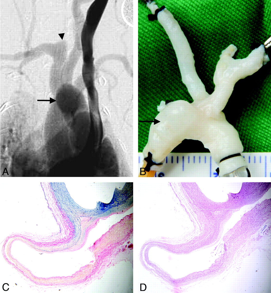
Elastase-induced rabbit aneurysm complicated by a TAA. A, Intravenous DSA shows an aneurysm (arrowhead) in the right subclavian artery and a TAA in the thoracic aorta (arrow). B, Macroscopically, the TAA (arrow) is located in the posterior part of the aortic arch and descending thoracic aorta (posteroanterior view). C, A specimen stained with Ponceau/Victoria Blue shows the normal elastic layers of the thoracic aorta as wavy blue lines. Abrupt termination of the elastic lamina at the dissection margins of the TAA is evident (original magnification, ×40). D, Hematoxylin-eosin staining shows a thin aneurysm wall, the absence of an inflammatory reaction, and marked loss of cellular elements (original magnification, ×40).

In contrast to traditional techniques, we used a temporary arcuated aneurysm clip rather than an occlusion balloon to achieve flow arrest in the right common carotid artery (RCCA). When we checked the primary records of the elastase-induction procedure in the 3 cases, we found that the stump of the RCCA had collapsed in the first few minutes after complete filling with elastase had been achieved. We deduced that when the origin of the RCCA was separated from the surrounding tissue in the initial procedure, ≥1 arterial branch originating from the proximal RCCA that was barely visible to the naked eye had split, allowing elastase to leak into the tissue surrounding the thoracic aorta during incubation, ultimately resulting in TAA formation. Thiex et al3 recommended avoiding elastase leaking into the branch originating from the proximal RCCA because of the danger of lethal hemorrhage of the trachea.3 We are unable to explain how the leaked elastase came into contact with the thoracic aorta and thereby induced TAA, because the TAAs we have reported here were in the pericardial cavity. It is possible that elastase permeated into the pericardial cavity and concentrated around the posterior part of thoracic aortic artery during the surgical procedure.

The TAAs we found here in rabbits reproduced key features of human TAAs in regard to site-specific phenotypes and pathology. The external surroundings of aneurysms in the pericardial cavity resembled those of the intracranial subarachnoid cavity, and the location is similar to that of human ophthalmic segment aneurysms.

This case report comes from an animal experimental study, which was supported by the China Postdoctoral Science Foundation (grant 20100481521). All procedures of the animal experimental study in this article were approved by the Institutional Animal Care and Use Committee at our hospital.

References

  1. 1.↵
    1. Zeng Z,
    2. Kallmes DF,
    3. Durka MJ,
    4. et al
    . Hemodynamics and anatomy of elastase- induced rabbit aneurysm models: similarity to human cerebral aneurysms? AJNR Am J Neuroradiol 2011;32:595–601
    Abstract/FREE Full Text
  2. 2.↵
    1. Wang K,
    2. Huang Q,
    3. Hong B,
    4. et al
    . Neck injury is critical to elastase-induced aneurysm model. AJNR Am J Neuroradiol 2009;30:1685–87
    Abstract/FREE Full Text
  3. 3.↵
    1. Thiex R,
    2. Hans FJ,
    3. Krings T,
    4. et al
    . Haemorrhagic tracheal necrosis as a lethal complication of an aneurysm model in rabbits via endoluminal incubation with elastase. Acta Neurochir (Wien) 2004;146:285–89, discussion 289
    CrossRefPubMed
  • © 2012 by American Journal of Neuroradiology
PreviousNext
Back to top

In this issue

American Journal of Neuroradiology: 33 (5)
American Journal of Neuroradiology
Vol. 33, Issue 5
1 May 2012
  • Table of Contents
  • Index by author
Advertisement
Print
Download PDF
Email Article

Thank you for your interest in spreading the word on American Journal of Neuroradiology.

NOTE: We only request your email address so that the person you are recommending the page to knows that you wanted them to see it, and that it is not junk mail. We do not capture any email address.

Enter multiple addresses on separate lines or separate them with commas.
Elastase-Induced Rabbit Aneurysms Model Complicated by Thoracic Aortic Aneurysms
(Your Name) has sent you a message from American Journal of Neuroradiology
(Your Name) thought you would like to see the American Journal of Neuroradiology web site.
CAPTCHA
This question is for testing whether or not you are a human visitor and to prevent automated spam submissions.
Cite this article
K. Wang, S. Yuan
Elastase-Induced Rabbit Aneurysms Model Complicated by Thoracic Aortic Aneurysms
American Journal of Neuroradiology May 2012, 33 (5) E76-E77; DOI: 10.3174/ajnr.A3095

Citation Manager Formats

  • BibTeX
  • Bookends
  • EasyBib
  • EndNote (tagged)
  • EndNote 8 (xml)
  • Medlars
  • Mendeley
  • Papers
  • RefWorks Tagged
  • Ref Manager
  • RIS
  • Zotero
0 Responses
Respond to this article
Share
Bookmark this article
Elastase-Induced Rabbit Aneurysms Model Complicated by Thoracic Aortic Aneurysms
K. Wang, S. Yuan
American Journal of Neuroradiology May 2012, 33 (5) E76-E77; DOI: 10.3174/ajnr.A3095
del.icio.us logo Twitter logo Facebook logo Mendeley logo
  • Tweet Widget
  • Facebook Like
  • Google Plus One
Purchase

Jump to section

  • Article
    • References
  • Figures & Data
  • Info & Metrics
  • Responses
  • References
  • PDF

Related Articles

  • No related articles found.
  • PubMed
  • Google Scholar

Cited By...

  • From bench to bedside: utility of the rabbit elastase aneurysm model in preclinical studies of intracranial aneurysm treatment
  • Crossref (2)
  • Google Scholar

This article has been cited by the following articles in journals that are participating in Crossref Cited-by Linking.

  • From bench to bedside: utility of the rabbit elastase aneurysm model in preclinical studies of intracranial aneurysm treatment
    Waleed Brinjikji, Yong H Ding, David F Kallmes, Ramanathan Kadirvel
    Journal of NeuroInterventional Surgery 2016 8 5
  • The development and understanding of intracranial aneurysm based on rabbit model
    Haishuang Tang, Zhiwen Lu, Gaici Xue, Sisi Li, Fengfeng Xu, Yazhou Yan, Jianmin Liu, Qiao Zuo, Yin Luo, Qinghai Huang
    Neuroradiology 2020 62 10

More in this TOC Section

  • Letter to the Editor regarding “Automated Volumetric Software in Dementia: Help or Hindrance to the Neuroradiologist?”
  • Reply:
  • Brain AVM’s Nidus: What if We Hadn’t Understood Anything?
Show more Letters

Similar Articles

Advertisement

Indexed Content

  • Current Issue
  • Accepted Manuscripts
  • Article Preview
  • Past Issues
  • Editorials
  • Editor's Choice
  • Fellows' Journal Club
  • Letters to the Editor
  • Video Articles

Cases

  • Case Collection
  • Archive - Case of the Week
  • Archive - Case of the Month
  • Archive - Classic Case

Special Collections

  • AJNR Awards
  • ASNR Foundation Special Collection
  • Most Impactful AJNR Articles
  • Photon-Counting CT
  • Spinal CSF Leak Articles (Jan 2020-June 2024)

More from AJNR

  • Trainee Corner
  • Imaging Protocols
  • MRI Safety Corner

Multimedia

  • AJNR Podcasts
  • AJNR Scantastics

Resources

  • Turnaround Time
  • Submit a Manuscript
  • Submit a Video Article
  • Submit an eLetter to the Editor/Response
  • Manuscript Submission Guidelines
  • Statistical Tips
  • Fast Publishing of Accepted Manuscripts
  • Graphical Abstract Preparation
  • Imaging Protocol Submission
  • Evidence-Based Medicine Level Guide
  • Publishing Checklists
  • Author Policies
  • Become a Reviewer/Academy of Reviewers
  • News and Updates

About Us

  • About AJNR
  • Editorial Board
  • Editorial Board Alumni
  • Alerts
  • Permissions
  • Not an AJNR Subscriber? Join Now
  • Advertise with Us
  • Librarian Resources
  • Feedback
  • Terms and Conditions
  • AJNR Editorial Board Alumni

American Society of Neuroradiology

  • Not an ASNR Member? Join Now

© 2025 by the American Society of Neuroradiology All rights, including for text and data mining, AI training, and similar technologies, are reserved.
Print ISSN: 0195-6108 Online ISSN: 1936-959X

Powered by HighWire