Advertisement

Main menu

  • Home
  • Content
    • Current Issue
    • Accepted Manuscripts
    • Article Preview
    • Past Issue Archive
    • Video Articles
    • AJNR Case Collection
    • Case of the Week Archive
    • Case of the Month Archive
    • Classic Case Archive
  • Special Collections
    • AJNR Awards
    • ASNR Foundation Special Collection
    • Most Impactful AJNR Articles
    • Photon-Counting CT
    • Spinal CSF Leak Articles (Jan 2020-June 2024)
  • Multimedia
    • AJNR Podcasts
    • AJNR SCANtastic
    • Trainee Corner
    • MRI Safety Corner
    • Imaging Protocols
  • For Authors
    • Submit a Manuscript
    • Submit a Video Article
    • Submit an eLetter to the Editor/Response
    • Manuscript Submission Guidelines
    • Statistical Tips
    • Fast Publishing of Accepted Manuscripts
    • Graphical Abstract Preparation
    • Imaging Protocol Submission
    • Author Policies
  • About Us
    • About AJNR
    • Editorial Board
    • Editorial Board Alumni
  • More
    • Become a Reviewer/Academy of Reviewers
    • Subscribers
    • Permissions
    • Alerts
    • Feedback
    • Advertisers
    • ASNR Home

User menu

  • Alerts
  • Log in

Search

  • Advanced search
American Journal of Neuroradiology

American Journal of Neuroradiology

ASHNR American Society of Functional Neuroradiology ASHNR American Society of Pediatric Neuroradiology ASSR
  • Alerts
  • Log in

Advanced Search

  • Home
  • Content
    • Current Issue
    • Accepted Manuscripts
    • Article Preview
    • Past Issue Archive
    • Video Articles
    • AJNR Case Collection
    • Case of the Week Archive
    • Case of the Month Archive
    • Classic Case Archive
  • Special Collections
    • AJNR Awards
    • ASNR Foundation Special Collection
    • Most Impactful AJNR Articles
    • Photon-Counting CT
    • Spinal CSF Leak Articles (Jan 2020-June 2024)
  • Multimedia
    • AJNR Podcasts
    • AJNR SCANtastic
    • Trainee Corner
    • MRI Safety Corner
    • Imaging Protocols
  • For Authors
    • Submit a Manuscript
    • Submit a Video Article
    • Submit an eLetter to the Editor/Response
    • Manuscript Submission Guidelines
    • Statistical Tips
    • Fast Publishing of Accepted Manuscripts
    • Graphical Abstract Preparation
    • Imaging Protocol Submission
    • Author Policies
  • About Us
    • About AJNR
    • Editorial Board
    • Editorial Board Alumni
  • More
    • Become a Reviewer/Academy of Reviewers
    • Subscribers
    • Permissions
    • Alerts
    • Feedback
    • Advertisers
    • ASNR Home
  • Follow AJNR on Twitter
  • Visit AJNR on Facebook
  • Follow AJNR on Instagram
  • Join AJNR on LinkedIn
  • RSS Feeds

AJNR is seeking candidates for the AJNR Podcast Editor. Read the position description.

Review ArticleReview Article
Open Access

Intracranial Dural Arteriovenous Fistulas: Classification, Imaging Findings, and Treatment

D. Gandhi, J. Chen, M. Pearl, J. Huang, J.J. Gemmete and S. Kathuria
American Journal of Neuroradiology June 2012, 33 (6) 1007-1013; DOI: https://doi.org/10.3174/ajnr.A2798
D. Gandhi
  • Find this author on Google Scholar
  • Find this author on PubMed
  • Search for this author on this site
J. Chen
  • Find this author on Google Scholar
  • Find this author on PubMed
  • Search for this author on this site
M. Pearl
  • Find this author on Google Scholar
  • Find this author on PubMed
  • Search for this author on this site
J. Huang
  • Find this author on Google Scholar
  • Find this author on PubMed
  • Search for this author on this site
J.J. Gemmete
  • Find this author on Google Scholar
  • Find this author on PubMed
  • Search for this author on this site
S. Kathuria
  • Find this author on Google Scholar
  • Find this author on PubMed
  • Search for this author on this site
  • Article
  • Figures & Data
  • Info & Metrics
  • Responses
  • References
  • PDF
Loading

Article Figures & Data

Figures

  • Tables
  • Fig 1.
    • Download figure
    • Open in new tab
    • Download powerpoint
    Fig 1.

    A 53-year-old man with a history of head trauma presented with seizures and right-arm weakness. MR imaging (not shown) demonstrated bilateral parietal hemorrhage and thrombosis of the sagittal sinus. A–C, Sequential sagittal maximum intensity projections from a 320-section dynamic CTA demonstrate a subtle DAVF of the superior sagittal sinus (SSS) supplied by the anterior division of the left middle meningeal artery (arrows). Note the early opacification of the SSS. Venous phase image (C) demonstrates multiple filling defects (asterisk) in the SSS, consistent with SSS thrombosis. D–F, Correlative DSA of the left external carotid artery confirms the DAVF.

  • Fig 2.
    • Download figure
    • Open in new tab
    • Download powerpoint
    Fig 2.

    Schematic overview of the Borden system of classification for DAVFs. A, Type 1 fistula with multiple communications between the occipital artery and transverse sinus. Note antegrade flow and no cortical venous reflux. B, Type 2 lesions are associated with cortical venous reflux. This illustration demonstrates the presence of a transverse sinus fistula with stenosis of the distal transverse and proximal sigmoid sinuses. There is retrograde blood flow into the proximal transverse sinus and the cortical vein (please also see Fig 1E, -F). C, Type 3 fistulas represent communication between the meningeal arteries and cortical vein (or an isolated segment of venous sinus). In this schematic drawing, the cortical vein harboring the fistula near the frontal convexity is tortuous and has multifocal stenoses in its pathway, 2011 Lydia Gregg.
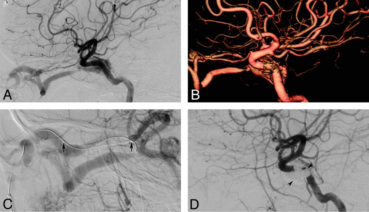
  • Fig 3.
    • Download figure
    • Open in new tab
    • Download powerpoint
    Fig 3.

    A 52-year-old man presented with severe headache, slurred speech, and acute left hemiparesis. A, Noncontrast CT reveals a large right frontoparietal hematoma with intraventricular extension. B–D, A DAVF was suspected on MR imaging (not shown), and DSA was performed. Right (B), left (C), and bilateral (D) external carotid artery injections confirm a convexity DAVF (Borden type 3) with arterial supply from the bilateral middle meningeal and superficial temporal arteries. Transarterial treatment was planned by using the right middle meningeal artery approach (D). A microcatheter was navigated close to the fistula, and the embolization was performed with 2.3 mL of Onyx (E). Note the penetration of Onyx into the arteriovenous junction and the proximal vein (asterisk) as well as arterioarterial reflux (arrows) into the contralateral feeders. F, Ipsilateral (not shown) and contralateral external carotid artery injections confirm complete occlusion of the fistula. The patient made a remarkable recovery during the next 3 months and has mild residual left-arm weakness.

  • Fig 4.
    • Download figure
    • Open in new tab
    • Download powerpoint
    Fig 4.

    A 43-year-old woman with Factor V Leiden mutation developed a spontaneous left-sided dural type (indirect) carotid cavernous fistula (CCF) with proptosis, chemosis, and left cranial nerve VI paralysis. A and B, Lateral view of the left internal carotid artery (ICA) injection and a left ICA 3D reconstruction demonstrates a moderate-flow CCF supplied from multiple left cavernous ICA branches. Additional supply also originated from left external carotid artery (ECA) and right ECA branches (not shown). Given the small size and multiplicity of feeders, transvenous treatment was favored. C and D, A microcatheter was navigated from the left facial vein into the left superior ophthalmic vein (arrows). The cavernous sinus was filled with coils (arrowhead), resulting in complete obliteration of the fistula (D). The proptosis and chemosis resolved the same day, and improvement of left cranial nerve VI function was already evident on postoperative day 1.
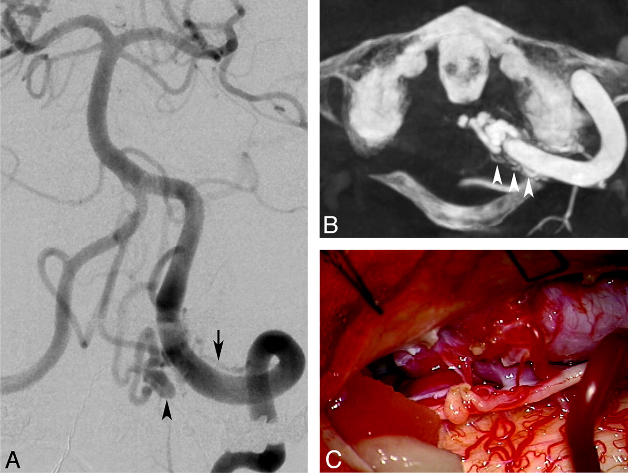
  • Fig 5.
    • Download figure
    • Open in new tab
    • Download powerpoint
    Fig 5.

    A 58-year-old male patient presented with thunderclap headache and a small subarachnoid hemorrhage centered at the foramen magnum. A, Left vertebral angiogram demonstrates a dural fistula at the foramen magnum adjacent to the V4 segment of the left vertebral artery. A dominant feeder is noted arising from the left vertebral artery (arrow), and there is a focal venous varix (arrowhead). The venous drainage was along the perimesencephalic vein into the left superior petrosal sinus (not shown). B, Axial DynaCT (Siemens, Erlangen, Germany) reconstruction reveals multiple additional feeders (arrows) that were difficult to appreciate on 2D DSA. The patient underwent an embolization of the larger feeder transarterially, but complete occlusion of the fistula was not accomplished. C, The residual fistula was surgically occluded. A partial C1 laminectomy and suboccipital craniotomy were fashioned. Surgical disconnection of the venous outflow was performed, resulting in complete obliteration of the fistula. The patient made an uncomplicated and complete recovery.

Tables

  • Figures
    • View popup
    Table 1:

    Borden classification of DAVFs12

    TypeVenous Drainage SiteCVD
    “Benign”
        IDural sinusNo
    “Aggressive”
        IIDural sinusYes
        IIICortical veinYes
    • View popup
    Table 2:

    Cognard classification of DAVFs13

    TypeVenous DrainageFlow Pattern in SinusCVD
    “Benign”
        IDural sinusAntegradeNo
        IIaDural sinusRetrogradeNo
    “Aggressive”
        IIbDural sinusAntegradeYes
        IIa+bDural sinusRetrogradeYes
        IIICortical veinYes
        IVCortical veinYes + venous ectasia
        VCortical vein with spinal perimedullary drainageYes
PreviousNext
Back to top

In this issue

American Journal of Neuroradiology: 33 (6)
American Journal of Neuroradiology
Vol. 33, Issue 6
1 Jun 2012
  • Table of Contents
  • Index by author
Advertisement
Print
Download PDF
Email Article

Thank you for your interest in spreading the word on American Journal of Neuroradiology.

NOTE: We only request your email address so that the person you are recommending the page to knows that you wanted them to see it, and that it is not junk mail. We do not capture any email address.

Enter multiple addresses on separate lines or separate them with commas.
Intracranial Dural Arteriovenous Fistulas: Classification, Imaging Findings, and Treatment
(Your Name) has sent you a message from American Journal of Neuroradiology
(Your Name) thought you would like to see the American Journal of Neuroradiology web site.
CAPTCHA
This question is for testing whether or not you are a human visitor and to prevent automated spam submissions.
Cite this article
D. Gandhi, J. Chen, M. Pearl, J. Huang, J.J. Gemmete, S. Kathuria
Intracranial Dural Arteriovenous Fistulas: Classification, Imaging Findings, and Treatment
American Journal of Neuroradiology Jun 2012, 33 (6) 1007-1013; DOI: 10.3174/ajnr.A2798

Citation Manager Formats

  • BibTeX
  • Bookends
  • EasyBib
  • EndNote (tagged)
  • EndNote 8 (xml)
  • Medlars
  • Mendeley
  • Papers
  • RefWorks Tagged
  • Ref Manager
  • RIS
  • Zotero
0 Responses
Respond to this article
Share
Bookmark this article
Intracranial Dural Arteriovenous Fistulas: Classification, Imaging Findings, and Treatment
D. Gandhi, J. Chen, M. Pearl, J. Huang, J.J. Gemmete, S. Kathuria
American Journal of Neuroradiology Jun 2012, 33 (6) 1007-1013; DOI: 10.3174/ajnr.A2798
del.icio.us logo Twitter logo Facebook logo Mendeley logo
  • Tweet Widget
  • Facebook Like
  • Google Plus One
Purchase

Jump to section

  • Article
    • Abstract
    • ABBREVIATIONS:
    • Etiopathogenesis
    • Clinical Presentation
    • Diagnosis
    • Treatment
    • Endovascular Therapy
    • TAE
    • TVE
    • Surgery
    • SRS
    • Conclusions
    • Acknowledgments
    • Footnotes
    • References
  • Figures & Data
  • Info & Metrics
  • Responses
  • References
  • PDF

Related Articles

  • No related articles found.
  • PubMed
  • Google Scholar

Cited By...

  • 'An eye for an eye' therapeutic strategy for cavernous sinus dural arteriovenous fistula: a single-center experience
  • Exploring the 'Visible Versus Invisible Paradigm in Cavernous Sinus Dural Arteriovenous Fistula
  • Long-term risk of hemorrhage and mortality after treatment of high-grade intracranial dural arteriovenous fistulas
  • Surgical Burr Hole Access for Direct Sinus Puncture and Transvenous Coil Embolization of a Complex Superior Sagittal Sinus Dural Arteriovenous Fistula
  • Unusual cause of treatable Parkinsonism
  • The VEBAS score: a practical scoring system for intracranial dural arteriovenous fistula obliteration
  • The 'corkscrew sign: an indirect MRI hint for intracranial venous hypertension
  • Clinical Reasoning: A 49-Year-Old Woman With Isolated Sinus Intracranial Dural Arteriovenous Fistula With Perimedullary Drainage
  • The VEBAS score: a practical scoring system for intracranial dural arteriovenous fistula obliteration
  • Association between Dural AVFs and Cerebral Venous Thrombosis
  • Embolization strategies for intracranial dural arteriovenous fistulas with an isolated sinus: a single-center experience in 20 patients
  • Spinal Dural Arteriovenous Fistula: The Missing-Piece Sign
  • Clinical Reasoning: Rapidly Progressive Thalamic Dementia
  • Dural arteriovenous fistula presenting with dementia and bulbar symptoms
  • The Influence of Angioarchitectural Features on the Success of Endovascular Embolization of Cranial Dural Arteriovenous Fistulas with Onyx
  • Catch me if you can: disappearing and reappearing posterior fossa dural arteriovenous malformation
  • Transvenous coil embolization with intra-operative cone beam CT assistance in the treatment of hypoglossal canal dural arteriovenous fistulae
  • Engorged medullary vein on CT angiography in patients with dural arteriovenous fistula: prevalence, types, and comparison between regional and extensive types
  • Giant arachnoid granulation with a thrombosed dural arteriovenous fistula
  • Concomitant conus medullaris arteriovenous shunts and sacral dural arteriovenous fistulas: pathophysiological links related to the venous drainage of the lesions in a series of five cases
  • 3D Deep Learning Angiography (3D-DLA) from C-arm Conebeam CT
  • Imaging features and prognostic factors in fetal and postnatal torcular dural sinus malformations, part II: synthesis of the literature and patient management
  • Pressure Mapping and Hemodynamic Assessment of Intracranial Dural Sinuses and Dural Arteriovenous Fistulas with 4D Flow MRI
  • 4D DSA for Dynamic Visualization of Cerebral Vasculature: A Single-Center Experience in 26 Cases
  • Anterior condylar confluence dural arteriovenous fistula: a rare cause of hoarseness
  • A woman with a red eye from a carotid-cavernous sinus fistula
  • Intracranial Arteriovenous Shunting: Detection with Arterial Spin-Labeling and Susceptibility-Weighted Imaging Combined
  • Embolization of Intracranial Dural Arteriovenous Fistulas Using PHIL Liquid Embolic Agent in 26 Patients: A Multicenter Study
  • Ocular Signs Caused by Dural Arteriovenous Fistula without Involvement of the Cavernous Sinus: A Case Series with Review of the Literature
  • Intracranial Dural Arteriovenous Fistulae: Clinical Presentation and Management Strategies
  • Clinical Reasoning: An unusual case of subacute encephalopathy
  • Posterior fossa dural arteriovenous fistula presenting clinically as a carotid-cavernous fistula treated by a direct access cavernous sinus approach
  • Dural arteriovenous fistula presenting with progressive dementia and parkinsonism
  • Transarterial treatment with Onyx of Cognard type IV anterior cranial fossa dural arteriovenous fistulas
  • Posterior fossa dural arteriovenous fistula presenting clinically as a carotid-cavernous fistula treated by a direct access cavernous sinus approach
  • Crossref (330)
  • Google Scholar

This article has been cited by the following articles in journals that are participating in Crossref Cited-by Linking.

  • Intracranial Dural Arteriovenous Fistulae
    Matthew R. Reynolds, Giuseppe Lanzino, Gregory J. Zipfel
    Stroke 2017 48 5
  • Intracranial Dural Arteriovenous Fistulae
    Timothy R. Miller, Dheeraj Gandhi
    Stroke 2015 46 7
  • ACR Appropriateness Criteria ® Cerebrovascular Disease
    Michael B. Salmela, Shabnam Mortazavi, Bharathi D. Jagadeesan, Daniel F. Broderick, Judah Burns, Tejaswini K. Deshmukh, H. Benjamin Harvey, Jenny Hoang, Christopher H. Hunt, Tabassum A. Kennedy, Alexander A. Khalessi, William Mack, Nandini D. Patel, Joel S. Perlmutter, Bruno Policeni, Jason W. Schroeder, Gavin Setzen, Matthew T. Whitehead, Rebecca S. Cornelius, Amanda S. Corey
    Journal of the American College of Radiology 2017 14 5
  • Dural arteriovenous fistulas of the hypoglossal canal: systematic review on imaging anatomy, clinical findings, and endovascular management
    Björn Spittau, Diego San Millán, Saad El-Sherifi, Claudia Hader, Tejinder Pal Singh, Edith Motschall, Werner Vach, Horst Urbach, Stephan Meckel
    Journal of Neurosurgery 2015 122 4
  • Interplay between VEGF and Nrf2 regulates angiogenesis due to intracranial venous hypertension
    Liwen Li, Hao Pan, Handong Wang, Xiang Li, Xiaomin Bu, Qiang Wang, Yongyue Gao, Guodao Wen, Yali Zhou, Zixiang Cong, Youqing Yang, Chao Tang, Zhengwei Liu
    Scientific Reports 2016 6 1
  • Diagnosis and management of dural arteriovenous fistulas: A 10 years single-center experience
    F. Signorelli, G.M. Della Pepa, G. Sabatino, E. Marchese, G. Maira, A. Puca, A. Albanese
    Clinical Neurology and Neurosurgery 2015 128
  • Intracranial Dural Arteriovenous Fistulas: Clinical Characteristics and Management Based on Location and Hemodynamics
    Jung Tae Oh, Seung Young Chung, Giuseppe Lanzino, Ki Seok Park, Seong Min Kim, Moon Sun Park, Han Kyu Kim
    Journal of Cerebrovascular and Endovascular Neurosurgery 2012 14 3
  • Embolization of Intracranial Dural Arteriovenous Fistulas Using PHIL Liquid Embolic Agent in 26 Patients: A Multicenter Study
    S. Lamin, H.S. Chew, S. Chavda, A. Thomas, M. Piano, L. Quilici, G. Pero, M. Holtmannspolter, M.E. Cronqvist, A. Casasco, L. Guimaraens, L. Paul, A. Gil Garcia, A. Aleu, R. Chapot
    American Journal of Neuroradiology 2017 38 1
  • 4D DSA for Dynamic Visualization of Cerebral Vasculature: A Single-Center Experience in 26 Cases
    S. Lang, P. Gölitz, T. Struffert, J. Rösch, K. Rössler, M. Kowarschik, C. Strother, A. Doerfler
    American Journal of Neuroradiology 2017 38 6
  • A pivotal role of the vascular endothelial growth factor signaling pathway in the formation of venous hypertension-induced dural arteriovenous fistulas
    QIANG LI, QI ZHANG, QING-HAI HUANG, YI-BIN FANG, ZHAO-LONG ZHANG, YI XU, JIAN-MIN LIU
    Molecular Medicine Reports 2014 9 5

More in this TOC Section

  • An Atlas of Neonatal Neurovascular Imaging Anatomy as Depicted with Microvascular Imaging: The Intracranial Arteries
  • An Atlas of Neonatal Neurovascular Imaging Anatomy as Depicted with Microvascular Imaging: The Intracranial Veins
  • Clinical Translation of Hyperpolarized 13C Metabolic Probes for Glioma Imaging
Show more Review Article

Similar Articles

Advertisement

Indexed Content

  • Current Issue
  • Accepted Manuscripts
  • Article Preview
  • Past Issues
  • Editorials
  • Editor's Choice
  • Fellows' Journal Club
  • Letters to the Editor
  • Video Articles

Cases

  • Case Collection
  • Archive - Case of the Week
  • Archive - Case of the Month
  • Archive - Classic Case

Special Collections

  • AJNR Awards
  • ASNR Foundation Special Collection
  • Most Impactful AJNR Articles
  • Photon-Counting CT
  • Spinal CSF Leak Articles (Jan 2020-June 2024)

More from AJNR

  • Trainee Corner
  • Imaging Protocols
  • MRI Safety Corner

Multimedia

  • AJNR Podcasts
  • AJNR Scantastics

Resources

  • Turnaround Time
  • Submit a Manuscript
  • Submit a Video Article
  • Submit an eLetter to the Editor/Response
  • Manuscript Submission Guidelines
  • Statistical Tips
  • Fast Publishing of Accepted Manuscripts
  • Graphical Abstract Preparation
  • Imaging Protocol Submission
  • Evidence-Based Medicine Level Guide
  • Publishing Checklists
  • Author Policies
  • Become a Reviewer/Academy of Reviewers
  • News and Updates

About Us

  • About AJNR
  • Editorial Board
  • Editorial Board Alumni
  • Alerts
  • Permissions
  • Not an AJNR Subscriber? Join Now
  • Advertise with Us
  • Librarian Resources
  • Feedback
  • Terms and Conditions
  • AJNR Editorial Board Alumni

American Society of Neuroradiology

  • Not an ASNR Member? Join Now

© 2025 by the American Society of Neuroradiology All rights, including for text and data mining, AI training, and similar technologies, are reserved.
Print ISSN: 0195-6108 Online ISSN: 1936-959X

Powered by HighWire