
Article Figures & Data
Figures
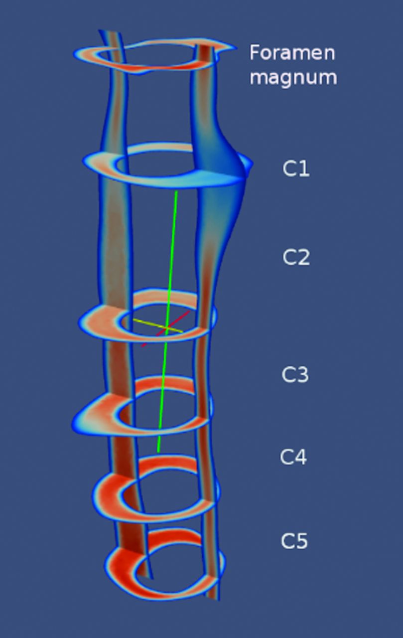
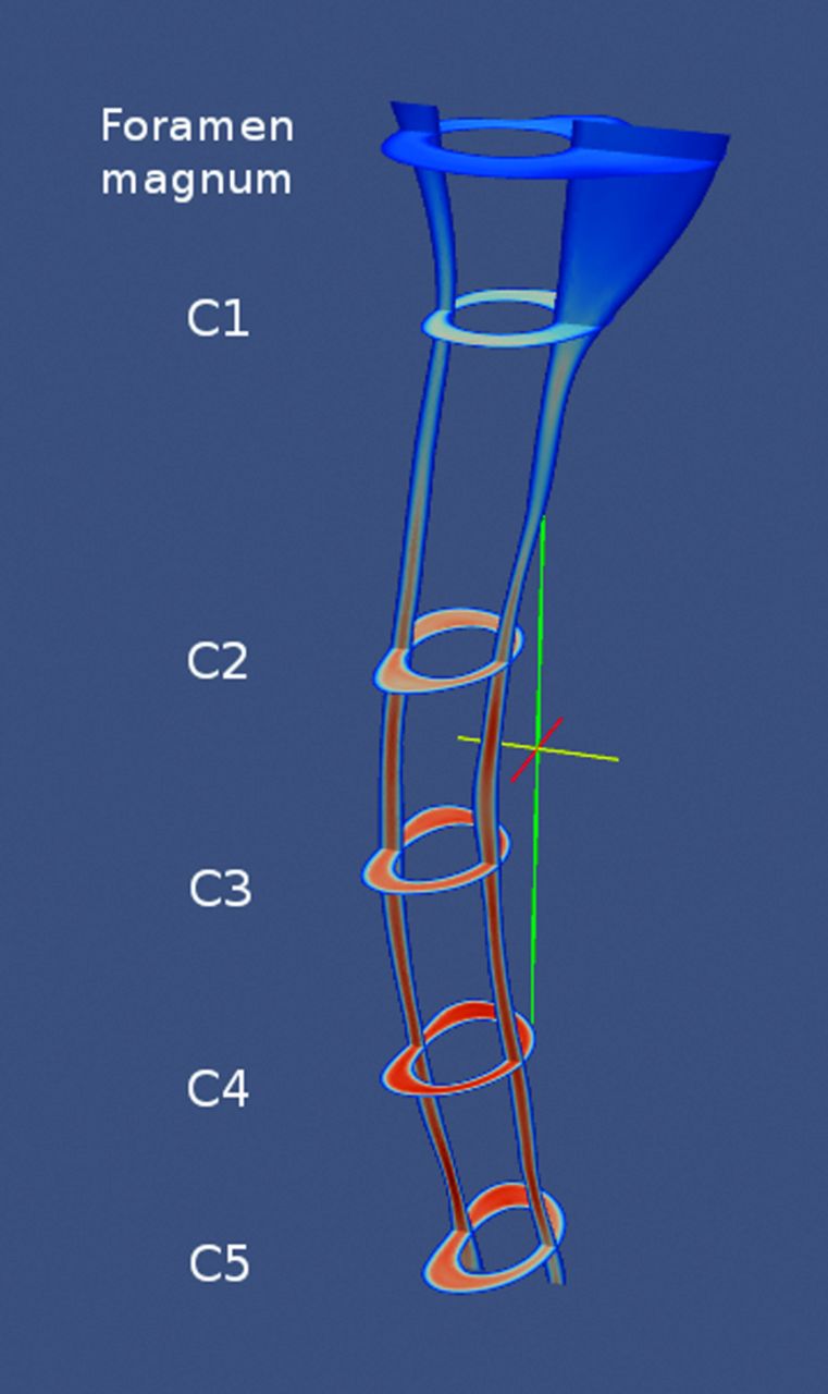
- Fig 1.
Model of the subarachnoid space for 1 subject.
- Fig 2.
Sagittal MR image (A) showing the axial image locations and an example of a Paraview image of the velocities computed for the sagittal and the axial locations (B).
- Fig 3.
Paraview image showing an example of systolic velocities in sagittal and multiple axial planes in a typical patient with a Chiari I malformation. Note that the C5 level has the fastest CSF velocities.
- Fig 4.
Paraview image showing an example of systolic velocities in sagittal and multiple axial planes in a volunteer. C4 has the peak CSF velocities.
- Fig 5.
Paraview snapshot image showing an example of velocities during systole in a patient with Chiari I post–craniovertebral decompression.
- Fig 6.
Paraview image showing an example of flow jets at multiple axial levels in a patient with Chiari I (A) and at the level of C4 in the same patient (B).
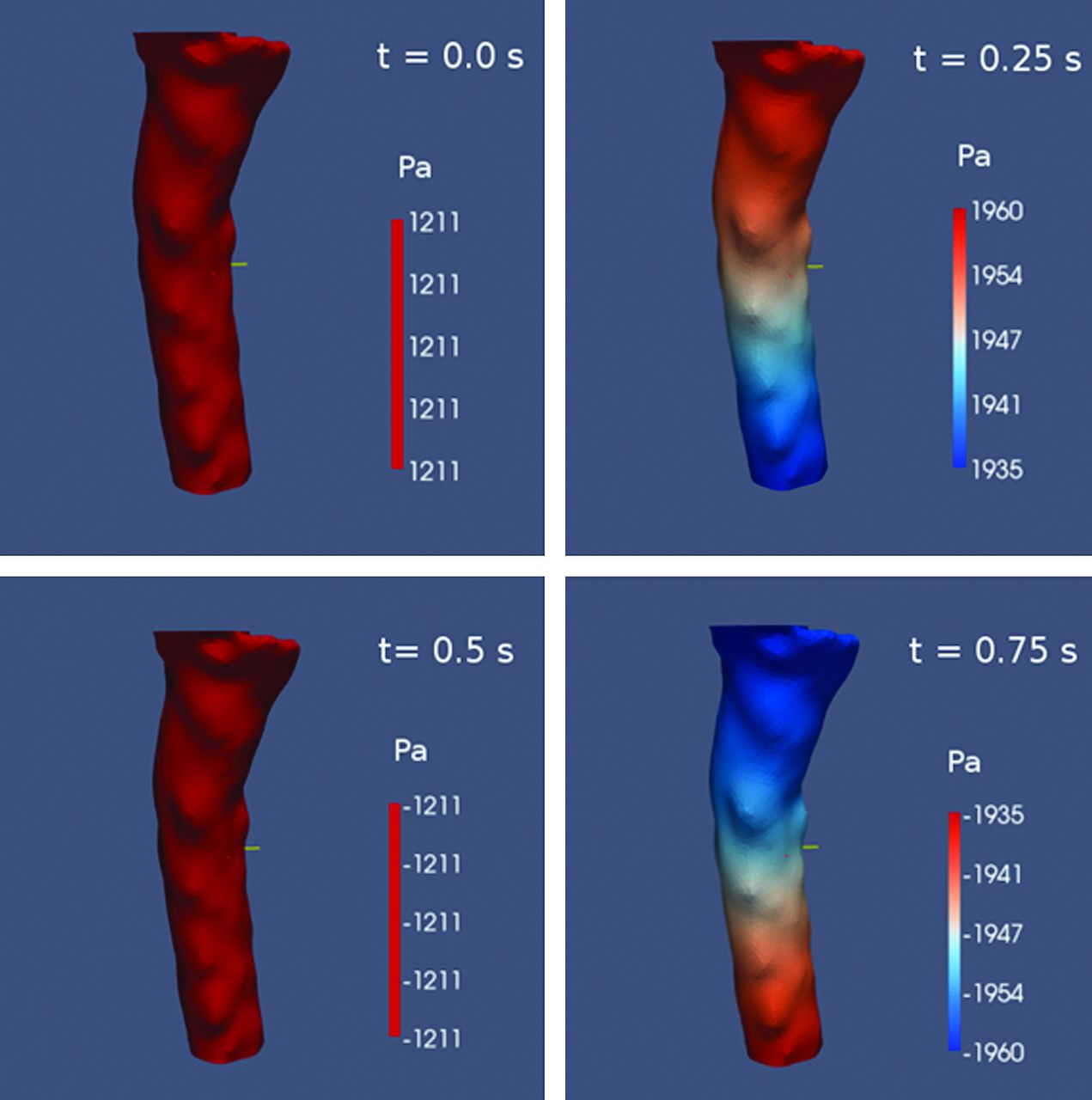
- Fig 7.
Paraview image of the pressure gradients at 4 times during the cardiac cycle. Pressure gradient has maximum value at t = 0.25 and 0.75 when flow is reversing and equals zero at t = 0 and 0.5 when CSF velocity is near maximum.
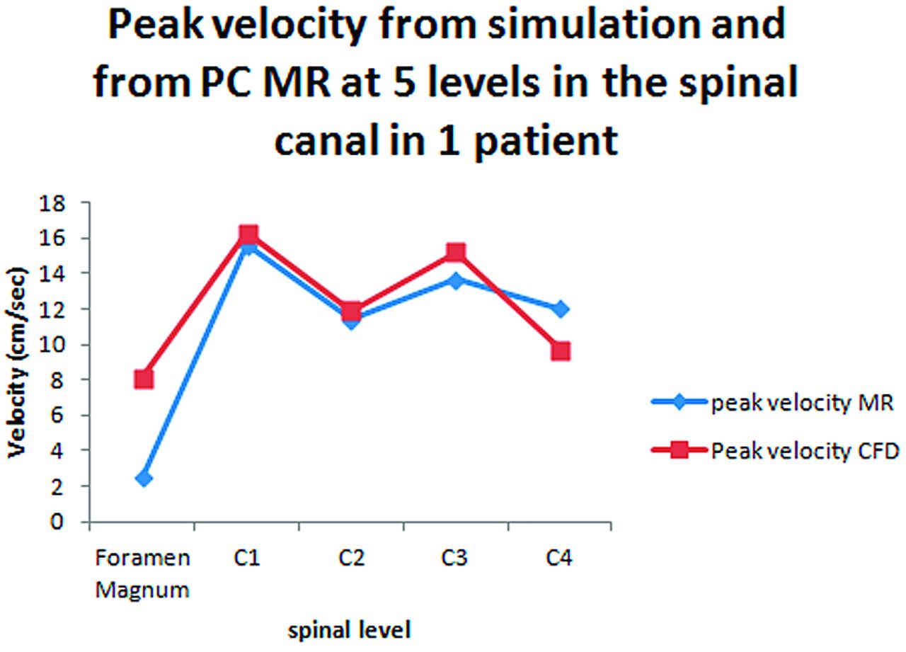
- Fig 8.
Comparison of velocities in simulations with velocities measured with PCMR in 5 patients (A) and in 5 locations in 1 patient (B).


Tables
- Table 1:
Gender, age, tonsilar herniation, and presence of syrinx in the patients with Chiari I
Chiari I Patients Sex Age Tonsilar Herniation (mm) Syrinx 1 M 2 12 No 2 M 4 9 No 3 F 5 13 No 4 F 30 6 No 5 F 40 5 No 6 M 10 5 No 7 F 16 5 No Controls Sex Age Status 8 NA Adult volunteer 9 NA Adult volunteer 10 F 13 Patient with no suspected CSF flow abnormality -
Note:—NA indicates not available.
-
Patients with Chiari I Post–Craniovertebral Decompression Sex Age Syrinx 11 M 54 C1 to T10 12 F 31 Cervical 13 M 3 No - Table 4:
Peak systolic and diastolic velocities and synchronous bidirectional flow in the patients with Chiari I, controls, and postoperative patients
Peak Systolic Velocity (cm/s) Peak Diastolic Velocity (cm/s) Magnitude of Bidirectional Flow (cm/s) Duration of Bidirectional Flow (sec) Patients with Chiari I 1 12.1 10.9 7.2 0.24 2 11.9 11.8 7.6 0.30 3 11.5 11.4 7.5 0.22 4 11.4 8.8 6.0 0.38 5 9.4 8.0 5.4 0.24 6 7.7 7.6 4.9 0.20 7 7.5 6.9 4.5 0.24 Average 10.2 9.3 6.2 0.26 Controls 8 6.8 6.5 3.8 0.22 9 7.5 7.4 3.2 0.22 10 7.9 7.5 5.3 0.18 Average 7.4 7.1 4.1 0.20 Postoperative patients with Chiari I 11 10.0 9.9 5.5 0.22 12 8.3 7.8 4.4 0.22 13 15.6 15.7 7.3 0.18 Average 11.3 11.1 5.7 0.21


