Advertisement

Main menu

  • Home
  • Content
    • Current Issue
    • Accepted Manuscripts
    • Article Preview
    • Past Issue Archive
    • Video Articles
    • AJNR Case Collection
    • Case of the Week Archive
    • Case of the Month Archive
    • Classic Case Archive
  • Special Collections
    • AJNR Awards
    • ASNR Foundation Special Collection
    • Most Impactful AJNR Articles
    • Photon-Counting CT
    • Spinal CSF Leak Articles (Jan 2020-June 2024)
  • Multimedia
    • AJNR Podcasts
    • AJNR SCANtastic
    • Trainee Corner
    • MRI Safety Corner
    • Imaging Protocols
  • For Authors
    • Submit a Manuscript
    • Submit a Video Article
    • Submit an eLetter to the Editor/Response
    • Manuscript Submission Guidelines
    • Statistical Tips
    • Fast Publishing of Accepted Manuscripts
    • Graphical Abstract Preparation
    • Imaging Protocol Submission
    • Author Policies
  • About Us
    • About AJNR
    • Editorial Board
    • Editorial Board Alumni
  • More
    • Become a Reviewer/Academy of Reviewers
    • Subscribers
    • Permissions
    • Alerts
    • Feedback
    • Advertisers
    • ASNR Home

User menu

  • Alerts
  • Log in

Search

  • Advanced search
American Journal of Neuroradiology

American Journal of Neuroradiology

ASHNR American Society of Functional Neuroradiology ASHNR American Society of Pediatric Neuroradiology ASSR
  • Alerts
  • Log in

Advanced Search

  • Home
  • Content
    • Current Issue
    • Accepted Manuscripts
    • Article Preview
    • Past Issue Archive
    • Video Articles
    • AJNR Case Collection
    • Case of the Week Archive
    • Case of the Month Archive
    • Classic Case Archive
  • Special Collections
    • AJNR Awards
    • ASNR Foundation Special Collection
    • Most Impactful AJNR Articles
    • Photon-Counting CT
    • Spinal CSF Leak Articles (Jan 2020-June 2024)
  • Multimedia
    • AJNR Podcasts
    • AJNR SCANtastic
    • Trainee Corner
    • MRI Safety Corner
    • Imaging Protocols
  • For Authors
    • Submit a Manuscript
    • Submit a Video Article
    • Submit an eLetter to the Editor/Response
    • Manuscript Submission Guidelines
    • Statistical Tips
    • Fast Publishing of Accepted Manuscripts
    • Graphical Abstract Preparation
    • Imaging Protocol Submission
    • Author Policies
  • About Us
    • About AJNR
    • Editorial Board
    • Editorial Board Alumni
  • More
    • Become a Reviewer/Academy of Reviewers
    • Subscribers
    • Permissions
    • Alerts
    • Feedback
    • Advertisers
    • ASNR Home
  • Follow AJNR on Twitter
  • Visit AJNR on Facebook
  • Follow AJNR on Instagram
  • Join AJNR on LinkedIn
  • RSS Feeds

AJNR is seeking candidates for the AJNR Podcast Editor. Read the position description.

Research ArticleBrain
Open Access

Effect of Age on MRI Phase Behavior in the Subcortical Deep Gray Matter of Healthy Individuals

J. Hagemeier, M.G. Dwyer, N. Bergsland, F. Schweser, C.R. Magnano, M. Heininen-Brown, D.P. Ramasamy, E. Carl, C. Kennedy, R. Melia, P. Polak, A. Deistung, J.J.G. Geurts, J.R. Reichenbach and R. Zivadinov
American Journal of Neuroradiology November 2013, 34 (11) 2144-2151; DOI: https://doi.org/10.3174/ajnr.A3569
J. Hagemeier
aFrom the Buffalo Neuroimaging Analysis Center (J.H., M.G.D., N.B., C.R.M., M.H.-B., D.P.R., E.C., C.K., R.M., P.P., R.Z.), Department of Neurology, University at Buffalo, Buffalo, New York
  • Find this author on Google Scholar
  • Find this author on PubMed
  • Search for this author on this site
M.G. Dwyer
aFrom the Buffalo Neuroimaging Analysis Center (J.H., M.G.D., N.B., C.R.M., M.H.-B., D.P.R., E.C., C.K., R.M., P.P., R.Z.), Department of Neurology, University at Buffalo, Buffalo, New York
  • Find this author on Google Scholar
  • Find this author on PubMed
  • Search for this author on this site
N. Bergsland
aFrom the Buffalo Neuroimaging Analysis Center (J.H., M.G.D., N.B., C.R.M., M.H.-B., D.P.R., E.C., C.K., R.M., P.P., R.Z.), Department of Neurology, University at Buffalo, Buffalo, New York
  • Find this author on Google Scholar
  • Find this author on PubMed
  • Search for this author on this site
F. Schweser
bMedical Physics Group (F.S., A.D., J.R.R.), Institute of Diagnostic and Interventional Radiology I, Jena University Hospital–Friedrich Schiller University, Jena, Germany
  • Find this author on Google Scholar
  • Find this author on PubMed
  • Search for this author on this site
C.R. Magnano
aFrom the Buffalo Neuroimaging Analysis Center (J.H., M.G.D., N.B., C.R.M., M.H.-B., D.P.R., E.C., C.K., R.M., P.P., R.Z.), Department of Neurology, University at Buffalo, Buffalo, New York
  • Find this author on Google Scholar
  • Find this author on PubMed
  • Search for this author on this site
M. Heininen-Brown
aFrom the Buffalo Neuroimaging Analysis Center (J.H., M.G.D., N.B., C.R.M., M.H.-B., D.P.R., E.C., C.K., R.M., P.P., R.Z.), Department of Neurology, University at Buffalo, Buffalo, New York
  • Find this author on Google Scholar
  • Find this author on PubMed
  • Search for this author on this site
D.P. Ramasamy
aFrom the Buffalo Neuroimaging Analysis Center (J.H., M.G.D., N.B., C.R.M., M.H.-B., D.P.R., E.C., C.K., R.M., P.P., R.Z.), Department of Neurology, University at Buffalo, Buffalo, New York
  • Find this author on Google Scholar
  • Find this author on PubMed
  • Search for this author on this site
E. Carl
aFrom the Buffalo Neuroimaging Analysis Center (J.H., M.G.D., N.B., C.R.M., M.H.-B., D.P.R., E.C., C.K., R.M., P.P., R.Z.), Department of Neurology, University at Buffalo, Buffalo, New York
  • Find this author on Google Scholar
  • Find this author on PubMed
  • Search for this author on this site
C. Kennedy
aFrom the Buffalo Neuroimaging Analysis Center (J.H., M.G.D., N.B., C.R.M., M.H.-B., D.P.R., E.C., C.K., R.M., P.P., R.Z.), Department of Neurology, University at Buffalo, Buffalo, New York
  • Find this author on Google Scholar
  • Find this author on PubMed
  • Search for this author on this site
R. Melia
aFrom the Buffalo Neuroimaging Analysis Center (J.H., M.G.D., N.B., C.R.M., M.H.-B., D.P.R., E.C., C.K., R.M., P.P., R.Z.), Department of Neurology, University at Buffalo, Buffalo, New York
  • Find this author on Google Scholar
  • Find this author on PubMed
  • Search for this author on this site
P. Polak
aFrom the Buffalo Neuroimaging Analysis Center (J.H., M.G.D., N.B., C.R.M., M.H.-B., D.P.R., E.C., C.K., R.M., P.P., R.Z.), Department of Neurology, University at Buffalo, Buffalo, New York
  • Find this author on Google Scholar
  • Find this author on PubMed
  • Search for this author on this site
A. Deistung
bMedical Physics Group (F.S., A.D., J.R.R.), Institute of Diagnostic and Interventional Radiology I, Jena University Hospital–Friedrich Schiller University, Jena, Germany
  • Find this author on Google Scholar
  • Find this author on PubMed
  • Search for this author on this site
J.J.G. Geurts
cDepartment of Anatomy and Neurosciences (J.J.G.G.), Section of Clinical Neuroscience, VU University Medical Center, Amsterdam, The Netherlands.
  • Find this author on Google Scholar
  • Find this author on PubMed
  • Search for this author on this site
J.R. Reichenbach
bMedical Physics Group (F.S., A.D., J.R.R.), Institute of Diagnostic and Interventional Radiology I, Jena University Hospital–Friedrich Schiller University, Jena, Germany
  • Find this author on Google Scholar
  • Find this author on PubMed
  • Search for this author on this site
R. Zivadinov
aFrom the Buffalo Neuroimaging Analysis Center (J.H., M.G.D., N.B., C.R.M., M.H.-B., D.P.R., E.C., C.K., R.M., P.P., R.Z.), Department of Neurology, University at Buffalo, Buffalo, New York
  • Find this author on Google Scholar
  • Find this author on PubMed
  • Search for this author on this site
  • Article
  • Figures & Data
  • Supplemental
  • Info & Metrics
  • Responses
  • References
  • PDF
Loading

Article Figures & Data

Figures

  • Tables
  • Fig 1.
    • Download figure
    • Open in new tab
    • Download powerpoint
    Fig 1.

    Phase maps (upper row) and thresholded low-phase voxels used in mean phase of low-phase voxel (MP-LPV) calculation (bottom row) of subcortical deep gray matter structures. MP-LPV was determined by thresholding the phase images to retain only those voxels with phase values lower than 2 standard deviations below reference group. Upper and lower rows show the same subjects, and each column contains a representative subject from each age group (left to right, <25 years, 25–39 years, 40–55 years, >55 years).
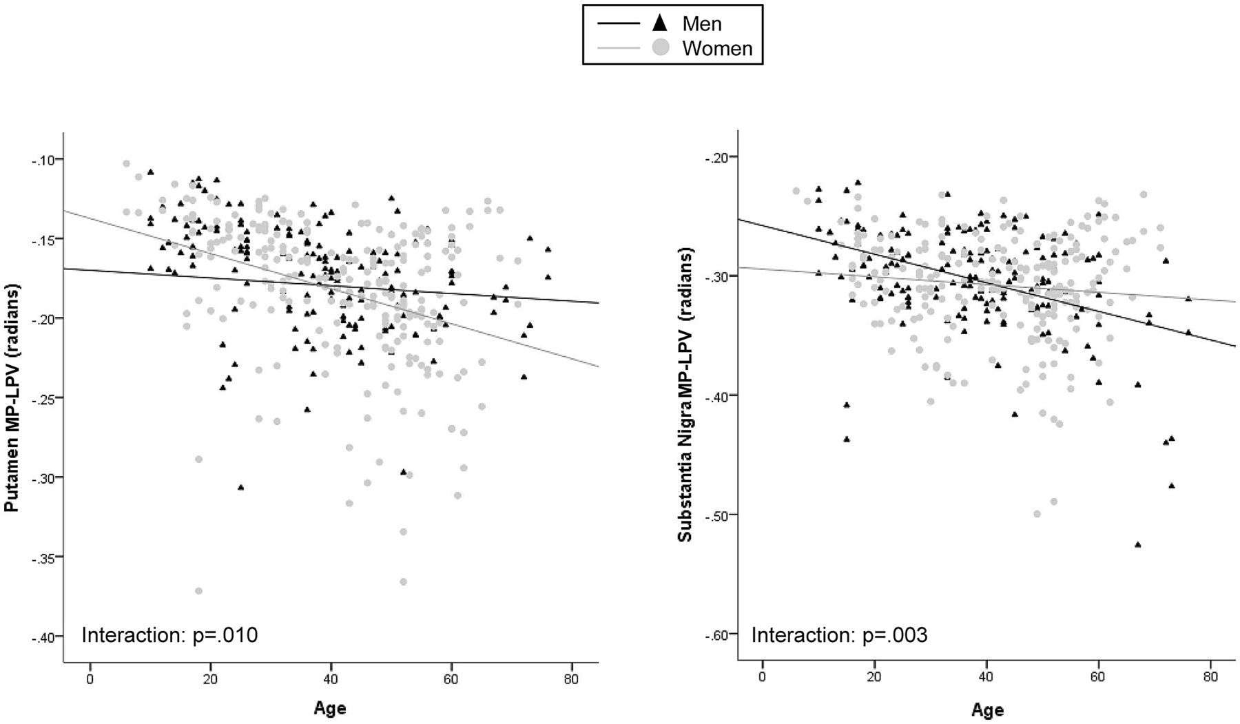
  • Fig 2.
    • Download figure
    • Open in new tab
    • Download powerpoint
    Fig 2.

    Scatterplots showing significant interaction effects between sex and the mean phase of low-phase voxels (MP-LPV) of the putamen and substantia nigra.

Tables

  • Figures
    • View popup
    Table 1:

    Demographic characteristics and global volumetric MRI measures of study participants

    TotalMaleFemaleP
    n (%)21089 (42.4)121 (57.6)
    Age (SD) median39.8 (15.5) 4138 (16.1) 3841.1 (14.9) 44.202
    Race, n (%) of available cases
        White139 (87.4)58 (87.9)81 (87.1).617
        African American13 (8.2)5 (7.6)8 (8.6)
        Other7 (4.4)3 (4.5)4 (4.3)
        Education, n (%) of available cases
        No high school15 (9.7)7 (11.3)8 (8.7).893
        High school24 (15.6)11 (17.7)13 (14.1)
        Some college32 (20.8)14 (22.6)18 (19.6)
        Associate/technical22 (14.3)7 (11.3)15 (16.3)
        Bachelor level36 (23.4)12 (19.4)24 (26.1)
        Graduate level23 (14.9)10 (16.1)13 (14.1)
        Postgraduate2 (1.3)1 (1.6)1 (1.1)
    MRI volumes in cm3, mean (SD)
        WM hyperintensities1.26 (4.47)1.74 (6.50)0.87 (1.29).160
        GM791.93 (65.56)787.64 (69.36)795.02 (62.81).525
        Cortical GM644.30 (56.33)637.10 (62.11)649.56 (51.36).181
        WM761.95 (41.39)771.66 (39.57)754.97 (41.41).008
        Lateral ventricles31.66 (12.70)33.46 (13.36)30.35 (12.07).136
    • Note:—Differences between men and women were assessed by means of the χ2 test (race and education) and Student t test (age and normalized volumes).

    • View popup
    Table 2:

    Sex differences of mean phase, mean phase of low-phase voxels, and normalized volume values of subcortical deep gray matter structures

    Mean PhaseMP-LPVNormalized Volume
    Male (n = 89)Female (n = 121)PMale (n = 89)Female (n = 121)PMale (n = 89)Female (n = 121)P
    Total SDGM−.012 (.014)−.016 (.014).036−.144 (.022)−.149 (.027).57349.209 (5.474)44.442 (3.551)<.001
    Caudate−.054 (.017)−.064 (.018).001−.170 (.016)−.167 (.014).5257.398 (1.079)6.832 (.890)<.001
    Putamen−.018 (.028)−.027 (.029).028−.175 (.039)−.183 (.047).48710.471 (1.386)9.426 (.914)<.001
    Globus pallidus−.016 (.025)−.015 (.024).643−.184 (.033)−.183 (.042).3483.809 (.442)3.431 (.399)<.001
    Thalamus.005 (.008).003 (.009).210−.095 (.015)−.091 (.013).15716.474 (1.923)14.661 (1.188)<.001
    Pulvinar−.042 (.030)−.042 (.028).657−.139 (.017)−.140 (.018).689.456 (.092).419 (.090).010
    Hippocampus.024 (.032).031 (.039).073−.161 (.044)−.165 (.049).8227.559 (.956)6.981 (.817)<.001
    Amygdala.011 (.039)−.007 (.066).086−.214 (.051)−.210 (.075).0862.634 (.424)2.326 (.307)<.001
    Accumbens−.179 (.140)−.230 (.163).035−.744 (.203)−.80 (.252).298.865 (.199).784 (.189).006
    Red nucleus−.050 (.057)−.059 (.051).331−.238 (.027)−.232 (.026).225.179 (.023).162 (.022)<.001
    Substantia nigra−.097 (.056)−.096 (.057).765−.305 (.039)−.308 (.041).574.305 (.048).282 (.053).005
    • Note:—Results are shown as mean (standard deviation); volume measurements are expressed in cubic centimeters. Mean phase and mean phase of low-phase tissue (MP-LPV) measurements are expressed in radians. Statistical analyses were carried out by use of independent-samples t test and Mann-Whitney U test.

    • View popup
    Table 3:

    Linear regression analyses assessing the association between age and mean phase, mean phase of low-phase tissue, and normalized volumes, adjusted for sex

    Mean phaseaMP-LPVaNormalized Volume
    βPβPβP
    Total SDGM−.102.057−.255<.001−.245<.001
    Caudate−.102.068−.350<.001−.247<.001
    Putamen−.208<.001−.193<.001−.239<.001
    lobus pallidus.210<.001−.273<.001.049.352
    Thalamus−.178<.001−.521<.001−.299<.001
    Pulvinar nucleus−.384<.001−.340<.001−.105.059
    Hippocampus−.027.645.033.589−.075.163
    Amygdala.039.525−.029.631.069.197
    Accumbens−.051.376.112.043−.271<.001
    Red nucleus−.301<.001−.252<.001−.195<.001
    Substantia nigra−.207.006−.256<.001.023.687
    • ↵a Adjusted for the effects of normalized volume.

    • View popup
    Table 4:

    Proportion of structural low-phase voxels versus structural normalized volume within age groups, adjusted for sex

    Age (years)P
    <25 n = 4325–39 n = 5640–55 n = 72≥55 n = 39
    Total SDGM.29 (.04).32 (.03).34 (.04).35 (.04)<.001
    Caudate.27 (.09).33 (.08).36 (.07).35 (.08)<.001
    Putamen.17 (.08).26 (.06).33 (.07).32 (.09)<.001
    Globus pallidus.42 (.07).40 (.06).38 (.06).35 (.06)<.001
    Thalamus.40 (.03).43 (.03).44 (.04).43 (.04)<.001
    Pulvinar nucleus.11 (.08).24 (.16).28 (.15).29 (.14)<.001
    Hippocampus.15 (.08).19 (.09).18 (.07).18 (.07).319
    Amygdala.17 (.14).18 (.10).17 (.10).16 (.08).738
    Accumbens.15 (.14).14 (.14).12 (.13).13 (.16).703
    Red nucleus.05 (.05).13 (.12).21 (.13).17 (.11)<.001
    Substantia nigra.17 (.11).23 (.11).27 (.11).24 (.14).002
    • General linear modeling comparing the proportion of structural low-phase voxels versus structural normalized volume between age groups. Results are presented as mean (standard deviation).

PreviousNext
Back to top

In this issue

American Journal of Neuroradiology: 34 (11)
American Journal of Neuroradiology
Vol. 34, Issue 11
1 Nov 2013
  • Table of Contents
  • Index by author
  • Complete Issue (PDF)
Advertisement
Print
Download PDF
Email Article

Thank you for your interest in spreading the word on American Journal of Neuroradiology.

NOTE: We only request your email address so that the person you are recommending the page to knows that you wanted them to see it, and that it is not junk mail. We do not capture any email address.

Enter multiple addresses on separate lines or separate them with commas.
Effect of Age on MRI Phase Behavior in the Subcortical Deep Gray Matter of Healthy Individuals
(Your Name) has sent you a message from American Journal of Neuroradiology
(Your Name) thought you would like to see the American Journal of Neuroradiology web site.
CAPTCHA
This question is for testing whether or not you are a human visitor and to prevent automated spam submissions.
Cite this article
J. Hagemeier, M.G. Dwyer, N. Bergsland, F. Schweser, C.R. Magnano, M. Heininen-Brown, D.P. Ramasamy, E. Carl, C. Kennedy, R. Melia, P. Polak, A. Deistung, J.J.G. Geurts, J.R. Reichenbach, R. Zivadinov
Effect of Age on MRI Phase Behavior in the Subcortical Deep Gray Matter of Healthy Individuals
American Journal of Neuroradiology Nov 2013, 34 (11) 2144-2151; DOI: 10.3174/ajnr.A3569

Citation Manager Formats

  • BibTeX
  • Bookends
  • EasyBib
  • EndNote (tagged)
  • EndNote 8 (xml)
  • Medlars
  • Mendeley
  • Papers
  • RefWorks Tagged
  • Ref Manager
  • RIS
  • Zotero
0 Responses
Respond to this article
Share
Bookmark this article
Effect of Age on MRI Phase Behavior in the Subcortical Deep Gray Matter of Healthy Individuals
J. Hagemeier, M.G. Dwyer, N. Bergsland, F. Schweser, C.R. Magnano, M. Heininen-Brown, D.P. Ramasamy, E. Carl, C. Kennedy, R. Melia, P. Polak, A. Deistung, J.J.G. Geurts, J.R. Reichenbach, R. Zivadinov
American Journal of Neuroradiology Nov 2013, 34 (11) 2144-2151; DOI: 10.3174/ajnr.A3569
del.icio.us logo Twitter logo Facebook logo Mendeley logo
  • Tweet Widget
  • Facebook Like
  • Google Plus One
Purchase

Jump to section

  • Article
    • Abstract
    • ABBREVIATIONS:
    • Materials and Methods
    • Results
    • Discussion
    • Conclusions
    • Acknowledgments
    • Footnotes
    • REFERENCES
  • Figures & Data
  • Supplemental
  • Info & Metrics
  • Responses
  • References
  • PDF

Related Articles

  • PubMed
  • Google Scholar

Cited By...

  • Iron and Volume in the Deep Gray Matter: Association with Cognitive Impairment in Multiple Sclerosis
  • Quantitative 7T Phase Imaging in Premanifest Huntington Disease
  • Crossref (28)
  • Google Scholar

This article has been cited by the following articles in journals that are participating in Crossref Cited-by Linking.

  • Age and sex related differences in subcortical brain iron concentrations among healthy adults
    Ninni Persson, Jianlin Wu, Qing Zhang, Ting Liu, Jing Shen, Ruyi Bao, Mingfei Ni, Tian Liu, Yi Wang, Pascal Spincemaille
    NeuroImage 2015 122
  • MRI of healthy brain aging: A review
    M. Ethan MacDonald, G. Bruce Pike
    NMR in Biomedicine 2021 34 9
  • Brain Iron at Quantitative MRI Is Associated with Disability in Multiple Sclerosis
    Robert Zivadinov, Eleonora Tavazzi, Niels Bergsland, Jesper Hagemeier, Fuchun Lin, Michael G. Dwyer, Ellen Carl, Channa Kolb, David Hojnacki, Deepa Ramasamy, Jacqueline Durfee, Bianca Weinstock-Guttman, Ferdinand Schweser
    Radiology 2018 289 2
  • Mapping of thalamic magnetic susceptibility in multiple sclerosis indicates decreasing iron with disease duration: A proposed mechanistic relationship between inflammation and oligodendrocyte vitality
    Ferdinand Schweser, Ana Luiza Raffaini Duarte Martins, Jesper Hagemeier, Fuchun Lin, Jannis Hanspach, Bianca Weinstock-Guttman, Simon Hametner, Niels Bergsland, Michael G. Dwyer, Robert Zivadinov
    NeuroImage 2018 167
  • Meta-analysis reveals a lack of sexual dimorphism in human amygdala volume
    Dhruv Marwha, Meha Halari, Lise Eliot
    NeuroImage 2017 147
  • MR‐based age‐related effects on the striatum, globus pallidus, and thalamus in healthy individuals across the adult lifespan
    Stephanie Tullo, Raihaan Patel, Gabriel A. Devenyi, Alyssa Salaciak, Saashi A. Bedford, Sarah Farzin, Nancy Wlodarski, Christine L. Tardif, John C. S. Breitner, M. Mallar Chakravarty
    Human Brain Mapping 2019 40 18
  • Changes of deep gray matter magnetic susceptibility over 2 years in multiple sclerosis and healthy control brain
    Jesper Hagemeier, Robert Zivadinov, Michael G. Dwyer, Paul Polak, Niels Bergsland, Bianca Weinstock-Guttman, Joshua Zalis, Andreas Deistung, Jürgen R. Reichenbach, Ferdinand Schweser
    NeuroImage: Clinical 2018 18
  • Decreasing brain iron in multiple sclerosis: The difference between concentration and content in iron MRI
    Ferdinand Schweser, Jesper Hagemeier, Michael G Dwyer, Niels Bergsland, Simon Hametner, Bianca Weinstock‐Guttman, Robert Zivadinov
    Human Brain Mapping 2021 42 5
  • Iron-related gene variants and brain iron in multiple sclerosis and healthy individuals
    Jesper Hagemeier, Murali Ramanathan, Ferdinand Schweser, Michael G. Dwyer, Fuchun Lin, Niels Bergsland, Bianca Weinstock-Guttman, Robert Zivadinov
    NeuroImage: Clinical 2018 17
  • An improved FSL-FIRST pipeline for subcortical gray matter segmentation to study abnormal brain anatomy using quantitative susceptibility mapping (QSM)
    Xiang Feng, Andreas Deistung, Michael G. Dwyer, Jesper Hagemeier, Paul Polak, Jessica Lebenberg, Frédérique Frouin, Robert Zivadinov, Jürgen R. Reichenbach, Ferdinand Schweser
    Magnetic Resonance Imaging 2017 39

More in this TOC Section

  • White Matter Alterations in the Brains of Patients with Active, Remitted, and Cured Cushing Syndrome: A DTI Study
  • Qualitative and Quantitative Analysis of MR Imaging Findings in Patients with Middle Cerebral Artery Stroke Implanted with Mesenchymal Stem Cells
  • Fast Contrast-Enhanced 4D MRA and 4D Flow MRI Using Constrained Reconstruction (HYPRFlow): Potential Applications for Brain Arteriovenous Malformations
Show more BRAIN

Similar Articles

Advertisement

Indexed Content

  • Current Issue
  • Accepted Manuscripts
  • Article Preview
  • Past Issues
  • Editorials
  • Editor's Choice
  • Fellows' Journal Club
  • Letters to the Editor
  • Video Articles

Cases

  • Case Collection
  • Archive - Case of the Week
  • Archive - Case of the Month
  • Archive - Classic Case

Special Collections

  • AJNR Awards
  • ASNR Foundation Special Collection
  • Most Impactful AJNR Articles
  • Photon-Counting CT
  • Spinal CSF Leak Articles (Jan 2020-June 2024)

More from AJNR

  • Trainee Corner
  • Imaging Protocols
  • MRI Safety Corner

Multimedia

  • AJNR Podcasts
  • AJNR Scantastics

Resources

  • Turnaround Time
  • Submit a Manuscript
  • Submit a Video Article
  • Submit an eLetter to the Editor/Response
  • Manuscript Submission Guidelines
  • Statistical Tips
  • Fast Publishing of Accepted Manuscripts
  • Graphical Abstract Preparation
  • Imaging Protocol Submission
  • Evidence-Based Medicine Level Guide
  • Publishing Checklists
  • Author Policies
  • Become a Reviewer/Academy of Reviewers
  • News and Updates

About Us

  • About AJNR
  • Editorial Board
  • Editorial Board Alumni
  • Alerts
  • Permissions
  • Not an AJNR Subscriber? Join Now
  • Advertise with Us
  • Librarian Resources
  • Feedback
  • Terms and Conditions
  • AJNR Editorial Board Alumni

American Society of Neuroradiology

  • Not an ASNR Member? Join Now

© 2025 by the American Society of Neuroradiology All rights, including for text and data mining, AI training, and similar technologies, are reserved.
Print ISSN: 0195-6108 Online ISSN: 1936-959X

Powered by HighWire