Advertisement

Main menu

  • Home
  • Content
    • Current Issue
    • Accepted Manuscripts
    • Article Preview
    • Past Issue Archive
    • Video Articles
    • AJNR Case Collection
    • Case of the Week Archive
    • Case of the Month Archive
    • Classic Case Archive
  • Special Collections
    • AJNR Awards
    • ASNR Foundation Special Collection
    • Most Impactful AJNR Articles
    • Photon-Counting CT
    • Spinal CSF Leak Articles (Jan 2020-June 2024)
  • Multimedia
    • AJNR Podcasts
    • AJNR SCANtastic
    • Trainee Corner
    • MRI Safety Corner
    • Imaging Protocols
  • For Authors
    • Submit a Manuscript
    • Submit a Video Article
    • Submit an eLetter to the Editor/Response
    • Manuscript Submission Guidelines
    • Statistical Tips
    • Fast Publishing of Accepted Manuscripts
    • Graphical Abstract Preparation
    • Imaging Protocol Submission
    • Author Policies
  • About Us
    • About AJNR
    • Editorial Board
    • Editorial Board Alumni
  • More
    • Become a Reviewer/Academy of Reviewers
    • Subscribers
    • Permissions
    • Alerts
    • Feedback
    • Advertisers
    • ASNR Home

User menu

  • Alerts
  • Log in

Search

  • Advanced search
American Journal of Neuroradiology

American Journal of Neuroradiology

ASHNR American Society of Functional Neuroradiology ASHNR American Society of Pediatric Neuroradiology ASSR
  • Alerts
  • Log in

Advanced Search

  • Home
  • Content
    • Current Issue
    • Accepted Manuscripts
    • Article Preview
    • Past Issue Archive
    • Video Articles
    • AJNR Case Collection
    • Case of the Week Archive
    • Case of the Month Archive
    • Classic Case Archive
  • Special Collections
    • AJNR Awards
    • ASNR Foundation Special Collection
    • Most Impactful AJNR Articles
    • Photon-Counting CT
    • Spinal CSF Leak Articles (Jan 2020-June 2024)
  • Multimedia
    • AJNR Podcasts
    • AJNR SCANtastic
    • Trainee Corner
    • MRI Safety Corner
    • Imaging Protocols
  • For Authors
    • Submit a Manuscript
    • Submit a Video Article
    • Submit an eLetter to the Editor/Response
    • Manuscript Submission Guidelines
    • Statistical Tips
    • Fast Publishing of Accepted Manuscripts
    • Graphical Abstract Preparation
    • Imaging Protocol Submission
    • Author Policies
  • About Us
    • About AJNR
    • Editorial Board
    • Editorial Board Alumni
  • More
    • Become a Reviewer/Academy of Reviewers
    • Subscribers
    • Permissions
    • Alerts
    • Feedback
    • Advertisers
    • ASNR Home
  • Follow AJNR on Twitter
  • Visit AJNR on Facebook
  • Follow AJNR on Instagram
  • Join AJNR on LinkedIn
  • RSS Feeds

AJNR is seeking candidates for the AJNR Podcast Editor. Read the position description.

Research ArticleInterventional
Open Access

Reduction of Coil Mass Artifacts in High-Resolution Flat Detector Conebeam CT of Cerebral Stent-Assisted Coiling

I.M.J. van der Bom, S.Y. Hou, A.S. Puri, G. Spilberg, D. Ruijters, P. van de Haar, B. Carelsen, S. Vedantham, M.J. Gounis and A.K. Wakhloo
American Journal of Neuroradiology November 2013, 34 (11) 2163-2170; DOI: https://doi.org/10.3174/ajnr.A3561
I.M.J. van der Bom
aFrom the Department of Radiology (I.M.J.v.d.B., S.V.H., A.P., G.S., S.V., M.J.G., A.K.W.), New England Center for Stroke Research, University of Massachusetts Medical School, Worcester, Massachusetts
  • Find this author on Google Scholar
  • Find this author on PubMed
  • Search for this author on this site
S.Y. Hou
aFrom the Department of Radiology (I.M.J.v.d.B., S.V.H., A.P., G.S., S.V., M.J.G., A.K.W.), New England Center for Stroke Research, University of Massachusetts Medical School, Worcester, Massachusetts
  • Find this author on Google Scholar
  • Find this author on PubMed
  • Search for this author on this site
A.S. Puri
aFrom the Department of Radiology (I.M.J.v.d.B., S.V.H., A.P., G.S., S.V., M.J.G., A.K.W.), New England Center for Stroke Research, University of Massachusetts Medical School, Worcester, Massachusetts
  • Find this author on Google Scholar
  • Find this author on PubMed
  • Search for this author on this site
G. Spilberg
aFrom the Department of Radiology (I.M.J.v.d.B., S.V.H., A.P., G.S., S.V., M.J.G., A.K.W.), New England Center for Stroke Research, University of Massachusetts Medical School, Worcester, Massachusetts
  • Find this author on Google Scholar
  • Find this author on PubMed
  • Search for this author on this site
D. Ruijters
bInterventional X-Ray (D.R., P.v.d.H., B.C.), Philips Healthcare, Best, The Netherlands.
  • Find this author on Google Scholar
  • Find this author on PubMed
  • Search for this author on this site
P. van de Haar
bInterventional X-Ray (D.R., P.v.d.H., B.C.), Philips Healthcare, Best, The Netherlands.
  • Find this author on Google Scholar
  • Find this author on PubMed
  • Search for this author on this site
B. Carelsen
bInterventional X-Ray (D.R., P.v.d.H., B.C.), Philips Healthcare, Best, The Netherlands.
  • Find this author on Google Scholar
  • Find this author on PubMed
  • Search for this author on this site
S. Vedantham
aFrom the Department of Radiology (I.M.J.v.d.B., S.V.H., A.P., G.S., S.V., M.J.G., A.K.W.), New England Center for Stroke Research, University of Massachusetts Medical School, Worcester, Massachusetts
  • Find this author on Google Scholar
  • Find this author on PubMed
  • Search for this author on this site
M.J. Gounis
aFrom the Department of Radiology (I.M.J.v.d.B., S.V.H., A.P., G.S., S.V., M.J.G., A.K.W.), New England Center for Stroke Research, University of Massachusetts Medical School, Worcester, Massachusetts
  • Find this author on Google Scholar
  • Find this author on PubMed
  • Search for this author on this site
A.K. Wakhloo
aFrom the Department of Radiology (I.M.J.v.d.B., S.V.H., A.P., G.S., S.V., M.J.G., A.K.W.), New England Center for Stroke Research, University of Massachusetts Medical School, Worcester, Massachusetts
  • Find this author on Google Scholar
  • Find this author on PubMed
  • Search for this author on this site
  • Article
  • Figures & Data
  • Info & Metrics
  • Responses
  • References
  • PDF
Loading

Article Figures & Data

Figures

  • Tables
  • Fig 1.
    • Download figure
    • Open in new tab
    • Download powerpoint
    Fig 1.

    Example x-ray image acquired by the flat panel angiographic C-arm system before (left) and after (right) removal and replacement of the high-absorption areas caused by coils and dental fillings (arrows).

  • Fig 2.
    • Download figure
    • Open in new tab
    • Download powerpoint
    Fig 2.

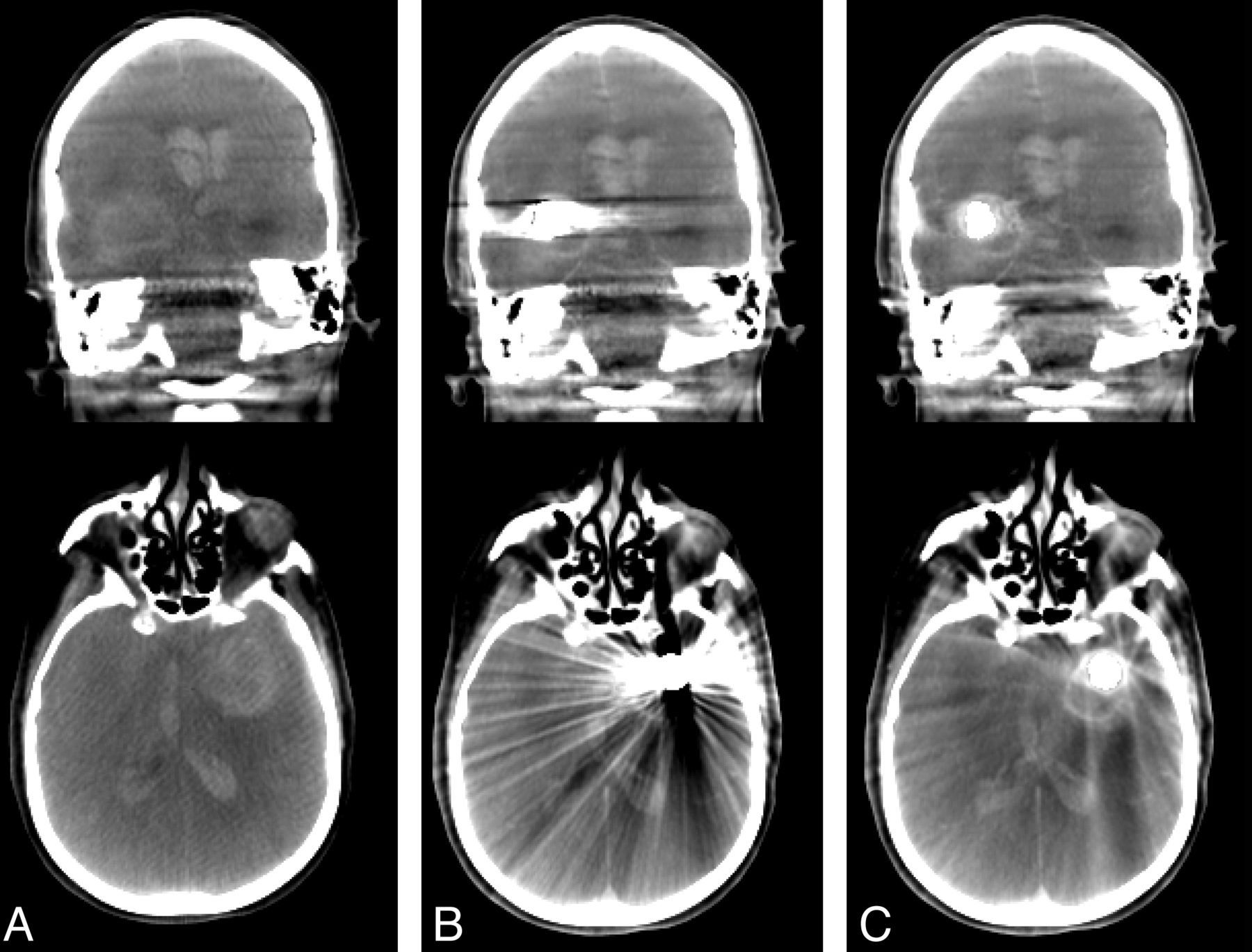
    Coronal (top row) and axial sections (bottom row) of conebeam CT data of a patient acquired before coil embolization (column A), after coil embolization without MAR (column B), and after coil embolization with MAR (column C).
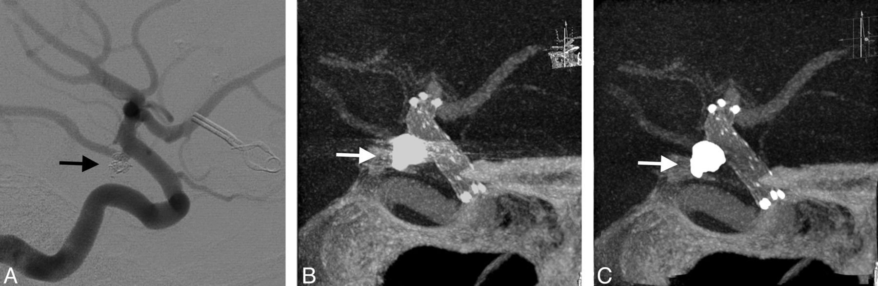
  • Fig 3.
    • Download figure
    • Open in new tab
    • Download powerpoint
    Fig 3.

    Corresponding axial sections of VasoCT data acquired after stent placement (A, stent indicated by asterisk) and after coil embolization (B and C). Streak artifacts generated by the coil mass visible in VasoCT data without MAR (B) are severely reduced with MAR (C). Because of the replacement of absent data in the raw projections, subtle new artifacts appear in VasoCT with MAR (C, arrows). Intensity profiles (yellow lines) were generated for all 3 images by use of the same physical coordinates (white lines). The intensity scale of the profile analysis is given on the left-hand side of the figure. Profile plots show that severe fluctuations outside the coil mass are reduced by MAR and the resulting profile in C is similar to the profile in A.

  • Fig 4.
    • Download figure
    • Open in new tab
    • Download powerpoint
    Fig 4.

    Conebeam CT data without (A) and with (B) MAR demonstrating the reduction of coil mass artifacts and the appearance of subtle streak caused by the algorithm, which is specifically well visualized in the indicated circular region of interest.

  • Fig 5.
    • Download figure
    • Open in new tab
    • Download powerpoint
    Fig 5.

    Illustrative case 1. DSA at 6-month follow-up shows no recanalization of the embolized posterior communicating artery aneurysm (A). Streak artifacts caused by the coil mass (arrows) in maximum intensity projection of VasoCT data without MAR (B) partially obscures visualization of stent and host artery. After MAR (C), streak artifacts in VasoCT data were removed, revealing the stent and host artery.

  • Fig 6.
    • Download figure
    • Open in new tab
    • Download powerpoint
    Fig 6.

    Illustrative case 2. Immediate DSA (A) maximum intensity projection of VasoCT data without MAR (B) and with MAR (C) of stent-assisted coil embolized aneurysm at the right A1 segment. Visibility is significantly affected by streak artifacts caused by the coil mass (arrows) and contralateral clip in VasoCT without MAR. With MAR, stent apposition to the vascular wall is fully appreciated.

  • Fig 7.
    • Download figure
    • Open in new tab
    • Download powerpoint
    Fig 7.

    Illustrative case 3. DSA (A), maximum intensity projections of VasoCT without (B), and with MAR (C) acquired immediately after stent-assisted coil (arrows) embolization procedure. Streak artifacts partially obscuring the host artery and side branches are removed by the MAR method.

  • Fig 8.
    • Download figure
    • Open in new tab
    • Download powerpoint
    Fig 8.

    Illustrative case 4. Immediate DSA (A), VasoCT without MAR (B), and VasoCT with MAR (C) after stent-assisted coiling of an unruptured middle cerebral artery aneurysm. Although streak artifacts caused by the coil mass (arrows) are significantly reduced, a small amount of streak remains after application of MAR.

Tables

  • Figures
  • Results of the observer study rating the visibility without and with MAR

    Without MARκWith MARκ
    Overall Agreement, %Score 1 Agreement, %Score ≥2 Agreement, %Overall Agreement, %Score 1 Agreement, %Score ≥2 Agreement, %
    Stent visibility
        P < .05
        OR = 7.87756120.667624400.64
        CI = 1.6–38.8
    Vessel visibility
        P < .05
        OR = 8.77652120.647220400.58
        CI = 1.7–45.2
    Relationship
        P < .05
        OR = 9.08160120.727020360.56
        CI = 1.7–47.0
    Overall Agreement, %Yes Agreement, %No Agreement, %κOverall Agreement, %Yes Agreement, %No Agreement, %κ
    Obscuring beyond
            coil mass?
        (P < .0001
        OR = 224.0735640.47784640.57
        CI = 12.8–3926.0)
    Overall Agreement, %Without MAR Agreement, %With MAR Agreement, %κ
    Overall best visibility940920.89
    • Note:—The rows “stent visibility,” “vessel visibility,” and “relationship” show the summarized results to the 3-point scale questions. Given are the percent overall agreement (ie, the number of cases that all reviewers agreed in total, calculated using the Fleiss method), the percent agreement for a score 1 (ie, the number of cases all reviewers agreed on giving a score of 1), and the percent agreement for a score ≥2 (ie, the number of cases all reviewers agreed on giving a score of 2 or 3). Similarly, agreements for the binary questions are indicated. For each observer question, the κ values, P value, odds ratio (OR), and 95% confidence intervals (CI) are given when applicable. OR represents the improvement of classification of 1 to ≥2 with MAR.

PreviousNext
Back to top

In this issue

American Journal of Neuroradiology: 34 (11)
American Journal of Neuroradiology
Vol. 34, Issue 11
1 Nov 2013
  • Table of Contents
  • Index by author
  • Complete Issue (PDF)
Advertisement
Print
Download PDF
Email Article

Thank you for your interest in spreading the word on American Journal of Neuroradiology.

NOTE: We only request your email address so that the person you are recommending the page to knows that you wanted them to see it, and that it is not junk mail. We do not capture any email address.

Enter multiple addresses on separate lines or separate them with commas.
Reduction of Coil Mass Artifacts in High-Resolution Flat Detector Conebeam CT of Cerebral Stent-Assisted Coiling
(Your Name) has sent you a message from American Journal of Neuroradiology
(Your Name) thought you would like to see the American Journal of Neuroradiology web site.
CAPTCHA
This question is for testing whether or not you are a human visitor and to prevent automated spam submissions.
Cite this article
I.M.J. van der Bom, S.Y. Hou, A.S. Puri, G. Spilberg, D. Ruijters, P. van de Haar, B. Carelsen, S. Vedantham, M.J. Gounis, A.K. Wakhloo
Reduction of Coil Mass Artifacts in High-Resolution Flat Detector Conebeam CT of Cerebral Stent-Assisted Coiling
American Journal of Neuroradiology Nov 2013, 34 (11) 2163-2170; DOI: 10.3174/ajnr.A3561

Citation Manager Formats

  • BibTeX
  • Bookends
  • EasyBib
  • EndNote (tagged)
  • EndNote 8 (xml)
  • Medlars
  • Mendeley
  • Papers
  • RefWorks Tagged
  • Ref Manager
  • RIS
  • Zotero
0 Responses
Respond to this article
Share
Bookmark this article
Reduction of Coil Mass Artifacts in High-Resolution Flat Detector Conebeam CT of Cerebral Stent-Assisted Coiling
I.M.J. van der Bom, S.Y. Hou, A.S. Puri, G. Spilberg, D. Ruijters, P. van de Haar, B. Carelsen, S. Vedantham, M.J. Gounis, A.K. Wakhloo
American Journal of Neuroradiology Nov 2013, 34 (11) 2163-2170; DOI: 10.3174/ajnr.A3561
del.icio.us logo Twitter logo Facebook logo Mendeley logo
  • Tweet Widget
  • Facebook Like
  • Google Plus One
Purchase

Jump to section

  • Article
    • Abstract
    • ABBREVIATIONS:
    • Materials and Methods
    • Results
    • Discussion
    • Conclusions
    • Acknowledgments
    • Footnotes
    • REFERENCES
  • Figures & Data
  • Info & Metrics
  • Responses
  • References
  • PDF

Related Articles

  • No related articles found.
  • PubMed
  • Google Scholar

Cited By...

  • Application of High-Resolution C-Arm CT Combined with Streak Metal Artifact Removal Technology for the Stent-Assisted Embolization of Intracranial Aneurysms
  • Combination of high-resolution cone beam computed tomography and metal artefact reduction software: a new image fusion technique for evaluating intracranial stent apposition after aneurysm treatment
  • Clinical evaluation of volume of interest imaging combined with metal artifact reduction reconstruction techniques in coiling and stent assisted coiling during neurointerventional procedures
  • A Patient Dose-Reduction Technique for Neuroendovascular Image-Guided Interventions: Image-Quality Comparison Study
  • Quantitative Analysis of Conebeam CT for Delineating Stents in Stent-Assisted Coil Embolization
  • Clinical Impact of Flat Panel Volume CT Angiography in Evaluating the Accurate Intraoperative Deployment of Flow-Diverter Stents
  • Flow changes in the posterior communicating artery related to flow-diverter stents in carotid siphon aneurysms
  • A novel reconstruction tool (syngo DynaCT Head Clear) in the post-processing of DynaCT images to reduce artefacts and improve image quality
  • Metal artifact reduction for flat panel detector intravenous CT angiography in patients with intracranial metallic implants after endovascular and surgical treatment
  • Low-Dose Volume-of-Interest C-Arm CT Imaging of Intracranial Stents and Flow Diverters
  • Intravenous C-Arm Conebeam CT Angiography following Long-Term Flow-Diverter Implantation: Technologic Evaluation and Preliminary Results
  • High-Resolution C-Arm CT and Metal Artifact Reduction Software: A Novel Imaging Modality for Analyzing Aneurysms Treated with Stent-Assisted Coil Embolization
  • Aneurysm permeability following coil embolization: packing density and coil distribution
  • Quantitative analysis of high-resolution, contrast-enhanced, cone-beam CT for the detection of intracranial in-stent hyperplasia
  • Evaluation of a Metal Artifacts Reduction Algorithm Applied to Postinterventional Flat Panel Detector CT Imaging
  • Role of C-Arm VasoCT in the Use of Endovascular WEB Flow Disruption in Intracranial Aneurysm Treatment
  • Artifact Reduction of Different Metallic Implants in Flat Detector C-Arm CT
  • Crossref (41)
  • Google Scholar

This article has been cited by the following articles in journals that are participating in Crossref Cited-by Linking.

  • Role of C-Arm VasoCT in the Use of Endovascular WEB Flow Disruption in Intracranial Aneurysm Treatment
    J. Caroff, C. Mihalea, H. Neki, D. Ruijters, L. Ikka, N. Benachour, J. Moret, L. Spelle
    American Journal of Neuroradiology 2014 35 7
  • Quantitative analysis of high-resolution, contrast-enhanced, cone-beam CT for the detection of intracranial in-stent hyperplasia
    Thomas F Flood, Imramsjah M J van der Bom, Lara Strittmatter, Ajit S Puri, Gregory M Hendricks, Ajay K Wakhloo, Matthew J Gounis
    Journal of NeuroInterventional Surgery 2015 7 2
  • Aneurysm permeability following coil embolization: packing density and coil distribution
    Ju-Yu Chueh, Srinivasan Vedantham, Ajay K Wakhloo, Sarena L Carniato, Ajit S Puri, Conrad Bzura, Spencer Coffin, Alexei A Bogdanov, Matthew J Gounis
    Journal of NeuroInterventional Surgery 2015 7 9
  • Metal artifact reduction for flat panel detector intravenous CT angiography in patients with intracranial metallic implants after endovascular and surgical treatment
    Rastislav Pjontek, Belgin Önenköprülü, Bernhard Scholz, Yiannis Kyriakou, Gerrit A Schubert, Omid Nikoubashman, Ahmed Othman, Martin Wiesmann, Marc A Brockmann
    Journal of NeuroInterventional Surgery 2016 8 8
  • High-Resolution C-Arm CT and Metal Artifact Reduction Software: A Novel Imaging Modality for Analyzing Aneurysms Treated with Stent-Assisted Coil Embolization
    I. Yuki, Y. Kambayashi, A. Ikemura, Y. Abe, I. Kan, A. Mohamed, C. Dahmani, T. Suzuki, T. Ishibashi, H. Takao, M. Urashima, Y. Murayama
    American Journal of Neuroradiology 2016 37 2
  • Evaluation of a Metal Artifacts Reduction Algorithm Applied to Postinterventional Flat Panel Detector CT Imaging
    D.A. Stidd, H. Theessen, Y. Deng, Y. Li, B. Scholz, C. Rohkohl, M.D. Jhaveri, R. Moftakhar, M. Chen, D.K. Lopes
    American Journal of Neuroradiology 2014 35 11
  • Artifact Reduction of Different Metallic Implants in Flat Detector C-Arm CT
    S.-C. Hung, C.-C. Wu, C.-J. Lin, W.-Y. Guo, C.-B. Luo, F.-C. Chang, C.-Y. Chang
    American Journal of Neuroradiology 2014 35 7
  • Flow changes in the posterior communicating artery related to flow-diverter stents in carotid siphon aneurysms
    Fernando Mota de Carvalho, Jildaz Caroff, Elizeu Pereira dos Santos Neto, Vanessa Chalumeau, Hazem Abdel Khalek, Hiroaki Neki, Guillaume Saliou, Aymeric Rouchaud, Jacques Moret, Laurent Spelle
    Journal of NeuroInterventional Surgery 2017 9 7
  • Fatal arterial rupture during angioplasty of a flow diverter in a recurrent, previously Y-stented giant MCA bifurcation aneurysm
    Jean-Christophe Gentric, Robert Fahed, Tim E Darsaut, Igor Salazkin, Daniel Roy, Jean Raymond
    Interventional Neuroradiology 2016 22 3
  • Clinical Impact of Flat Panel Volume CT Angiography in Evaluating the Accurate Intraoperative Deployment of Flow-Diverter Stents
    F. Clarençon, F. Di Maria, J. Gabrieli, E. Shotar, V. Degos, A. Nouet, A. Biondi, N.-A. Sourour
    American Journal of Neuroradiology 2017 38 10

More in this TOC Section

  • SAVE vs. Solumbra Techniques for Thrombectomy
  • Contrast-Induced Encephalopathy after NeuroIR
  • CT Perfusion&Reperfusion in Acute Ischemic Stroke
Show more Interventional

Similar Articles

Advertisement

Indexed Content

  • Current Issue
  • Accepted Manuscripts
  • Article Preview
  • Past Issues
  • Editorials
  • Editor's Choice
  • Fellows' Journal Club
  • Letters to the Editor
  • Video Articles

Cases

  • Case Collection
  • Archive - Case of the Week
  • Archive - Case of the Month
  • Archive - Classic Case

Special Collections

  • AJNR Awards
  • ASNR Foundation Special Collection
  • Most Impactful AJNR Articles
  • Photon-Counting CT
  • Spinal CSF Leak Articles (Jan 2020-June 2024)

More from AJNR

  • Trainee Corner
  • Imaging Protocols
  • MRI Safety Corner

Multimedia

  • AJNR Podcasts
  • AJNR Scantastics

Resources

  • Turnaround Time
  • Submit a Manuscript
  • Submit a Video Article
  • Submit an eLetter to the Editor/Response
  • Manuscript Submission Guidelines
  • Statistical Tips
  • Fast Publishing of Accepted Manuscripts
  • Graphical Abstract Preparation
  • Imaging Protocol Submission
  • Evidence-Based Medicine Level Guide
  • Publishing Checklists
  • Author Policies
  • Become a Reviewer/Academy of Reviewers
  • News and Updates

About Us

  • About AJNR
  • Editorial Board
  • Editorial Board Alumni
  • Alerts
  • Permissions
  • Not an AJNR Subscriber? Join Now
  • Advertise with Us
  • Librarian Resources
  • Feedback
  • Terms and Conditions
  • AJNR Editorial Board Alumni

American Society of Neuroradiology

  • Not an ASNR Member? Join Now

© 2025 by the American Society of Neuroradiology All rights, including for text and data mining, AI training, and similar technologies, are reserved.
Print ISSN: 0195-6108 Online ISSN: 1936-959X

Powered by HighWire