Advertisement

Main menu

  • Home
  • Content
    • Current Issue
    • Accepted Manuscripts
    • Article Preview
    • Past Issue Archive
    • Video Articles
    • AJNR Case Collection
    • Case of the Week Archive
    • Case of the Month Archive
    • Classic Case Archive
  • Special Collections
    • AJNR Awards
    • ASNR Foundation Special Collection
    • Most Impactful AJNR Articles
    • Photon-Counting CT
    • Spinal CSF Leak Articles (Jan 2020-June 2024)
  • Multimedia
    • AJNR Podcasts
    • AJNR SCANtastic
    • Trainee Corner
    • MRI Safety Corner
    • Imaging Protocols
  • For Authors
    • Submit a Manuscript
    • Submit a Video Article
    • Submit an eLetter to the Editor/Response
    • Manuscript Submission Guidelines
    • Statistical Tips
    • Fast Publishing of Accepted Manuscripts
    • Graphical Abstract Preparation
    • Imaging Protocol Submission
    • Author Policies
  • About Us
    • About AJNR
    • Editorial Board
    • Editorial Board Alumni
  • More
    • Become a Reviewer/Academy of Reviewers
    • Subscribers
    • Permissions
    • Alerts
    • Feedback
    • Advertisers
    • ASNR Home

User menu

  • Alerts
  • Log in

Search

  • Advanced search
American Journal of Neuroradiology

American Journal of Neuroradiology

ASHNR American Society of Functional Neuroradiology ASHNR American Society of Pediatric Neuroradiology ASSR
  • Alerts
  • Log in

Advanced Search

  • Home
  • Content
    • Current Issue
    • Accepted Manuscripts
    • Article Preview
    • Past Issue Archive
    • Video Articles
    • AJNR Case Collection
    • Case of the Week Archive
    • Case of the Month Archive
    • Classic Case Archive
  • Special Collections
    • AJNR Awards
    • ASNR Foundation Special Collection
    • Most Impactful AJNR Articles
    • Photon-Counting CT
    • Spinal CSF Leak Articles (Jan 2020-June 2024)
  • Multimedia
    • AJNR Podcasts
    • AJNR SCANtastic
    • Trainee Corner
    • MRI Safety Corner
    • Imaging Protocols
  • For Authors
    • Submit a Manuscript
    • Submit a Video Article
    • Submit an eLetter to the Editor/Response
    • Manuscript Submission Guidelines
    • Statistical Tips
    • Fast Publishing of Accepted Manuscripts
    • Graphical Abstract Preparation
    • Imaging Protocol Submission
    • Author Policies
  • About Us
    • About AJNR
    • Editorial Board
    • Editorial Board Alumni
  • More
    • Become a Reviewer/Academy of Reviewers
    • Subscribers
    • Permissions
    • Alerts
    • Feedback
    • Advertisers
    • ASNR Home
  • Follow AJNR on Twitter
  • Visit AJNR on Facebook
  • Follow AJNR on Instagram
  • Join AJNR on LinkedIn
  • RSS Feeds

AJNR is seeking candidates for the AJNR Podcast Editor. Read the position description.

Research ArticleInterventional

Thrombosis Heralding Aneurysmal Rupture: An Exploration of Potential Mechanisms in a Novel Giant Swine Aneurysm Model

J. Raymond, T.E. Darsaut, M. Kotowski, A. Makoyeva, G. Gevry, F. Berthelet and I. Salazkin
American Journal of Neuroradiology February 2013, 34 (2) 346-353; DOI: https://doi.org/10.3174/ajnr.A3407
J. Raymond
aFrom the Department of Radiology (J.R.)
  • Find this author on Google Scholar
  • Find this author on PubMed
  • Search for this author on this site
T.E. Darsaut
bInterventional Neuroradiology Research Laboratory (T.E.D., M.K., A.M., G.G., I.S.)
dDivision of Neurosurgery (T.E.D.), Department of Surgery, University of Alberta Hospital, Mackenzie Health Sciences Centre, Edmonton, Alberta, Canada.
  • Find this author on Google Scholar
  • Find this author on PubMed
  • Search for this author on this site
M. Kotowski
bInterventional Neuroradiology Research Laboratory (T.E.D., M.K., A.M., G.G., I.S.)
  • Find this author on Google Scholar
  • Find this author on PubMed
  • Search for this author on this site
A. Makoyeva
bInterventional Neuroradiology Research Laboratory (T.E.D., M.K., A.M., G.G., I.S.)
  • Find this author on Google Scholar
  • Find this author on PubMed
  • Search for this author on this site
G. Gevry
bInterventional Neuroradiology Research Laboratory (T.E.D., M.K., A.M., G.G., I.S.)
  • Find this author on Google Scholar
  • Find this author on PubMed
  • Search for this author on this site
F. Berthelet
cDepartment of Pathology (F.B.), Centre Hospitalier de l'Université de Montréal, Notre-Dame Hospital, Montreal, Quebec, Canada
  • Find this author on Google Scholar
  • Find this author on PubMed
  • Search for this author on this site
I. Salazkin
bInterventional Neuroradiology Research Laboratory (T.E.D., M.K., A.M., G.G., I.S.)
  • Find this author on Google Scholar
  • Find this author on PubMed
  • Search for this author on this site
  • Article
  • Figures & Data
  • Info & Metrics
  • Responses
  • References
  • PDF
Loading

Article Figures & Data

Figures

  • Tables
  • Fig 1.
    • Download figure
    • Open in new tab
    • Download powerpoint
    Fig 1.

    Untreated ruptured giant aneurysm. Angiography (A), sagittal sections (B), and microscopic sections (C and D) of a control giant aneurysm 7 days after construction and rupture. Note the thrombosed caudal third (T), the flowing cephalad third (P), and the partially thrombosed middle third of the fundus. While there is neointimal hypertrophy of the wall of the cephalad third (C), there is thrombosis with recanalization of the caudal section and hemorrhagic transformation of the wall (asterisk, B and D). To demonstrate patency of the aneurysm, one must catheterize the aneurysm and inject contrast directly into its lumen. Note recanalization between the clot and wall (arrows in A and D). Movat pentachrome, original magnification ×20. N indicates aneurysm neck.
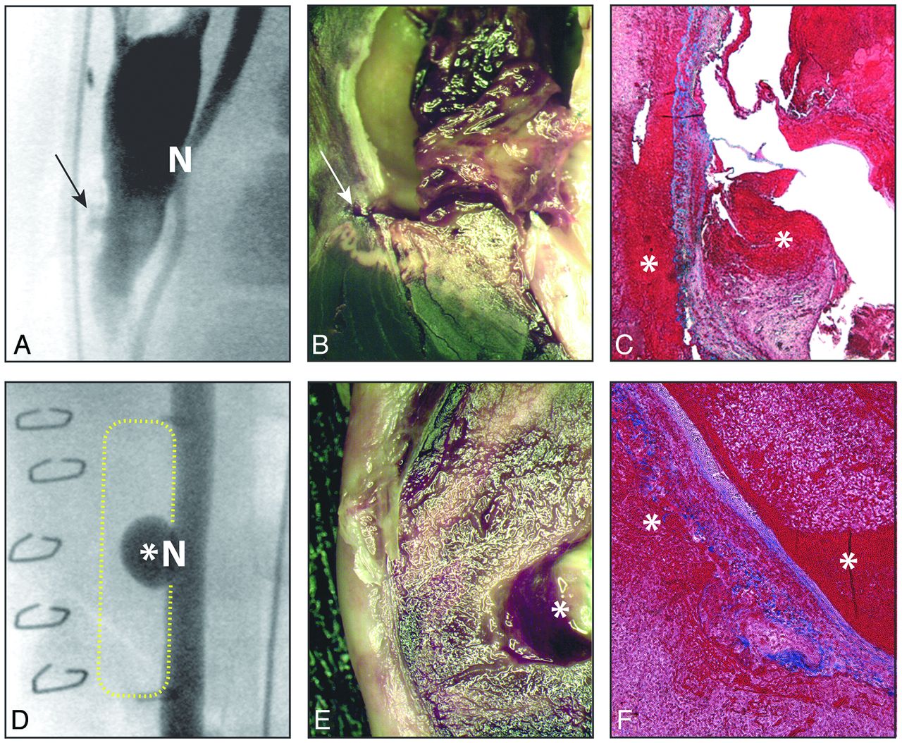
  • Fig 2.
    • Download figure
    • Open in new tab
    • Download powerpoint
    Fig 2.

    Ruptured and unruptured group 3 aneurysms. Angiography (A and D), sagittal section (B and E), and microscopic sections (C and F) 7 days after surgery. The arrows in A and B show the rupture site in a control partially thrombosed aneurysm (N indicates aneurysm neck). The asterisks in D and E show the recurrent aneurysm in a thrombosed lesion lacking an endothelial lining. Note recanalization between the clot and the wall of the control aneurysm, absent in the aneurysm lacking an endothelial lining, and the presence of blood on both sides of the attenuated aneurysm wall media in both cases (asterisks, C and F). C and F, Movat pentachrome, original magnification ×20.

  • Fig 3.
    • Download figure
    • Open in new tab
    • Download powerpoint
    Fig 3.

    Aneurysm wall. Microscopic section of the wall of the aneurysms, 4 (A and D) and 7 (B and C) days after surgery. Note normal wall (A), thickened, attenuated wall in a thrombosed aneurysm with hemorrhagic infiltration of the adventitia (B and C), with (C) or without (B) recanalization. Note extrusion of endoaneurysmal clot (C) through complete rupture of the wall (arrow, D). HPS (A and D) and Movat pentachrome (B and C), original magnification ×20 (A and D), ×50 (B and C).
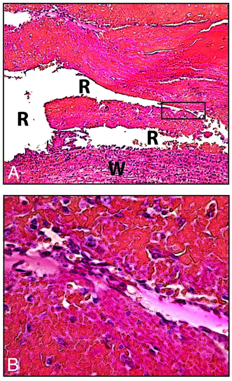
  • Fig 4.
    • Download figure
    • Open in new tab
    • Download powerpoint
    Fig 4.

    Recanalization and rupture. Recanalized endothelialized crescents can be found in thrombosed aneurysms as early as 4 days after aneurysm construction and rupture. A, W indicates venous wall; R, recanalization. HPS, original magnification ×50 (A). Original magnification ×100 (B).
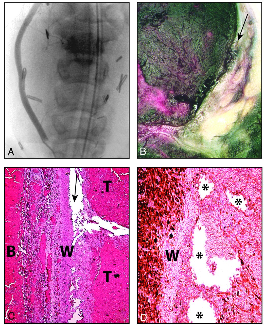
  • Fig 5.
    • Download figure
    • Open in new tab
    • Download powerpoint
    Fig 5.

    Bleeding despite clipping. Angiography (A), sagittal section (B), and microscopic pathology (C and D) of giant aneurysms constructed and clipped 7 days before. Note severe displacement of the right carotid artery by hematoma (A), but the aneurysm is completely clipped. Recanalization between the clot and the aneurysm wall can be seen (B and C). The attenuated wall (W) with intraluminal thrombosis (T) and perianeurysmal blood (B) is shown (C). D, Microscopic sections immune-stained with α-actin show infiltration of the clot with myofibroblasts and the formation of endothelialized vascular spaces (asterisk) between the clot and the wall (W).

Tables

  • Figures
  • Characteristics of the various groups of aneurysmsa

    No.Long Axis (mm)Short Axis (mm)Neck (mm)Aspect Ratio
    Group
        11211.3 ± 2.66.7 ± 2.15.8 ± 0.62.1 ± 0.6
        2816.9 ± 4.08.1 ± 1.34.8 ± 1.13.6 ± 1.0
        32826.1 ± 10.99.4 ± 1.45.8 ± 1.04.6 ± 1.8
    P value.000.01.84.001
    • ↵a Data are means.

PreviousNext
Back to top

In this issue

American Journal of Neuroradiology: 34 (2)
American Journal of Neuroradiology
Vol. 34, Issue 2
1 Feb 2013
  • Table of Contents
  • Index by author
Advertisement
Print
Download PDF
Email Article

Thank you for your interest in spreading the word on American Journal of Neuroradiology.

NOTE: We only request your email address so that the person you are recommending the page to knows that you wanted them to see it, and that it is not junk mail. We do not capture any email address.

Enter multiple addresses on separate lines or separate them with commas.
Thrombosis Heralding Aneurysmal Rupture: An Exploration of Potential Mechanisms in a Novel Giant Swine Aneurysm Model
(Your Name) has sent you a message from American Journal of Neuroradiology
(Your Name) thought you would like to see the American Journal of Neuroradiology web site.
CAPTCHA
This question is for testing whether or not you are a human visitor and to prevent automated spam submissions.
Cite this article
J. Raymond, T.E. Darsaut, M. Kotowski, A. Makoyeva, G. Gevry, F. Berthelet, I. Salazkin
Thrombosis Heralding Aneurysmal Rupture: An Exploration of Potential Mechanisms in a Novel Giant Swine Aneurysm Model
American Journal of Neuroradiology Feb 2013, 34 (2) 346-353; DOI: 10.3174/ajnr.A3407

Citation Manager Formats

  • BibTeX
  • Bookends
  • EasyBib
  • EndNote (tagged)
  • EndNote 8 (xml)
  • Medlars
  • Mendeley
  • Papers
  • RefWorks Tagged
  • Ref Manager
  • RIS
  • Zotero
0 Responses
Respond to this article
Share
Bookmark this article
Thrombosis Heralding Aneurysmal Rupture: An Exploration of Potential Mechanisms in a Novel Giant Swine Aneurysm Model
J. Raymond, T.E. Darsaut, M. Kotowski, A. Makoyeva, G. Gevry, F. Berthelet, I. Salazkin
American Journal of Neuroradiology Feb 2013, 34 (2) 346-353; DOI: 10.3174/ajnr.A3407
del.icio.us logo Twitter logo Facebook logo Mendeley logo
  • Tweet Widget
  • Facebook Like
  • Google Plus One
Purchase

Jump to section

  • Article
    • Abstract
    • ABBREVIATIONS:
    • Materials and Methods
    • Results
    • Discussion
    • Conclusions
    • Footnotes
    • References
  • Figures & Data
  • Info & Metrics
  • Responses
  • References
  • PDF

Related Articles

  • No related articles found.
  • PubMed
  • Google Scholar

Cited By...

  • In situ decellularization of a large animal saccular aneurysm model: sustained inflammation and active aneurysm wall remodeling
  • Reduced Activity of von Willebrand Factor after Flow-Diverting Stent Implantation for Intracranial Aneurysms: A Link to Acquired von Willebrand Disease?
  • MR Imaging of Myeloperoxidase Activity in a Model of the Inflamed Aneurysm Wall
  • Intraluminal Cell Transplantation Prevents Growth and Rupture in a Model of Rupture-Prone Saccular Aneurysms
  • Advances in Stroke: Advances in Interventional Radiology 2013
  • Crossref (30)
  • Google Scholar

This article has been cited by the following articles in journals that are participating in Crossref Cited-by Linking.

  • Flow Diversion in Middle Cerebral Artery Aneurysms: Is It Really an All-Purpose Treatment?
    Osman Melih Topcuoglu, Erol Akgul, Ergun Daglioglu, Elif Dilara Topcuoglu, Ahmet Peker, Ilkay Akmangit, Deniz Belen, Anil Arat
    World Neurosurgery 2016 87
  • Recurrence of endovascularly and microsurgically treated intracranial aneurysms—review of the putative role of aneurysm wall biology
    Serge Marbacher, Mika Niemelä, Juha Hernesniemi, Juhana Frösén
    Neurosurgical Review 2019 42 1
  • In vivo cerebral aneurysm models
    John W. Thompson, Omar Elwardany, David J. McCarthy, Dallas L. Sheinberg, Carlos M. Alvarez, Ahmed Nada, Brian M. Snelling, Stephanie H. Chen, Samir Sur, Robert M. Starke
    Neurosurgical Focus 2019 47 1
  • Testing flow diversion in animal models: a systematic review
    Robert Fahed, Jean Raymond, Célina Ducroux, Jean-Christophe Gentric, Igor Salazkin, Daniela Ziegler, Guylaine Gevry, Tim E. Darsaut
    Neuroradiology 2016 58 4
  • Aneurysm Rupture after Endovascular Flow Diversion: The Possible Role of Persistent Flows through the Transition Zone Associated with Device Deformation
    T.E. Darsaut, E. Rayner-Hartley, A. Makoyeva, I. Salazkin, F. Berthelet, J. Raymond
    Interventional Neuroradiology 2013 19 2
  • Intraluminal Cell Transplantation Prevents Growth and Rupture in a Model of Rupture-Prone Saccular Aneurysms
    Serge Marbacher, Juhana Frösén, Johan Marjamaa, Andrey Anisimov, Petri Honkanen, Michael von Gunten, Usama Abo-Ramadan, Juha Hernesniemi, Mika Niemelä
    Stroke 2014 45 12
  • Trends in Cerebrovascular Surgery
    Takayuki Hara, Shintaro Arai, Yoshiaki Goto, Tsuguhito Takizawa, Tatsuya Uchida
    2016 123
  • Usefulness of the Pipeline Embolic Device for Large and Giant Carotid Cavernous Aneurysms
    Shigeru Miyachi, Ryo Hiramatsu, Hiroyuki Ohnishi, Ryokichi Yagi, Toshihiko Kuroiwa
    Neurointervention 2017 12 2
  • Flow Diversion of Giant Curved Sidewall and Bifurcation Experimental Aneurysms with Very-Low-Porosity Devices
    Tim E. Darsaut, Fabrice Bing, Alina Makoyeva, Guylaine Gevry, Igor Salazkin, Jean Raymond
    World Neurosurgery 2014 82 6
  • Innovations in Endovascular Treatment Strategies for Large Carotid Cavernous Aneurysms—The Safety and Efficacy of a Flow Diverter
    Shigeru Miyachi, Hiroyuki Ohnishi, Ryo Hiramatsu, Takashi Izumi, Noriaki Matsubara, Toshihiko Kuroiwa
    Journal of Stroke and Cerebrovascular Diseases 2017 26 5

More in this TOC Section

  • SAVE vs. Solumbra Techniques for Thrombectomy
  • Contrast-Induced Encephalopathy after NeuroIR
  • CT Perfusion&Reperfusion in Acute Ischemic Stroke
Show more Interventional

Similar Articles

Advertisement

Indexed Content

  • Current Issue
  • Accepted Manuscripts
  • Article Preview
  • Past Issues
  • Editorials
  • Editor's Choice
  • Fellows' Journal Club
  • Letters to the Editor
  • Video Articles

Cases

  • Case Collection
  • Archive - Case of the Week
  • Archive - Case of the Month
  • Archive - Classic Case

Special Collections

  • AJNR Awards
  • ASNR Foundation Special Collection
  • Most Impactful AJNR Articles
  • Photon-Counting CT
  • Spinal CSF Leak Articles (Jan 2020-June 2024)

More from AJNR

  • Trainee Corner
  • Imaging Protocols
  • MRI Safety Corner

Multimedia

  • AJNR Podcasts
  • AJNR Scantastics

Resources

  • Turnaround Time
  • Submit a Manuscript
  • Submit a Video Article
  • Submit an eLetter to the Editor/Response
  • Manuscript Submission Guidelines
  • Statistical Tips
  • Fast Publishing of Accepted Manuscripts
  • Graphical Abstract Preparation
  • Imaging Protocol Submission
  • Evidence-Based Medicine Level Guide
  • Publishing Checklists
  • Author Policies
  • Become a Reviewer/Academy of Reviewers
  • News and Updates

About Us

  • About AJNR
  • Editorial Board
  • Editorial Board Alumni
  • Alerts
  • Permissions
  • Not an AJNR Subscriber? Join Now
  • Advertise with Us
  • Librarian Resources
  • Feedback
  • Terms and Conditions
  • AJNR Editorial Board Alumni

American Society of Neuroradiology

  • Not an ASNR Member? Join Now

© 2025 by the American Society of Neuroradiology All rights, including for text and data mining, AI training, and similar technologies, are reserved.
Print ISSN: 0195-6108 Online ISSN: 1936-959X

Powered by HighWire