Advertisement

Main menu

  • Home
  • Content
    • Current Issue
    • Accepted Manuscripts
    • Article Preview
    • Past Issue Archive
    • Video Articles
    • AJNR Case Collection
    • Case of the Week Archive
    • Case of the Month Archive
    • Classic Case Archive
  • Special Collections
    • AJNR Awards
    • ASNR Foundation Special Collection
    • Most Impactful AJNR Articles
    • Photon-Counting CT
    • Spinal CSF Leak Articles (Jan 2020-June 2024)
  • Multimedia
    • AJNR Podcasts
    • AJNR SCANtastic
    • Trainee Corner
    • MRI Safety Corner
    • Imaging Protocols
  • For Authors
    • Submit a Manuscript
    • Submit a Video Article
    • Submit an eLetter to the Editor/Response
    • Manuscript Submission Guidelines
    • Statistical Tips
    • Fast Publishing of Accepted Manuscripts
    • Graphical Abstract Preparation
    • Imaging Protocol Submission
    • Author Policies
  • About Us
    • About AJNR
    • Editorial Board
    • Editorial Board Alumni
  • More
    • Become a Reviewer/Academy of Reviewers
    • Subscribers
    • Permissions
    • Alerts
    • Feedback
    • Advertisers
    • ASNR Home

User menu

  • Alerts
  • Log in

Search

  • Advanced search
American Journal of Neuroradiology

American Journal of Neuroradiology

ASHNR American Society of Functional Neuroradiology ASHNR American Society of Pediatric Neuroradiology ASSR
  • Alerts
  • Log in

Advanced Search

  • Home
  • Content
    • Current Issue
    • Accepted Manuscripts
    • Article Preview
    • Past Issue Archive
    • Video Articles
    • AJNR Case Collection
    • Case of the Week Archive
    • Case of the Month Archive
    • Classic Case Archive
  • Special Collections
    • AJNR Awards
    • ASNR Foundation Special Collection
    • Most Impactful AJNR Articles
    • Photon-Counting CT
    • Spinal CSF Leak Articles (Jan 2020-June 2024)
  • Multimedia
    • AJNR Podcasts
    • AJNR SCANtastic
    • Trainee Corner
    • MRI Safety Corner
    • Imaging Protocols
  • For Authors
    • Submit a Manuscript
    • Submit a Video Article
    • Submit an eLetter to the Editor/Response
    • Manuscript Submission Guidelines
    • Statistical Tips
    • Fast Publishing of Accepted Manuscripts
    • Graphical Abstract Preparation
    • Imaging Protocol Submission
    • Author Policies
  • About Us
    • About AJNR
    • Editorial Board
    • Editorial Board Alumni
  • More
    • Become a Reviewer/Academy of Reviewers
    • Subscribers
    • Permissions
    • Alerts
    • Feedback
    • Advertisers
    • ASNR Home
  • Follow AJNR on Twitter
  • Visit AJNR on Facebook
  • Follow AJNR on Instagram
  • Join AJNR on LinkedIn
  • RSS Feeds

AJNR is seeking candidates for the AJNR Podcast Editor. Read the position description.

Research ArticleSpineF

Spinal Meningeal Diverticula in Spontaneous Intracranial Hypotension: Analysis of Prevalence and Myelographic Appearance

P.G. Kranz, S.S. Stinnett, K.T. Huang and L. Gray
American Journal of Neuroradiology June 2013, 34 (6) 1284-1289; DOI: https://doi.org/10.3174/ajnr.A3359
P.G. Kranz
aFrom the Departments of Radiology (P.G.K., K.T.H., L.G.)
  • Find this author on Google Scholar
  • Find this author on PubMed
  • Search for this author on this site
S.S. Stinnett
bBiostatistics and Bioinformatics (S.S.S.)
cOphthalmology (S.S.S.), Duke University Medical Center, Durham, North Carolina.
  • Find this author on Google Scholar
  • Find this author on PubMed
  • Search for this author on this site
K.T. Huang
aFrom the Departments of Radiology (P.G.K., K.T.H., L.G.)
  • Find this author on Google Scholar
  • Find this author on PubMed
  • Search for this author on this site
L. Gray
aFrom the Departments of Radiology (P.G.K., K.T.H., L.G.)
  • Find this author on Google Scholar
  • Find this author on PubMed
  • Search for this author on this site
  • Article
  • Figures & Data
  • Info & Metrics
  • Responses
  • References
  • PDF
Loading

Article Figures & Data

Figures

  • Tables
  • Fig 1.
    • Download figure
    • Open in new tab
    • Download powerpoint
    Fig 1.

    A 58-year-old woman with positional headaches and tinnitus. A, Curved plane reformatted image from a CT myelogram demonstrates numerous spinal meningeal diverticula of varying sizes (arrowheads); no CSF leak was directly visualized. B, Coronal T1 postcontrast image from an MR image obtained before treatment shows diffuse smooth dural enhancement (arrows) and pituitary enlargement, compatible with SIH. C, Coronal T1 postcontrast image obtained following blood patching targeting the diverticula seen in A shows resolution of the imaging findings of SIH. The patient's symptoms completely resolved following treatment.
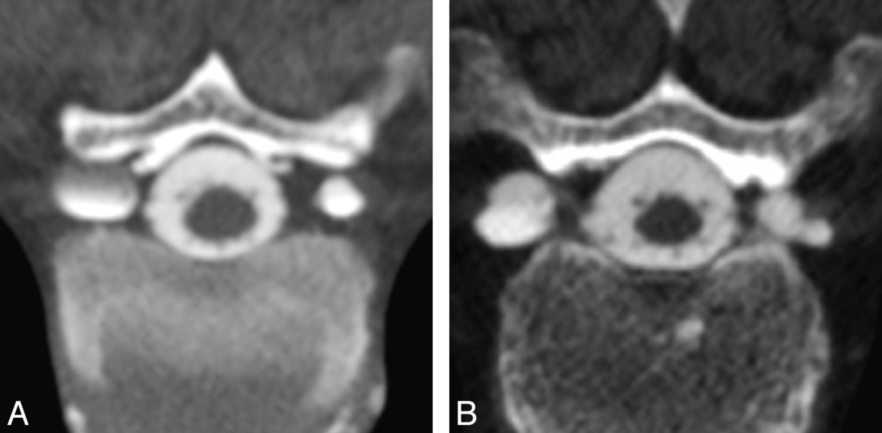
  • Fig 2.
    • Download figure
    • Open in new tab
    • Download powerpoint
    Fig 2.

    Classification of diverticular morphology. Lesions were categorized as either round (A) or multilobulated (B) on the basis of analysis of axial images from CT myelography.
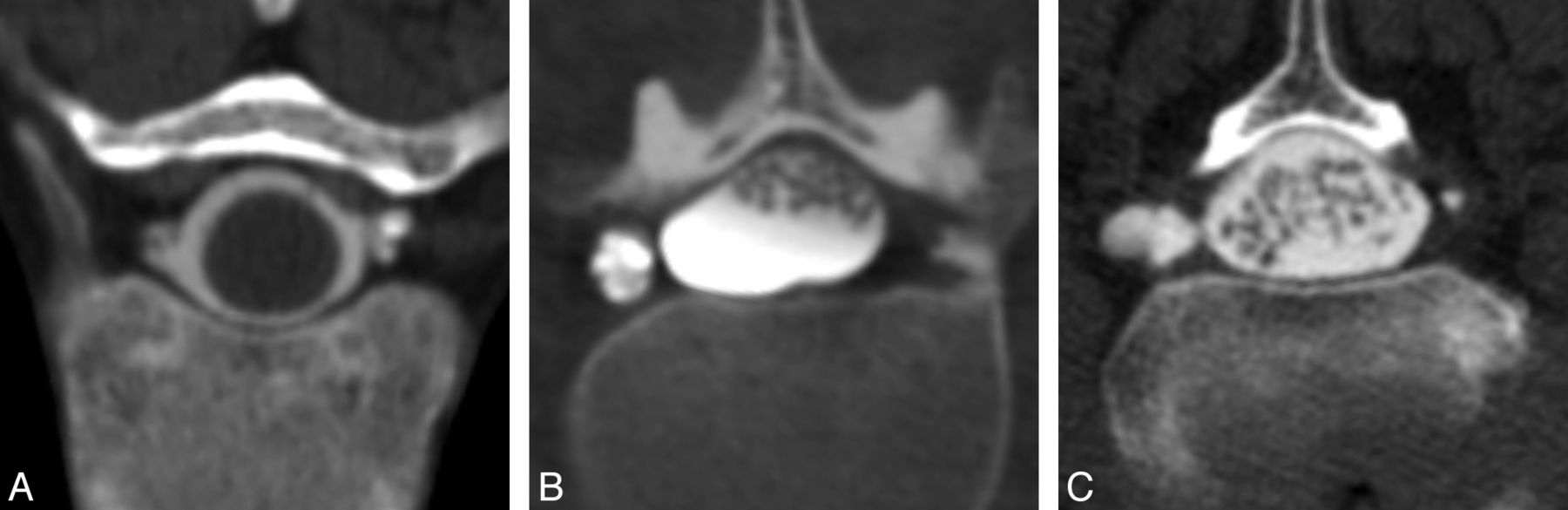
  • Fig 3.
    • Download figure
    • Open in new tab
    • Download powerpoint
    Fig 3.

    Classification of diverticular size. On the basis of the maximum diameter of the diverticulum on an axial CT myelogram, lesions were trichotomized into 3 groups: 0–3 (A), 4–6 (B), or >6 mm (C).

  • Fig 4.
    • Download figure
    • Open in new tab
    • Download powerpoint
    Fig 4.

    Axial CT myelogram obtained immediately following intrathecal contrast administration in a patient with SIH shows an extrathecal contrast collection. These rapidly filling collections indicate the presence of a high-flow CSF leak.
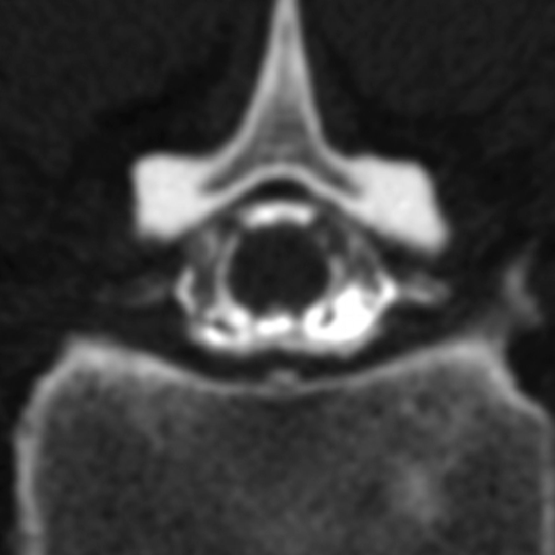
  • Fig 5.
    • Download figure
    • Open in new tab
    • Download powerpoint
    Fig 5.

    Axial CT myelogram shows filling of the proximal nerve root sheaths bilaterally, findings seen intermittently in both controls and patients with SIH that we classified as “prominent” nerve root sheaths.
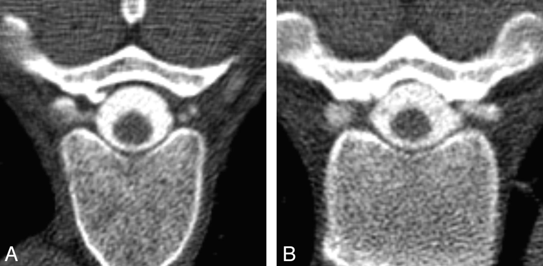
  • Fig 6.
    • Download figure
    • Open in new tab
    • Download powerpoint
    Fig 6.

    Incidental spinal diverticula in a control patient. Axial CT myelograms demonstrate multiple thoracic spinal diverticula in a 48-year-old woman. The patient underwent the myelography for postoperative back pain; she had no history of headache or other symptoms suggestive of SIH.

Tables

  • Figures
    • View popup
    Table 1:

    Demographic data

    ControlSIHP Value
    Age (mean) (SD) (yr)55.56 (13.85)50.37 (13.27)
        Min/median/max35/58/7730/52/81.186a
    Sex
        Male (No.) (%)12 (67)5 (26)
        Female (No.) (%)6 (33)14 (74).014b
    • Note:—Min indicates minimum; max, maximum.

    • ↵a P value based on the Wilcoxon rank sum test of the difference between medians.

    • ↵b P value based on a χ2 test of the difference in proportions.

    • View popup
    Table 2:

    Prevalence of spinal meningeal diverticula by sex

    Diverticula per PatientMaleFemaleP Valuea
    Mean (SD)1.6 (2.3)6.5 (7.9).166
    Min/median/max0/1/70/1.5/23
    • Note:—Min indicates minimum; max, maximum.

    • ↵a P value based on the Wilcoxon rank sum test of the difference between medians.

    • View popup
    Table 3:

    Prevalence of spinal meningeal diverticula and prominent nerve sheaths

    ControlSIHP Value
    Patients with diverticula present (No.) (%)8 (44)13 (68).141a
    Diverticula per patient (mean)2.2 (3.3)6.3 (8.0).099b
        Min/median/max0/0/100/2/23
    Patients with prominent nerve sheaths present (No.) (%)14 (78)17 (89).405c
    Prominent nerve sheaths per patient (mean)2.6 (3.1)6.1 (4.2).004c
        Min/median/max0/1.5/130/5/15
    • Note:—Min indicates minimum; max, maximum.

    • ↵a P value based on a χ2 test of the difference between proportions.

    • ↵b P value based on the Wilcoxon rank sum test of the difference between medians.

    • ↵c P value based on the Fisher exact test of the difference between proportions.

    • View popup
    Table 4:

    Comparison of myelographic appearance of spinal meningeal diverticula

    ControlSIHP Valuea
    Morphology
        Round (No.) (%)20 (51)62 (52).946
        Multilobulated (No.) (%)19 (49)57 (48)
    Size
        0–3 mm (No.) (%)9 (23)22 (18).711
        4–6 mm (No.) (%)23 (59)71 (60)
        >6 mm (No.) (%)7 (18)26 (22)
    Location
        Cervical (No.) (%)2 (5)4 (3)0.050
        Upper thoracic (No.) (%)6 (15)33 (28)
        Lower thoracic (No.) (%)29 (75)65 (55)
        Lumbar (No.) (%)2 (5)17 (14)
    • ↵a P value based on the generalized estimating equations test of the difference between groups in proportions in morphologic categories while accounting for multiple observations per patient.

PreviousNext
Back to top

In this issue

American Journal of Neuroradiology: 34 (6)
American Journal of Neuroradiology
Vol. 34, Issue 6
1 Jun 2013
  • Table of Contents
  • Index by author
Advertisement
Print
Download PDF
Email Article

Thank you for your interest in spreading the word on American Journal of Neuroradiology.

NOTE: We only request your email address so that the person you are recommending the page to knows that you wanted them to see it, and that it is not junk mail. We do not capture any email address.

Enter multiple addresses on separate lines or separate them with commas.
Spinal Meningeal Diverticula in Spontaneous Intracranial Hypotension: Analysis of Prevalence and Myelographic Appearance
(Your Name) has sent you a message from American Journal of Neuroradiology
(Your Name) thought you would like to see the American Journal of Neuroradiology web site.
CAPTCHA
This question is for testing whether or not you are a human visitor and to prevent automated spam submissions.
Cite this article
P.G. Kranz, S.S. Stinnett, K.T. Huang, L. Gray
Spinal Meningeal Diverticula in Spontaneous Intracranial Hypotension: Analysis of Prevalence and Myelographic Appearance
American Journal of Neuroradiology Jun 2013, 34 (6) 1284-1289; DOI: 10.3174/ajnr.A3359

Citation Manager Formats

  • BibTeX
  • Bookends
  • EasyBib
  • EndNote (tagged)
  • EndNote 8 (xml)
  • Medlars
  • Mendeley
  • Papers
  • RefWorks Tagged
  • Ref Manager
  • RIS
  • Zotero
0 Responses
Respond to this article
Share
Bookmark this article
Spinal Meningeal Diverticula in Spontaneous Intracranial Hypotension: Analysis of Prevalence and Myelographic Appearance
P.G. Kranz, S.S. Stinnett, K.T. Huang, L. Gray
American Journal of Neuroradiology Jun 2013, 34 (6) 1284-1289; DOI: 10.3174/ajnr.A3359
del.icio.us logo Twitter logo Facebook logo Mendeley logo
  • Tweet Widget
  • Facebook Like
  • Google Plus One
Purchase

Jump to section

  • Article
    • Abstract
    • ABBREVIATIONS:
    • Materials and Methods
    • Results
    • Discussion
    • Conclusions
    • REFERENCES
  • Figures & Data
  • Info & Metrics
  • Responses
  • References
  • PDF

Related Articles

  • No related articles found.
  • PubMed
  • Google Scholar

Cited By...

  • Prevalence of Spinal Meningeal Diverticula in Autosomal Dominant Polycystic Kidney Disease
  • Monro-Kellie Hypothesis: Increase of Ventricular CSF Volume after Surgical Closure of a Spinal Dural Leak in Patients with Spontaneous Intracranial Hypotension
  • Spontaneous Intracranial Hypotension: Atypical Radiologic Appearances, Imaging Mimickers, and Clinical Look-Alikes
  • Diskogenic microspurs as a major cause of intractable spontaneous intracranial hypotension
  • A classification system of spontaneous spinal CSF leaks
  • Crossref (60)
  • Google Scholar

This article has been cited by the following articles in journals that are participating in Crossref Cited-by Linking.

  • A classification system of spontaneous spinal CSF leaks
    Wouter I. Schievink, M. Marcel Maya, Stacey Jean-Pierre, Miriam Nuño, Ravi S. Prasad, Franklin G. Moser
    Neurology 2016 87 7
  • Diskogenic microspurs as a major cause of intractable spontaneous intracranial hypotension
    Jürgen Beck, Christian T. Ulrich, Christian Fung, Jens Fichtner, Kathleen Seidel, Michael Fiechter, Kety Hsieh, Michael Murek, David Bervini, Niklaus Meier, Marie-Luise Mono, Pasquale Mordasini, Ekkehard Hewer, Werner J. Z'Graggen, Jan Gralla, Andreas Raabe
    Neurology 2016 87 12
  • CSF Venous Fistulas in Spontaneous Intracranial Hypotension: Imaging Characteristics on Dynamic and CT Myelography
    Peter G. Kranz, Timothy J. Amrhein, Linda Gray
    American Journal of Roentgenology 2017 209 6
  • Myelographic Techniques for the Detection of Spinal CSF Leaks in Spontaneous Intracranial Hypotension
    Peter G. Kranz, Patrick H. Luetmer, Felix E. Diehn, Timothy J. Amrhein, Teerath Peter Tanpitukpongse, Linda Gray
    American Journal of Roentgenology 2016 206 1
  • Spontaneous Intracranial Hypotension: 10 Myths and Misperceptions
    Peter G. Kranz, Linda Gray, Timothy J. Amrhein
    Headache: The Journal of Head and Face Pain 2018 58 7
  • Spontaneous Intracranial Hypotension
    Peter G. Kranz, Linda Gray, Michael D. Malinzak, Timothy J. Amrhein
    Neuroimaging Clinics of North America 2019 29 4
  • Spontaneous intracranial hypotension: searching for the CSF leak
    Tomas Dobrocky, Patrick Nicholson, Levin Häni, Pasquale Mordasini, Timo Krings, Waleed Brinjikji, Jeremy K Cutsforth-Gregory, Ralph Schär, Christoph Schankin, Jan Gralla, Vitor M Pereira, Andreas Raabe, Richard Farb, Jürgen Beck, Eike I Piechowiak
    The Lancet Neurology 2022 21 4
  • Update on the Diagnosis and Treatment of Spontaneous Intracranial Hypotension
    Peter G. Kranz, Michael D. Malinzak, Timothy J. Amrhein, Linda Gray
    Current Pain and Headache Reports 2017 21 8
  • CSF–Venous Fistulas: Anatomy and Diagnostic Imaging
    Peter G. Kranz, Linda Gray, Michael D. Malinzak, Jessica L. Houk, Dong Kun Kim, Timothy J. Amrhein
    American Journal of Roentgenology 2021 217 6
  • Spontaneous Intracranial Hypotension
    Timothy J. Amrhein, Peter G. Kranz
    Radiologic Clinics of North America 2019 57 2

More in this TOC Section

  • Bern Score Validity for SIH
  • MP2RAGE 7T in MS Lesions of the Cervical Spine
  • Resisted Inspiration for CSF-Venous Fistula
Show more Spine

Similar Articles

Advertisement

Indexed Content

  • Current Issue
  • Accepted Manuscripts
  • Article Preview
  • Past Issues
  • Editorials
  • Editor's Choice
  • Fellows' Journal Club
  • Letters to the Editor
  • Video Articles

Cases

  • Case Collection
  • Archive - Case of the Week
  • Archive - Case of the Month
  • Archive - Classic Case

Special Collections

  • AJNR Awards
  • ASNR Foundation Special Collection
  • Most Impactful AJNR Articles
  • Photon-Counting CT
  • Spinal CSF Leak Articles (Jan 2020-June 2024)

More from AJNR

  • Trainee Corner
  • Imaging Protocols
  • MRI Safety Corner

Multimedia

  • AJNR Podcasts
  • AJNR Scantastics

Resources

  • Turnaround Time
  • Submit a Manuscript
  • Submit a Video Article
  • Submit an eLetter to the Editor/Response
  • Manuscript Submission Guidelines
  • Statistical Tips
  • Fast Publishing of Accepted Manuscripts
  • Graphical Abstract Preparation
  • Imaging Protocol Submission
  • Evidence-Based Medicine Level Guide
  • Publishing Checklists
  • Author Policies
  • Become a Reviewer/Academy of Reviewers
  • News and Updates

About Us

  • About AJNR
  • Editorial Board
  • Editorial Board Alumni
  • Alerts
  • Permissions
  • Not an AJNR Subscriber? Join Now
  • Advertise with Us
  • Librarian Resources
  • Feedback
  • Terms and Conditions
  • AJNR Editorial Board Alumni

American Society of Neuroradiology

  • Not an ASNR Member? Join Now

© 2025 by the American Society of Neuroradiology All rights, including for text and data mining, AI training, and similar technologies, are reserved.
Print ISSN: 0195-6108 Online ISSN: 1936-959X

Powered by HighWire