Advertisement

Main menu

  • Home
  • Content
    • Current Issue
    • Accepted Manuscripts
    • Article Preview
    • Past Issue Archive
    • Video Articles
    • AJNR Case Collection
    • Case of the Week Archive
    • Case of the Month Archive
    • Classic Case Archive
  • Special Collections
    • AJNR Awards
    • ASNR Foundation Special Collection
    • Most Impactful AJNR Articles
    • Photon-Counting CT
    • Spinal CSF Leak Articles (Jan 2020-June 2024)
  • Multimedia
    • AJNR Podcasts
    • AJNR SCANtastic
    • Trainee Corner
    • MRI Safety Corner
    • Imaging Protocols
  • For Authors
    • Submit a Manuscript
    • Submit a Video Article
    • Submit an eLetter to the Editor/Response
    • Manuscript Submission Guidelines
    • Statistical Tips
    • Fast Publishing of Accepted Manuscripts
    • Graphical Abstract Preparation
    • Imaging Protocol Submission
    • Author Policies
  • About Us
    • About AJNR
    • Editorial Board
    • Editorial Board Alumni
  • More
    • Become a Reviewer/Academy of Reviewers
    • Subscribers
    • Permissions
    • Alerts
    • Feedback
    • Advertisers
    • ASNR Home

User menu

  • Alerts
  • Log in

Search

  • Advanced search
American Journal of Neuroradiology

American Journal of Neuroradiology

ASHNR American Society of Functional Neuroradiology ASHNR American Society of Pediatric Neuroradiology ASSR
  • Alerts
  • Log in

Advanced Search

  • Home
  • Content
    • Current Issue
    • Accepted Manuscripts
    • Article Preview
    • Past Issue Archive
    • Video Articles
    • AJNR Case Collection
    • Case of the Week Archive
    • Case of the Month Archive
    • Classic Case Archive
  • Special Collections
    • AJNR Awards
    • ASNR Foundation Special Collection
    • Most Impactful AJNR Articles
    • Photon-Counting CT
    • Spinal CSF Leak Articles (Jan 2020-June 2024)
  • Multimedia
    • AJNR Podcasts
    • AJNR SCANtastic
    • Trainee Corner
    • MRI Safety Corner
    • Imaging Protocols
  • For Authors
    • Submit a Manuscript
    • Submit a Video Article
    • Submit an eLetter to the Editor/Response
    • Manuscript Submission Guidelines
    • Statistical Tips
    • Fast Publishing of Accepted Manuscripts
    • Graphical Abstract Preparation
    • Imaging Protocol Submission
    • Author Policies
  • About Us
    • About AJNR
    • Editorial Board
    • Editorial Board Alumni
  • More
    • Become a Reviewer/Academy of Reviewers
    • Subscribers
    • Permissions
    • Alerts
    • Feedback
    • Advertisers
    • ASNR Home
  • Follow AJNR on Twitter
  • Visit AJNR on Facebook
  • Follow AJNR on Instagram
  • Join AJNR on LinkedIn
  • RSS Feeds

AJNR is seeking candidates for the AJNR Podcast Editor. Read the position description.

Research ArticleHead & Neck
Open Access

Visualization of the Trochlear Nerve in the Cistern with Use of High-Resolution Turbo Spin-Echo Multisection Motion-Sensitized Driven Equilibrium

M. Kanoto, Y. Toyoguchi, T. Hosoya, A. Oda and Y. Sugai
American Journal of Neuroradiology July 2013, 34 (7) 1434-1437; DOI: https://doi.org/10.3174/ajnr.A3403
M. Kanoto
aFrom the Department of Diagnostic Radiology, Yamagata University Faculty of Medicine, Yamagata, Japan.
  • Find this author on Google Scholar
  • Find this author on PubMed
  • Search for this author on this site
Y. Toyoguchi
aFrom the Department of Diagnostic Radiology, Yamagata University Faculty of Medicine, Yamagata, Japan.
  • Find this author on Google Scholar
  • Find this author on PubMed
  • Search for this author on this site
T. Hosoya
aFrom the Department of Diagnostic Radiology, Yamagata University Faculty of Medicine, Yamagata, Japan.
  • Find this author on Google Scholar
  • Find this author on PubMed
  • Search for this author on this site
A. Oda
aFrom the Department of Diagnostic Radiology, Yamagata University Faculty of Medicine, Yamagata, Japan.
  • Find this author on Google Scholar
  • Find this author on PubMed
  • Search for this author on this site
Y. Sugai
aFrom the Department of Diagnostic Radiology, Yamagata University Faculty of Medicine, Yamagata, Japan.
  • Find this author on Google Scholar
  • Find this author on PubMed
  • Search for this author on this site
  • Article
  • Figures & Data
  • Info & Metrics
  • Responses
  • References
  • PDF
Loading

Article Figures & Data

Figures

  • Tables
  • Fig 1.
    • Download figure
    • Open in new tab
    • Download powerpoint
    Fig 1.

    Segmentation of the trochlear nerve. The trochlear nerve in the cistern was divided into 2 segments: the perimesencephalic segment and the tentorial segment, by use of the line that links the mesencephalic tegmentum-cerebral peduncle junction to the cerebellar anterior border in the same section. 1. Perimesencephalic segment. 2. Tentorial segment.
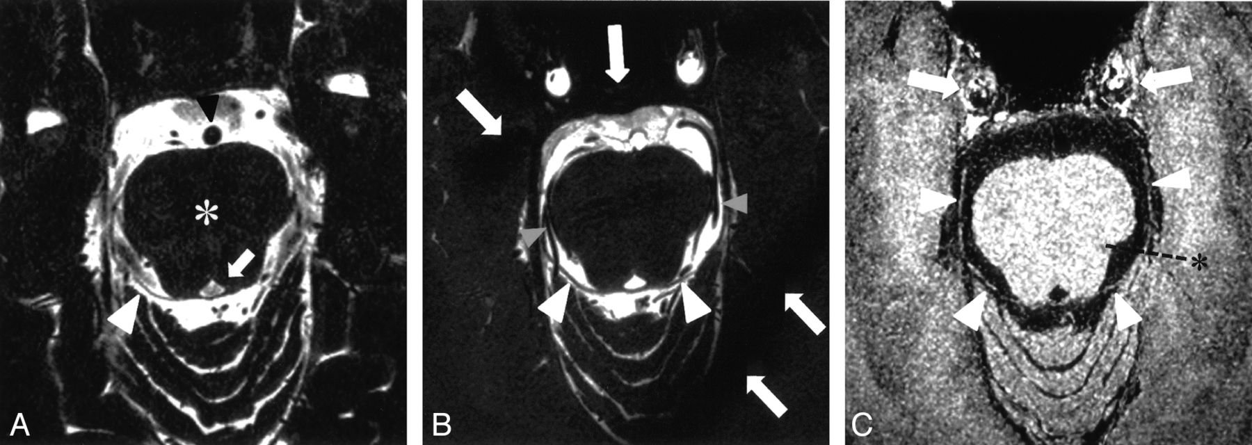
  • Fig 2.
    • Download figure
    • Open in new tab
    • Download powerpoint
    Fig 2.

    MR images of each sequence. A, HR-MRC: Perimesencephalic segment of the right trochlear nerve is observed (white arrowhead). Marked CSF flow-related artifacts in the cistern are apparent. The basilar artery (black arrowhead), midbrain (*), and cerebral aqueduct (white arrow) are noted. B, BS-MRC: Bilateral trochlear nerves are apparent in the cistern (arrowheads). The bilateral posterior cerebral artery is observed around the trochlear nerve (gray arrowheads). Band artifacts are also evident (white arrows). C, HR-MSDE: Trochlear nerves are visualized as curvilinear structures in full length (arrowheads). The line for segmentation (*) and the carotid artery (white arrows) are observed.

Tables

  • Figures
    • View popup
    Table 1:

    Visibility of 20 trochlear nerves in full length

    VisibilityHR-MRCBS-MRCHR-MSDE
    Excellent61318
    Good521
    Fair941
    Not010
    • View popup
    Table 2:

    Visibility of 20 trochlear nerves in each segment between 2 neuroradiologists

    Segment of the Trochlear NerveHR-MRCBS-MRCHR-MSDE
    ABABAB
    Perimesencephalic191419182020
    Tentorial*7213141918
    • A indicates neuroradiologist A; B, neuroradiologist B.

    • ↵* Significant differences among 3 MRI sequences by the χ2 test (P < .01).

    • View popup
    Table 3:

    Concordance in each MR imaging sequence between 2 neuroradiologists

    Segment of the Trochlear Nerveκ Coefficient
    HR-MRCBS-MRCHR-MSDE
    Perimesencephalic0.2860.643−*
    Tentorial0.2190.6590.643
    • ↵* The κ coefficient could not be calculated because both neuroradiologists evaluated the trochlear nerve as “visible” in all cases.

PreviousNext
Back to top

In this issue

American Journal of Neuroradiology: 34 (7)
American Journal of Neuroradiology
Vol. 34, Issue 7
1 Jul 2013
  • Table of Contents
  • Index by author
Advertisement
Print
Download PDF
Email Article

Thank you for your interest in spreading the word on American Journal of Neuroradiology.

NOTE: We only request your email address so that the person you are recommending the page to knows that you wanted them to see it, and that it is not junk mail. We do not capture any email address.

Enter multiple addresses on separate lines or separate them with commas.
Visualization of the Trochlear Nerve in the Cistern with Use of High-Resolution Turbo Spin-Echo Multisection Motion-Sensitized Driven Equilibrium
(Your Name) has sent you a message from American Journal of Neuroradiology
(Your Name) thought you would like to see the American Journal of Neuroradiology web site.
CAPTCHA
This question is for testing whether or not you are a human visitor and to prevent automated spam submissions.
Cite this article
M. Kanoto, Y. Toyoguchi, T. Hosoya, A. Oda, Y. Sugai
Visualization of the Trochlear Nerve in the Cistern with Use of High-Resolution Turbo Spin-Echo Multisection Motion-Sensitized Driven Equilibrium
American Journal of Neuroradiology Jul 2013, 34 (7) 1434-1437; DOI: 10.3174/ajnr.A3403

Citation Manager Formats

  • BibTeX
  • Bookends
  • EasyBib
  • EndNote (tagged)
  • EndNote 8 (xml)
  • Medlars
  • Mendeley
  • Papers
  • RefWorks Tagged
  • Ref Manager
  • RIS
  • Zotero
0 Responses
Respond to this article
Share
Bookmark this article
Visualization of the Trochlear Nerve in the Cistern with Use of High-Resolution Turbo Spin-Echo Multisection Motion-Sensitized Driven Equilibrium
M. Kanoto, Y. Toyoguchi, T. Hosoya, A. Oda, Y. Sugai
American Journal of Neuroradiology Jul 2013, 34 (7) 1434-1437; DOI: 10.3174/ajnr.A3403
del.icio.us logo Twitter logo Facebook logo Mendeley logo
  • Tweet Widget
  • Facebook Like
  • Google Plus One
Purchase

Jump to section

  • Article
    • Abstract
    • ABBREVIATIONS:
    • Materials and Methods
    • Results
    • Discussion
    • Conclusions
    • Footnotes
    • REFERENCES
  • Figures & Data
  • Info & Metrics
  • Responses
  • References
  • PDF

Related Articles

  • No related articles found.
  • PubMed
  • Google Scholar

Cited By...

  • Improved Blood Suppression of Motion-Sensitized Driven Equilibrium in High-Resolution Whole-Brain Vessel Wall Imaging: Comparison of Contrast-Enhanced 3D T1-Weighted FSE with Motion-Sensitized Driven Equilibrium and Delay Alternating with Nutation for Tailored Excitation
  • Coregistration and Fusion: An Easy and Reliable Method for Identifying Cranial Nerve IV on MRI
  • Trochlear Groove and Trochlear Cistern: Useful Anatomic Landmarks for Identifying the Tentorial Segment of Cranial Nerve IV on MRI
  • MRI Findings in Patients with a History of Failed Prior Microvascular Decompression for Hemifacial Spasm: How to Image and Where to Look
  • Crossref (11)
  • Google Scholar

This article has been cited by the following articles in journals that are participating in Crossref Cited-by Linking.

  • MRI Findings in Patients with a History of Failed Prior Microvascular Decompression for Hemifacial Spasm: How to Image and Where to Look
    M.A. Hughes, B.F. Branstetter, C.T. Taylor, S. Fakhran, W.T. Delfyett, A.M. Frederickson, R.F. Sekula
    American Journal of Neuroradiology 2015 36 4
  • Diagnosis of Intracranial Artery Dissection
    Masafumi KANOTO, Takaaki HOSOYA
    Neurologia medico-chirurgica 2016 56 9
  • A preliminary study of deep learning-based reconstruction specialized for denoising in high-frequency domain: usefulness in high-resolution three-dimensional magnetic resonance cisternography of the cerebellopontine angle
    Hiroyuki Uetani, Takeshi Nakaura, Mika Kitajima, Yuichi Yamashita, Tadashi Hamasaki, Machiko Tateishi, Kosuke Morita, Akira Sasao, Seitaro Oda, Osamu Ikeda, Yasuyuki Yamashita
    Neuroradiology 2021 63 1
  • Thin‐slice T2 MRI imaging predicts vascular pathology in hemifacial spasm: A case‐control study
    Raymond F. Sekula, Andrew M. Frederickson, Barton F. Branstetter, James E. Oskin, Dale R. Stevens, Nathan T. Zwagerman, Ramesh Grandhi, Marion A. Hughes
    Movement Disorders 2014 29 10
  • Trochlear Groove and Trochlear Cistern: Useful Anatomic Landmarks for Identifying the Tentorial Segment of Cranial Nerve IV on MRI
    P.M. Bunch, H.R. Kelly, D.A. Zander, H.D. Curtin
    American Journal of Neuroradiology 2017 38 5
  • Improved Blood Suppression of Motion-Sensitized Driven Equilibrium in High-Resolution Whole-Brain Vessel Wall Imaging: Comparison of Contrast-Enhanced 3D T1-Weighted FSE with Motion-Sensitized Driven Equilibrium and Delay Alternating with Nutation for Tailored Excitation
    D.J. Kim, H.-J. Lee, J. Baik, M.J. Hwang, M. Miyoshi, Y. Kang
    American Journal of Neuroradiology 2022 43 12
  • Effect of deep learning-based reconstruction on high-resolution three-dimensional T2-weighted fast asymmetric spin-echo imaging in the preoperative evaluation of cerebellopontine angle tumors
    Masamichi Hokamura, Hiroyuki Uetani, Tadashi Hamasaki, Takeshi Nakaura, Kosuke Morita, Yuichi Yamashita, Mika Kitajima, Aki Sugitani, Akitake Mukasa, Toshinori Hirai
    Neuroradiology 2024 66 7
  • Trochlear cistern of the cavernous sinus: an anatomical study using magnetic resonance imaging
    Satoshi Tsutsumi, Hideo Ono, Hisato Ishii
    Surgical and Radiologic Anatomy 2021 43 8
  • Root exit zone of the trochlear nerve on the dorsal brainstem: an MRI study
    Satoshi Tsutsumi, Hideo Ono, Hisato Ishii
    Surgical and Radiologic Anatomy 2022 44 3
  • Coregistration and Fusion: An Easy and Reliable Method for Identifying Cranial Nerve IV on MRI
    A. Lecler, J. Savatovsky, F. Audren
    American Journal of Neuroradiology 2017 38 10

More in this TOC Section

  • Chondrosarcoma vs Synovial Chondromatosis: Imaging
  • WHO Classification Update: Nasal&Skull Base Tumors
  • Peritumoral Signal in Vestibular Schwannomas
Show more HEAD & NECK

Similar Articles

Advertisement

Indexed Content

  • Current Issue
  • Accepted Manuscripts
  • Article Preview
  • Past Issues
  • Editorials
  • Editor's Choice
  • Fellows' Journal Club
  • Letters to the Editor
  • Video Articles

Cases

  • Case Collection
  • Archive - Case of the Week
  • Archive - Case of the Month
  • Archive - Classic Case

Special Collections

  • AJNR Awards
  • ASNR Foundation Special Collection
  • Most Impactful AJNR Articles
  • Photon-Counting CT
  • Spinal CSF Leak Articles (Jan 2020-June 2024)

More from AJNR

  • Trainee Corner
  • Imaging Protocols
  • MRI Safety Corner

Multimedia

  • AJNR Podcasts
  • AJNR Scantastics

Resources

  • Turnaround Time
  • Submit a Manuscript
  • Submit a Video Article
  • Submit an eLetter to the Editor/Response
  • Manuscript Submission Guidelines
  • Statistical Tips
  • Fast Publishing of Accepted Manuscripts
  • Graphical Abstract Preparation
  • Imaging Protocol Submission
  • Evidence-Based Medicine Level Guide
  • Publishing Checklists
  • Author Policies
  • Become a Reviewer/Academy of Reviewers
  • News and Updates

About Us

  • About AJNR
  • Editorial Board
  • Editorial Board Alumni
  • Alerts
  • Permissions
  • Not an AJNR Subscriber? Join Now
  • Advertise with Us
  • Librarian Resources
  • Feedback
  • Terms and Conditions
  • AJNR Editorial Board Alumni

American Society of Neuroradiology

  • Not an ASNR Member? Join Now

© 2025 by the American Society of Neuroradiology All rights, including for text and data mining, AI training, and similar technologies, are reserved.
Print ISSN: 0195-6108 Online ISSN: 1936-959X

Powered by HighWire