
Article Figures & Data
Figures
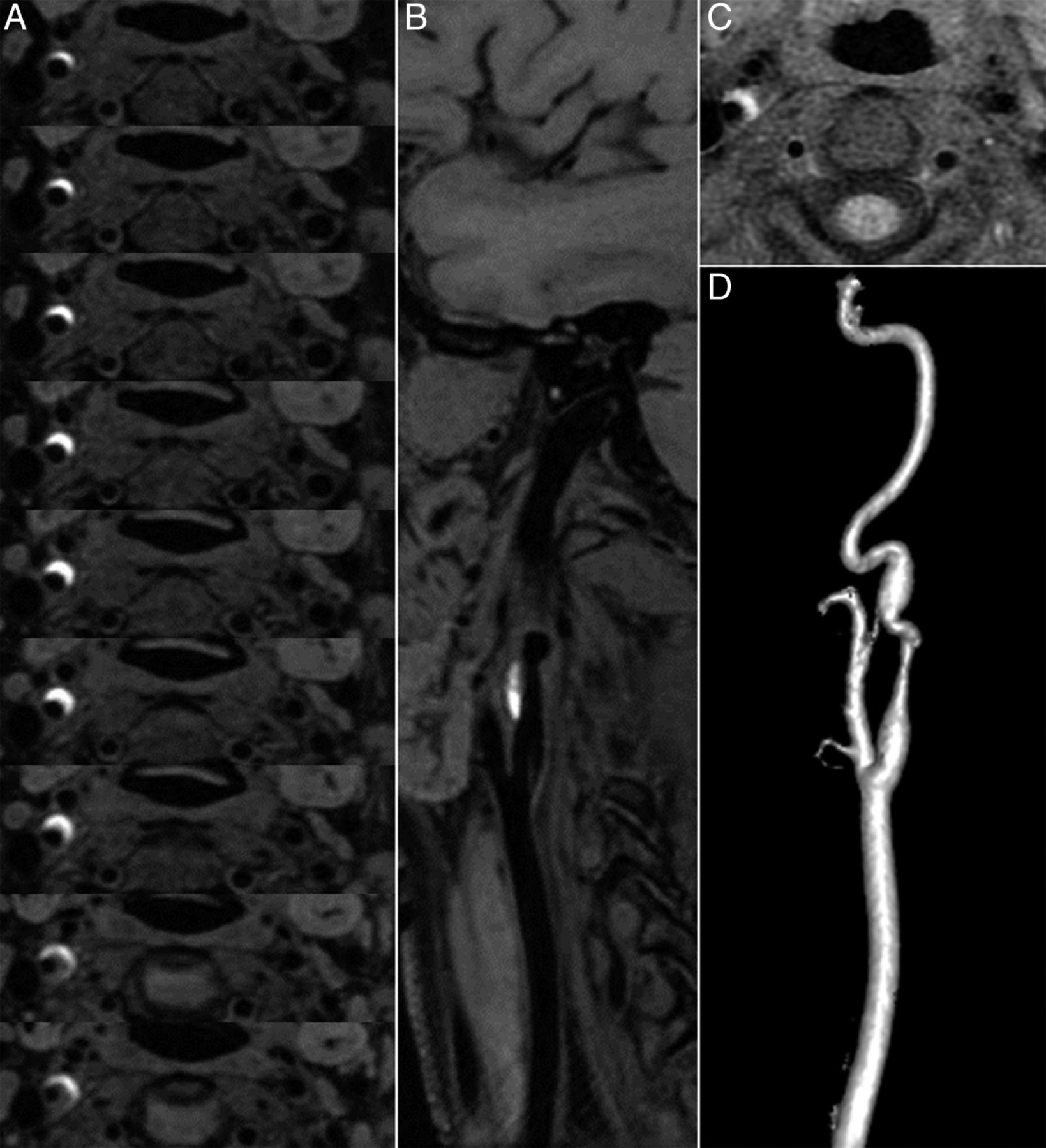
- Fig 1.
A, CUBE T1 consecutive axial views (thickness, 0.5 mm) covering the V3 vertebral segment. A bright crescentic signal (mural hematoma) surrounds the dark arterial lumen of the right vertebral artery. B and D, CUBE T1 coronal and sagittal views show the head and neck coverage and the mural hematoma. C, CUBE T1 oblique view centered on the V3-V4 junction shows that the vertebral dissection thinly extends intradurally. E, Contrast-enhanced MRA (CE-MRA) shows irregularity without stenosis of the right V3 segment lumen. F, CUBE T1 sagittal view fused with CE-MRA shows the bright T1 hematoma surrounding an irregular arterial lumen. G, Standard axial T1 BB image with fat saturation shows that the hematoma is also seen surrounding the right V3 segment.
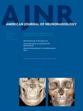
- Fig 2.
A, CUBE T1 consecutive axial views (thickness, 0.5 mm) covering the dissected right carotid artery show a bright crescentic signal surrounding the dark arterial lumen of the internal carotid artery. B, CUBE T1 oblique view oriented along the common carotid axis shows the dark lumen of the artery and the bright crescent signal of the hematoma. C, Standard axial T1 BB image with fat saturation shows that the hematoma is also seen surrounding the right internal carotid artery, but only on 2 sections. D, Contrast-enhanced MRA shows irregularity with stenosis of the suprabulbar internal carotid artery.
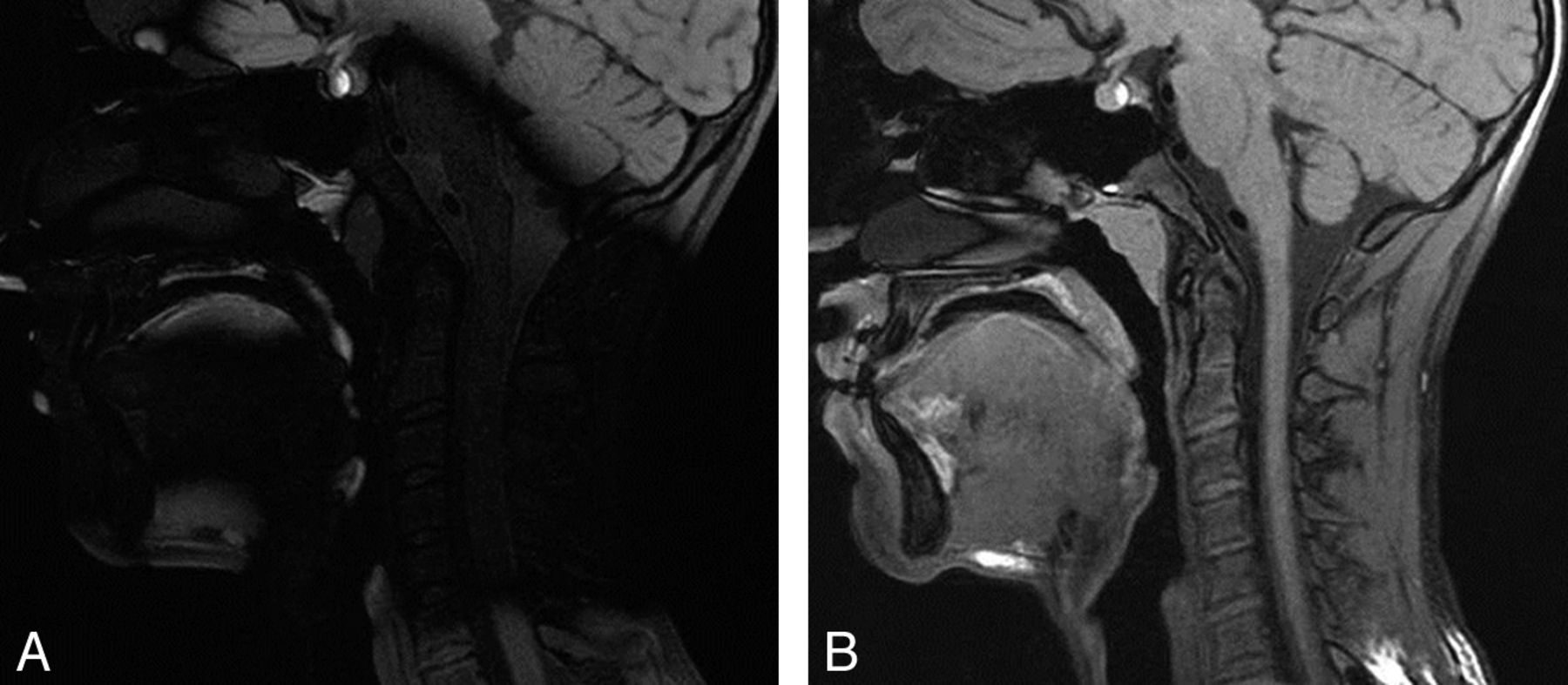
- Fig 3.
3D T1 sagittal views of 2 MR imaging acquisitions in the same patient without (A) and with (B) a dielectric pad.


Jump to section
Related Articles
Cited By...
- Mechanical disorders of the cervicocerebral circulation in children and young adults
- Standard Diffusion-Weighted Imaging in the Brain Can Detect Cervical Internal Carotid Artery Dissections
- 3D T1-weighted black blood sequence at 3.0 Tesla for the diagnosis of cervical artery dissection
- High-resolution intracranial vessel wall imaging: imaging beyond the lumen
- Characterization of Craniocervical Artery Dissection by Simultaneous MR Noncontrast Angiography and Intraplaque Hemorrhage Imaging at 3T
- Does Aneurysmal Wall Enhancement on Vessel Wall MRI Help to Distinguish Stable From Unstable Intracranial Aneurysms?