
Article Figures & Data
Figures
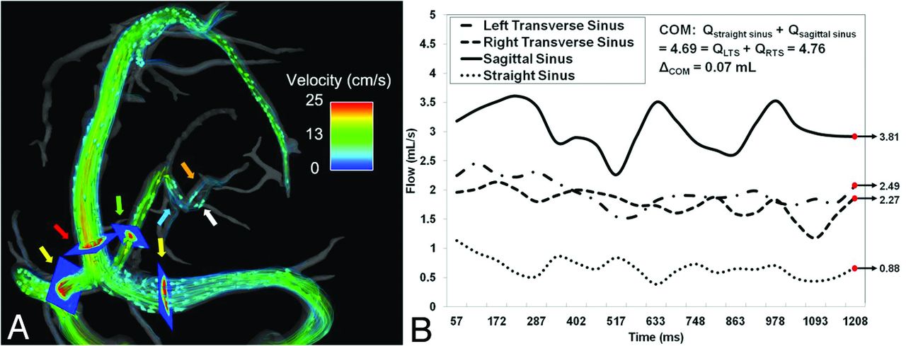
- Fig 1.
Example of flow visualization for COM (A). Arrows: yellow indicates left/right transverse sinus; red, sagittal sinus; green, straight sinus; blue, vein of Galen; internal cerebral veins: left (orange), right (white). Planes mark location of flow measurement. Blood flow waveforms (B) exemplify COM at the torcular herophili, with total flow measurements differing by only 0.37%.
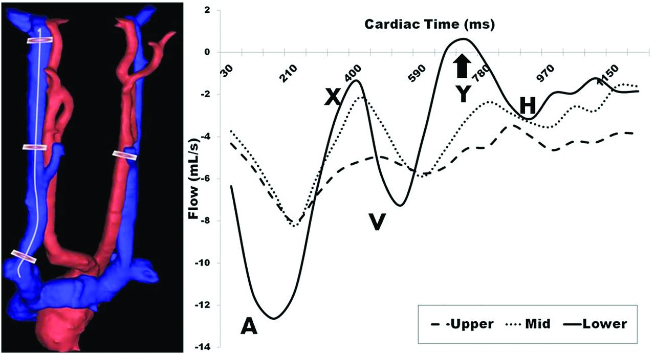
- Fig 2.
Left: Example placement of measurement planes in the right IJV down a centerline cubic spline and in the left common carotid artery. Right: Blood flow waveforms over the cardiac cycle indicate increasing pulsatility proximal to the heart, with a portion of the lower waveform showing retrograde flow (arrow). A indicates atrial systole; X, atrial relaxation; V, ventricular systole; Y, tricuspid reopening; H, atrial refilling.
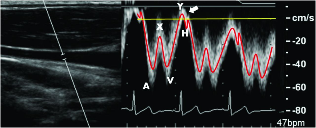
- Fig 3.
Left: B-mode anatomic location of right IJV blood flow. Right: Doppler ultrasonography displays triphasic IJV blood flow waveform as indicated in Fig 2. Arrow indicates minor normal reflux during the tricuspid valve reopening.
- Fig 4.
Interscan Bland-Altman plots for cerebral vein analysis within individual veins. Small biases and LOA indicate reproducibility of PC-VIPR in assessment of cerebral venous flow.
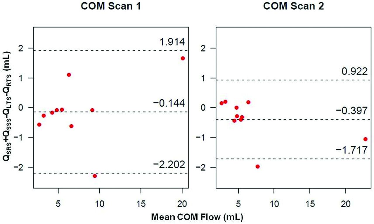
- Fig 5.
Bland-Altman plots of COM analysis for scan 1 (left) and scan 2 (right), showing small biases and LOA.
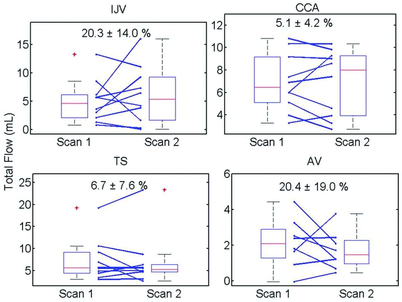
- Fig 6.
Boxplot results for all measurement locations. Individual changes (blue lines) show high variation in both the IJV and AV from scan to scan. No differences were considered significant (P < .05).
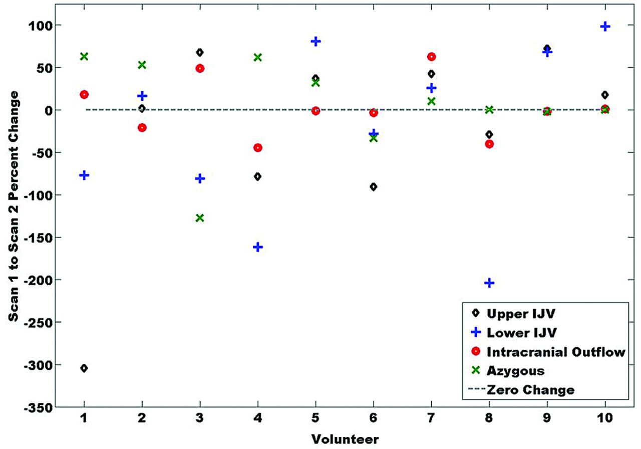
- Fig 7.
Percent change in total flow from scan 1 to scan 2 across volunteers. Volunteers 5–10 have similar directional changes in all measurements.


Tables
- Table 1:
Total flow (mL/cardiac cycle), delineated by side, measurement level, and scan, in all volunteers
Volunteer Left IJV Right IJV Upper Mid Lower Upper Mid Lower S1 S2 S1 S2 S1 S2 S1 S2 S1 S2 S1 S2 1 0 0 0 0 0 0 1.90 8.36 2.00 7.15 3.95 3.07 2 4.36 4.42 4.45 2.80 4.63 4.11 6.03 6.40 4.56 3.34 9.59 8.47 3 3.35 2.63 4.39 1.41 1.99 0.31 0 0 0 0 0 0 4 2.09 2.82 0.80 3.34 2.14 3.70 3.40 3.02 2.99 6.53 2.78 8.75 5 3.89 2.03 1.84 1.74 2.46 1.09 2.37 0.46 2.73 1.86 1.47 0.37 6 1.00 1.68 0.98 1.44 2.26 2.82 3.53 5.37 2.90 4.27 1.39 3.48 7 0.49 0.46 0.82 0.33 0.57 0.50 6.35 4.31 4.91 2.82 5.25 3.63 8 4.18 4.77 3.60 4.01 2.95 4.24 4.54 5.39 3.42 5.72 2.69 5.53 9 2.87 0.74 2.54 1.14 0.64 0.34 0 0 0 0 0 0 10 3.09 3.25 2.60 2.59 1.03 0.99 3.99 3.33 3.75 2.35 1.28 1.19 Average/Max %RF 0.79/6.15 2.98/14.39 6.02/24.59 0.30/4.83 1.19/12.31 3.52/29.46 Note:—Average and maximum retrograde flow percentages are presented (bottom row).
- Table 2:
Average (± standard deviation) scores from CE-MRA from both radiologists and across all scans
IJV Image Quality Left IJV Morphology Right IJV Morphology AV Image Quality AV Morphology Scoring, average ± standard deviation 3.70 ± 0.56 2.93 ± 1.00 3.40 ± 1.26 3.08 ± 0.89 2.95 ± 0.22 Interscan κ −0.064 0.474 0.366 0.202 −0.053 Interrater κ −0.042 −0.055 0.281 0.104 − Note:—Interscan and interrater agreement is slight or nonexistent across scoring; κ <0 indicates no agreement; 0–0.20, slight; 0.21–0.40, fair; 0.41–0.60, moderate; 0.61–0.80, substantial; 0.81–1, almost perfect.


