Advertisement

Main menu

  • Home
  • Content
    • Current Issue
    • Accepted Manuscripts
    • Article Preview
    • Past Issue Archive
    • Video Articles
    • AJNR Case Collection
    • Case of the Week Archive
    • Case of the Month Archive
    • Classic Case Archive
  • Special Collections
    • AJNR Awards
    • ASNR Foundation Special Collection
    • Most Impactful AJNR Articles
    • Photon-Counting CT
    • Spinal CSF Leak Articles (Jan 2020-June 2024)
  • Multimedia
    • AJNR Podcasts
    • AJNR SCANtastic
    • Trainee Corner
    • MRI Safety Corner
    • Imaging Protocols
  • For Authors
    • Submit a Manuscript
    • Submit a Video Article
    • Submit an eLetter to the Editor/Response
    • Manuscript Submission Guidelines
    • Statistical Tips
    • Fast Publishing of Accepted Manuscripts
    • Graphical Abstract Preparation
    • Imaging Protocol Submission
    • Author Policies
  • About Us
    • About AJNR
    • Editorial Board
    • Editorial Board Alumni
  • More
    • Become a Reviewer/Academy of Reviewers
    • Subscribers
    • Permissions
    • Alerts
    • Feedback
    • Advertisers
    • ASNR Home

User menu

  • Alerts
  • Log in

Search

  • Advanced search
American Journal of Neuroradiology

American Journal of Neuroradiology

ASHNR American Society of Functional Neuroradiology ASHNR American Society of Pediatric Neuroradiology ASSR
  • Alerts
  • Log in

Advanced Search

  • Home
  • Content
    • Current Issue
    • Accepted Manuscripts
    • Article Preview
    • Past Issue Archive
    • Video Articles
    • AJNR Case Collection
    • Case of the Week Archive
    • Case of the Month Archive
    • Classic Case Archive
  • Special Collections
    • AJNR Awards
    • ASNR Foundation Special Collection
    • Most Impactful AJNR Articles
    • Photon-Counting CT
    • Spinal CSF Leak Articles (Jan 2020-June 2024)
  • Multimedia
    • AJNR Podcasts
    • AJNR SCANtastic
    • Trainee Corner
    • MRI Safety Corner
    • Imaging Protocols
  • For Authors
    • Submit a Manuscript
    • Submit a Video Article
    • Submit an eLetter to the Editor/Response
    • Manuscript Submission Guidelines
    • Statistical Tips
    • Fast Publishing of Accepted Manuscripts
    • Graphical Abstract Preparation
    • Imaging Protocol Submission
    • Author Policies
  • About Us
    • About AJNR
    • Editorial Board
    • Editorial Board Alumni
  • More
    • Become a Reviewer/Academy of Reviewers
    • Subscribers
    • Permissions
    • Alerts
    • Feedback
    • Advertisers
    • ASNR Home
  • Follow AJNR on Twitter
  • Visit AJNR on Facebook
  • Follow AJNR on Instagram
  • Join AJNR on LinkedIn
  • RSS Feeds

AJNR is seeking candidates for the AJNR Podcast Editor. Read the position description.

Research ArticleHead & Neck

Detection and Grading of Endolymphatic Hydrops in Menière Disease Using MR Imaging

K. Baráth, B. Schuknecht, A. Monge Naldi, T. Schrepfer, C.J. Bockisch and S.C.A. Hegemann
American Journal of Neuroradiology July 2014, 35 (7) 1387-1392; DOI: https://doi.org/10.3174/ajnr.A3856
K. Baráth
aFrom the Medizinisches Radiologisches Institut (MRI Bethanien/Bahnhofplatz/Stadelhofen) Zurich (K.B., B.S.), Zurich, Switzerland
  • Find this author on Google Scholar
  • Find this author on PubMed
  • Search for this author on this site
B. Schuknecht
aFrom the Medizinisches Radiologisches Institut (MRI Bethanien/Bahnhofplatz/Stadelhofen) Zurich (K.B., B.S.), Zurich, Switzerland
  • Find this author on Google Scholar
  • Find this author on PubMed
  • Search for this author on this site
A. Monge Naldi
bDepartment of Oto-Rhino-Laryngology (A.M.N., T.S.), University Hospital of Zurich, Zurich, Switzerland
  • Find this author on Google Scholar
  • Find this author on PubMed
  • Search for this author on this site
T. Schrepfer
bDepartment of Oto-Rhino-Laryngology (A.M.N., T.S.), University Hospital of Zurich, Zurich, Switzerland
  • Find this author on Google Scholar
  • Find this author on PubMed
  • Search for this author on this site
C.J. Bockisch
cDepartments of Neurology, Ophthalmology and Oto-Rhino-Laryngology (C.J.B.)
  • Find this author on Google Scholar
  • Find this author on PubMed
  • Search for this author on this site
S.C.A. Hegemann
dOto-Rhino-Laryngology, Interdisciplinary Center for Vertigo and Balance Disorders (S.C.A.H.), University Hospital of Zurich, Zurich, Switzerland.
  • Find this author on Google Scholar
  • Find this author on PubMed
  • Search for this author on this site
  • Article
  • Figures & Data
  • Info & Metrics
  • Responses
  • References
  • PDF
Loading

Article Figures & Data

Figures

  • Fig 1.
    • Download figure
    • Open in new tab
    • Download powerpoint
    Fig 1.

    A, Normal labyrinth: interscalar septum (thin arrow), scala tympani (large arrowhead), osseous spiral lamina/cochlear duct (thick arrow), scala vestibuli (small arrowhead), saccule (dashed arrow), and utricle (dotted arrow). B, Cochlear hydrops grade I with irregular dilation and partial obstruction of the scala vestibuli (arrows). In vestibular hydrops grade I, dilation of the endolymphatic space (dotted arrow) encompasses >50% of the vestibulum. A circular perilymphatic space (dashed arrow) remains visible. C, Cochlear hydrops grade II with total obliteration of the scala vestibuli (arrows). In vestibular hydrops grade II, dilation of the endolymphatic space leads to effacement of the perilymphatic space (dotted arrow).

  • Fig 2.
    • Download figure
    • Open in new tab
    • Download powerpoint
    Fig 2.

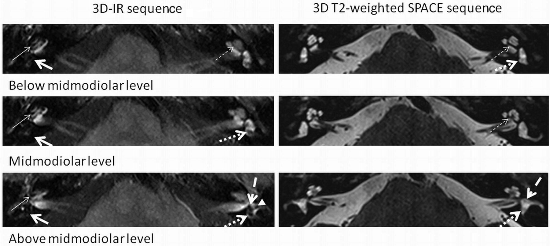
    A 3D-IR sequence depicts cochlear EH grade II (thin arrow) and vestibular EH grade II (thick arrow) on the right. EH is not visible on the corresponding 3D T2-weighted spatial and chemical-shift encoded excitation (SPACE) sequence. No EH on the normal left side is seen. Normal anatomy is shown at different levels (below the midmodiolar, midmodiolar, and above the midmodiolar sections) on the 3D-IR (0.8 mm) and 3D heavily T2-weighted SPACE (0.4 mm) sequence: interscalar septum (thin dashed arrow), anterior ampulla (thick dashed arrow), utricle/common crus (thick dotted arrow), and the lateral ampulla (arrowhead).

  • Fig 3.
    • Download figure
    • Open in new tab
    • Download powerpoint
    Fig 3.

    EH is present in 22% (10/45) of clinically normal ears and in 90% (55/61) of clinically diseased ears (irrespective of clinical score) (A) and in 73% of ears with possible (11/15), in 100% in ears with probable (3/3), and in 95% (41/43) of ears with definite MD (B).

  • Fig 4.
    • Download figure
    • Open in new tab
    • Download powerpoint
    Fig 4.

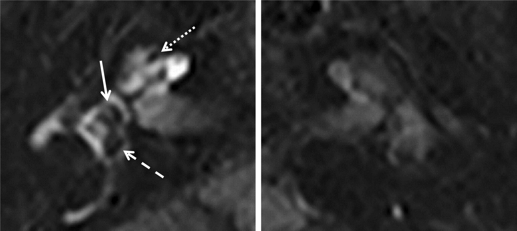
    Predominant saccular dilation: 3D-IR sequence (right and left side of the same patient). The right side shows a dilated saccule (arrow) and a slightly distended utricle (dashed arrow) with grade I cochlear hydrops (dotted arrow). Note increased contrast enhancement of the perilymph on the symptomatic right side compared with the normal left labyrinth.

  • Fig 5.
    • Download figure
    • Open in new tab
    • Download powerpoint
    Fig 5.

    Predominant utricular dilation: 3D-IR sequence left side. Marked distention of the utricle (dashed arrow) and sparing of the normal-sized saccule (arrow) leave the perilymphatic space visible (grade I) with no cochlear hydrops. The interscalar septum (dotted arrow) should not be mistaken for a slight cochlear hydrops.

PreviousNext
Back to top

In this issue

American Journal of Neuroradiology: 35 (7)
American Journal of Neuroradiology
Vol. 35, Issue 7
1 Jul 2014
  • Table of Contents
  • Index by author
  • Complete Issue (PDF)
Advertisement
Print
Download PDF
Email Article

Thank you for your interest in spreading the word on American Journal of Neuroradiology.

NOTE: We only request your email address so that the person you are recommending the page to knows that you wanted them to see it, and that it is not junk mail. We do not capture any email address.

Enter multiple addresses on separate lines or separate them with commas.
Detection and Grading of Endolymphatic Hydrops in Menière Disease Using MR Imaging
(Your Name) has sent you a message from American Journal of Neuroradiology
(Your Name) thought you would like to see the American Journal of Neuroradiology web site.
CAPTCHA
This question is for testing whether or not you are a human visitor and to prevent automated spam submissions.
Cite this article
K. Baráth, B. Schuknecht, A. Monge Naldi, T. Schrepfer, C.J. Bockisch, S.C.A. Hegemann
Detection and Grading of Endolymphatic Hydrops in Menière Disease Using MR Imaging
American Journal of Neuroradiology Jul 2014, 35 (7) 1387-1392; DOI: 10.3174/ajnr.A3856

Citation Manager Formats

  • BibTeX
  • Bookends
  • EasyBib
  • EndNote (tagged)
  • EndNote 8 (xml)
  • Medlars
  • Mendeley
  • Papers
  • RefWorks Tagged
  • Ref Manager
  • RIS
  • Zotero
0 Responses
Respond to this article
Share
Bookmark this article
Detection and Grading of Endolymphatic Hydrops in Menière Disease Using MR Imaging
K. Baráth, B. Schuknecht, A. Monge Naldi, T. Schrepfer, C.J. Bockisch, S.C.A. Hegemann
American Journal of Neuroradiology Jul 2014, 35 (7) 1387-1392; DOI: 10.3174/ajnr.A3856
del.icio.us logo Twitter logo Facebook logo Mendeley logo
  • Tweet Widget
  • Facebook Like
  • Google Plus One
Purchase

Jump to section

  • Article
    • Abstract
    • ABBREVIATIONS:
    • Materials and Methods
    • Results
    • Discussion
    • Conclusions
    • Acknowledgments
    • Footnotes
    • References
  • Figures & Data
  • Info & Metrics
  • Responses
  • References
  • PDF

Related Articles

  • No related articles found.
  • PubMed
  • Google Scholar

Cited By...

  • Morphometric Evaluation of the Facial and Vestibulocochlear Nerves Using MR Imaging in Patients with Meniere Disease
  • A Novel MR Imaging Sequence of 3D-ZOOMit Real Inversion-Recovery Imaging Improves Endolymphatic Hydrops Detection in Patients with Meniere Disease
  • A Novel MR Imaging Sequence of 3D-ZOOMit Real Inversion-Recovery Imaging Improves Endolymphatic Hydrops Detection in Patients with Meniere Disease
  • Effectiveness of endolymphatic duct blockage versus endolymphatic sac decompression in patients with intractable Menieres disease: study protocol for a double-blinded, randomised controlled trial
  • Comparison of Enhancement of the Vestibular Perilymph between Variable and Constant Flip Angle-Delayed 3D-FLAIR Sequences in Meniere Disease
  • Value of Endolymphatic Hydrops and Perilymph Signal Intensity in Suspected Meniere Disease
  • Blood-Labyrinth Barrier Permeability in Meniere Disease and Idiopathic Sudden Sensorineural Hearing Loss: Findings on Delayed Postcontrast 3D-FLAIR MRI
  • Endolymphatic Hydrops Reversal following Acetazolamide Therapy: Demonstration with Delayed Intravenous Contrast-Enhanced 3D-FLAIR MRI
  • Intratympanic Contrast in the Evaluation of Meniere Disease: Understanding the Limits
  • 3D Real Inversion Recovery MR Imaging for the Visualization of Endolymphatic Hydrops
  • Crossref (193)
  • Google Scholar

This article has been cited by the following articles in journals that are participating in Crossref Cited-by Linking.

  • Clinical Practice Guideline: Ménière’s Disease
    Gregory J. Basura, Meredith E. Adams, Ashkan Monfared, Seth R. Schwartz, Patrick J. Antonelli, Robert Burkard, Matthew L. Bush, Julie Bykowski, Maria Colandrea, Jennifer Derebery, Elizabeth A. Kelly, Kevin A. Kerber, Charles F. Koopman, Amy Angie Kuch, Evie Marcolini, Brian J. McKinnon, Michael J. Ruckenstein, Carla V. Valenzuela, Alexis Vosooney, Sandra A. Walsh, Lorraine C. Nnacheta, Nui Dhepyasuwan, Erin M. Buchanan
    Otolaryngology–Head and Neck Surgery 2020 162 S2
  • The value of four stage vestibular hydrops grading and asymmetric perilymphatic enhancement in the diagnosis of Menière’s disease on MRI
    Anja Bernaerts, Robby Vanspauwen, Cathérine Blaivie, Joost van Dinther, Andrzej Zarowski, Floris L. Wuyts, Stephanie Vanden Bossche, Erwin Offeciers, Jan W. Casselman, Bert De Foer
    Neuroradiology 2019 61 4
  • MRI of endolymphatic hydrops in patients with Meniere’s disease: a case-controlled study with a simplified classification based on saccular morphology
    Arnaud Attyé, Michael Eliezer, Naïla Boudiaf, Irène Tropres, David Chechin, Sébastien Schmerber, Georges Dumas, Alexandre Krainik
    European Radiology 2017 27 8
  • The blood labyrinthine barrier in the human normal and Meniere’s disease macula utricle
    Gail Ishiyama, Ivan A. Lopez, Paul Ishiyama, Harry V. Vinters, Akira Ishiyama
    Scientific Reports 2017 7 1
  • What does the head impulse test versus caloric dissociation reveal about vestibular dysfunction in Ménière's disease?
    Leigh A. McGarvie, Ian S. Curthoys, Hamish G. MacDougall, G. Michael Halmagyi
    Annals of the New York Academy of Sciences 2015 1343 1
  • Blood-Labyrinth Barrier Permeability in Menière Disease and Idiopathic Sudden Sensorineural Hearing Loss: Findings on Delayed Postcontrast 3D-FLAIR MRI
    M.N. Pakdaman, G. Ishiyama, A. Ishiyama, K.A. Peng, H.J. Kim, W.B. Pope, A.R. Sepahdari
    American Journal of Neuroradiology 2016 37 10
  • Meniere's disease: histopathology, cytochemistry, and imaging
    Gail Ishiyama, Ivan A. Lopez, Ali R. Sepahdari, Akira Ishiyama
    Annals of the New York Academy of Sciences 2015 1343 1
  • In vivo imaging of saccular hydrops in humans reflects sensorineural hearing loss rather than Meniere’s disease symptoms
    Arnaud Attyé, Michael Eliezer, Maud Medici, Irène Tropres, Georges Dumas, Alexandre Krainik, Sébastien Schmerber
    European Radiology 2018 28 7
  • Value of Endolymphatic Hydrops and Perilymph Signal Intensity in Suspected Ménière Disease
    J.M. van Steekelenburg, A. van Weijnen, L.M.H. de Pont, O.D. Vijlbrief, C.C. Bommeljé, J.P. Koopman, B.M. Verbist, H.M. Blom, S. Hammer
    American Journal of Neuroradiology 2020 41 3
  • Endolymphatic Hydrops Reversal following Acetazolamide Therapy: Demonstration with Delayed Intravenous Contrast-Enhanced 3D-FLAIR MRI
    A.R. Sepahdari, N. Vorasubin, G. Ishiyama, A. Ishiyama
    American Journal of Neuroradiology 2016 37 1

More in this TOC Section

  • Chondrosarcoma vs Synovial Chondromatosis: Imaging
  • WHO Classification Update: Nasal&Skull Base Tumors
  • Peritumoral Signal in Vestibular Schwannomas
Show more HEAD & NECK

Similar Articles

Advertisement

Indexed Content

  • Current Issue
  • Accepted Manuscripts
  • Article Preview
  • Past Issues
  • Editorials
  • Editor's Choice
  • Fellows' Journal Club
  • Letters to the Editor
  • Video Articles

Cases

  • Case Collection
  • Archive - Case of the Week
  • Archive - Case of the Month
  • Archive - Classic Case

Special Collections

  • AJNR Awards
  • ASNR Foundation Special Collection
  • Most Impactful AJNR Articles
  • Photon-Counting CT
  • Spinal CSF Leak Articles (Jan 2020-June 2024)

More from AJNR

  • Trainee Corner
  • Imaging Protocols
  • MRI Safety Corner

Multimedia

  • AJNR Podcasts
  • AJNR Scantastics

Resources

  • Turnaround Time
  • Submit a Manuscript
  • Submit a Video Article
  • Submit an eLetter to the Editor/Response
  • Manuscript Submission Guidelines
  • Statistical Tips
  • Fast Publishing of Accepted Manuscripts
  • Graphical Abstract Preparation
  • Imaging Protocol Submission
  • Evidence-Based Medicine Level Guide
  • Publishing Checklists
  • Author Policies
  • Become a Reviewer/Academy of Reviewers
  • News and Updates

About Us

  • About AJNR
  • Editorial Board
  • Editorial Board Alumni
  • Alerts
  • Permissions
  • Not an AJNR Subscriber? Join Now
  • Advertise with Us
  • Librarian Resources
  • Feedback
  • Terms and Conditions
  • AJNR Editorial Board Alumni

American Society of Neuroradiology

  • Not an ASNR Member? Join Now

© 2025 by the American Society of Neuroradiology All rights, including for text and data mining, AI training, and similar technologies, are reserved.
Print ISSN: 0195-6108 Online ISSN: 1936-959X

Powered by HighWire