Advertisement

Main menu

  • Home
  • Content
    • Current Issue
    • Accepted Manuscripts
    • Article Preview
    • Past Issue Archive
    • Video Articles
    • AJNR Case Collection
    • Case of the Week Archive
    • Case of the Month Archive
    • Classic Case Archive
  • Special Collections
    • AJNR Awards
    • ASNR Foundation Special Collection
    • Most Impactful AJNR Articles
    • Photon-Counting CT
    • Spinal CSF Leak Articles (Jan 2020-June 2024)
  • Multimedia
    • AJNR Podcasts
    • AJNR SCANtastic
    • Trainee Corner
    • MRI Safety Corner
    • Imaging Protocols
  • For Authors
    • Submit a Manuscript
    • Submit a Video Article
    • Submit an eLetter to the Editor/Response
    • Manuscript Submission Guidelines
    • Statistical Tips
    • Fast Publishing of Accepted Manuscripts
    • Graphical Abstract Preparation
    • Imaging Protocol Submission
    • Author Policies
  • About Us
    • About AJNR
    • Editorial Board
    • Editorial Board Alumni
  • More
    • Become a Reviewer/Academy of Reviewers
    • Subscribers
    • Permissions
    • Alerts
    • Feedback
    • Advertisers
    • ASNR Home

User menu

  • Alerts
  • Log in

Search

  • Advanced search
American Journal of Neuroradiology

American Journal of Neuroradiology

ASHNR American Society of Functional Neuroradiology ASHNR American Society of Pediatric Neuroradiology ASSR
  • Alerts
  • Log in

Advanced Search

  • Home
  • Content
    • Current Issue
    • Accepted Manuscripts
    • Article Preview
    • Past Issue Archive
    • Video Articles
    • AJNR Case Collection
    • Case of the Week Archive
    • Case of the Month Archive
    • Classic Case Archive
  • Special Collections
    • AJNR Awards
    • ASNR Foundation Special Collection
    • Most Impactful AJNR Articles
    • Photon-Counting CT
    • Spinal CSF Leak Articles (Jan 2020-June 2024)
  • Multimedia
    • AJNR Podcasts
    • AJNR SCANtastic
    • Trainee Corner
    • MRI Safety Corner
    • Imaging Protocols
  • For Authors
    • Submit a Manuscript
    • Submit a Video Article
    • Submit an eLetter to the Editor/Response
    • Manuscript Submission Guidelines
    • Statistical Tips
    • Fast Publishing of Accepted Manuscripts
    • Graphical Abstract Preparation
    • Imaging Protocol Submission
    • Author Policies
  • About Us
    • About AJNR
    • Editorial Board
    • Editorial Board Alumni
  • More
    • Become a Reviewer/Academy of Reviewers
    • Subscribers
    • Permissions
    • Alerts
    • Feedback
    • Advertisers
    • ASNR Home
  • Follow AJNR on Twitter
  • Visit AJNR on Facebook
  • Follow AJNR on Instagram
  • Join AJNR on LinkedIn
  • RSS Feeds

AJNR is seeking candidates for the AJNR Podcast Editor. Read the position description.

Research ArticlePediatrics

Midbrain-Hindbrain Involvement in Septo-Optic Dysplasia

M. Severino, A.E.M. Allegri, A. Pistorio, B. Roviglione, N. Di Iorgi, M. Maghnie and A. Rossi
American Journal of Neuroradiology August 2014, 35 (8) 1586-1592; DOI: https://doi.org/10.3174/ajnr.A3959
M. Severino
aFrom the Neuroradiology Unit (M.S., A.R.)
  • Find this author on Google Scholar
  • Find this author on PubMed
  • Search for this author on this site
A.E.M. Allegri
bPediatric Department (A.E.M.A., N.D.I., M.M.)
  • Find this author on Google Scholar
  • Find this author on PubMed
  • Search for this author on this site
A. Pistorio
cEpidemiology and Biostatistics Unit (A.P.), Istituto Giannina Gaslini, Università di Genova, Genoa, Italy
  • Find this author on Google Scholar
  • Find this author on PubMed
  • Search for this author on this site
B. Roviglione
dUniversità di Genova (B.R.), Genoa, Italy.
  • Find this author on Google Scholar
  • Find this author on PubMed
  • Search for this author on this site
N. Di Iorgi
bPediatric Department (A.E.M.A., N.D.I., M.M.)
  • Find this author on Google Scholar
  • Find this author on PubMed
  • Search for this author on this site
M. Maghnie
bPediatric Department (A.E.M.A., N.D.I., M.M.)
  • Find this author on Google Scholar
  • Find this author on PubMed
  • Search for this author on this site
A. Rossi
aFrom the Neuroradiology Unit (M.S., A.R.)
  • Find this author on Google Scholar
  • Find this author on PubMed
  • Search for this author on this site
  • Article
  • Figures & Data
  • Supplemental
  • Info & Metrics
  • Responses
  • References
  • PDF
Loading

Article Figures & Data

Figures

  • Fig 1.
    • Download figure
    • Open in new tab
    • Download powerpoint
    Fig 1.

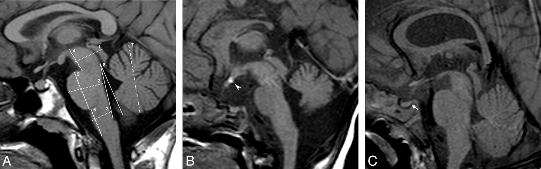
    A, Midsagittal T1-weighted image in a healthy child demonstrates how the measurements were performed: dotted lines indicate the AP diameter of the midbrain (1), pons (2), and medulla (3), dashed lines indicate the height of the midbrain (4), pons (5), and medulla (6), dash-dot line indicates the height of the vermis (7), and solid lines indicates the tegmento-vermian angle (8). B, Midsagittal T1-weighted image in a patient with SOD with visible brain stem abnormalities (group A) reveals hypoplasia of the pons and cerebellar vermis. Note the hypoplasia of the pituitary gland with ectopic posterior lobe (arrowhead) and associated small corpus callosum. C, Midsagittal T1-weighted image in a patient with SOD without visible brain stem abnormalities (group B) reveals grossly normal brain stem and vermis. Note the absence of the normal posterior pituitary bright spot (arrow).

  • Fig 2.
    • Download figure
    • Open in new tab
    • Download powerpoint
    Fig 2.

    Boxplots of MH measurements in patients with SOD (group A and B) and healthy controls. AP diameter of the pons (A) and medulla (B), midbrain to pons ratio (C), height of the pons (D), and vermis (E) are shown.

  • Fig 3.
    • Download figure
    • Open in new tab
    • Download powerpoint
    Fig 3.

    Scatterplots of MH measurements for age categories in patients with SOD and healthy controls. AP diameter of the pons (A) and medulla (B), midbrain to pons ratio (C), height of the pons (D), and vermis (E) are shown. In healthy controls, the AP diameters and height of brain stem structures and vermis showed an exponential growth curve. The growth spurt was steep until 16 months of age. Thereafter, it became less steep, reaching the adult level at about 8 years of age. On the other hand, the M/P ratios remained stable in controls over time, with a mean value of 0.82. In patients of group A, the AP diameter of the pons and medulla and the pontine and vermian height were lower compared with the values observed in control subjects of the same age category; in patients of group B, pontine and vermis height values tended to be lower compared with the values of control subjects of the same age category but these differences did not reach statistical significance. C, Note the lower M/P ratio in the patient harboring the 14q22.1-q23.1 deletion (arrow) compared with the corresponding values in age-matched controls.

  • Fig 4.
    • Download figure
    • Open in new tab
    • Download powerpoint
    Fig 4.

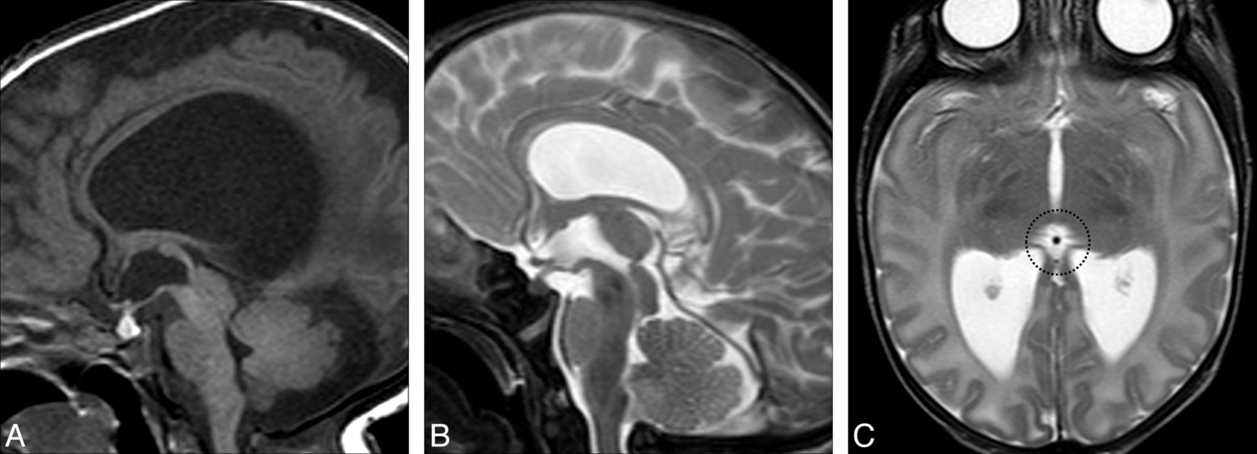
    A, Midline sagittal T1-weighted image demonstrates hypoplasia of the pons and small vermis, and enlarged quadrigeminal plate with secondary aqueductal stenosis and triventricular hydrocephalus. Midline sagittal (B) and axial (C) T2-weighted images in another patient reveal agenesis of the epithalamus including agenesis of the pineal gland (dotted circle), stria medullaris, and posterior commissure.

  • Fig 5.
    • Download figure
    • Open in new tab
    • Download powerpoint
    Fig 5.

    Midsagittal T1-weighted image of the patient harboring the 14q22.1-q23.1 deletion shows a small and short midbrain with elongated pons and relatively larger superior portion of the cerebellar vermis. Note the small anterior pituitary gland with ectopic posterior lobe along the pituitary stalk (arrowhead).

PreviousNext
Back to top

In this issue

American Journal of Neuroradiology: 35 (8)
American Journal of Neuroradiology
Vol. 35, Issue 8
1 Aug 2014
  • Table of Contents
  • Index by author
  • Complete Issue (PDF)
Advertisement
Print
Download PDF
Email Article

Thank you for your interest in spreading the word on American Journal of Neuroradiology.

NOTE: We only request your email address so that the person you are recommending the page to knows that you wanted them to see it, and that it is not junk mail. We do not capture any email address.

Enter multiple addresses on separate lines or separate them with commas.
Midbrain-Hindbrain Involvement in Septo-Optic Dysplasia
(Your Name) has sent you a message from American Journal of Neuroradiology
(Your Name) thought you would like to see the American Journal of Neuroradiology web site.
CAPTCHA
This question is for testing whether or not you are a human visitor and to prevent automated spam submissions.
Cite this article
M. Severino, A.E.M. Allegri, A. Pistorio, B. Roviglione, N. Di Iorgi, M. Maghnie, A. Rossi
Midbrain-Hindbrain Involvement in Septo-Optic Dysplasia
American Journal of Neuroradiology Aug 2014, 35 (8) 1586-1592; DOI: 10.3174/ajnr.A3959

Citation Manager Formats

  • BibTeX
  • Bookends
  • EasyBib
  • EndNote (tagged)
  • EndNote 8 (xml)
  • Medlars
  • Mendeley
  • Papers
  • RefWorks Tagged
  • Ref Manager
  • RIS
  • Zotero
0 Responses
Respond to this article
Share
Bookmark this article
Midbrain-Hindbrain Involvement in Septo-Optic Dysplasia
M. Severino, A.E.M. Allegri, A. Pistorio, B. Roviglione, N. Di Iorgi, M. Maghnie, A. Rossi
American Journal of Neuroradiology Aug 2014, 35 (8) 1586-1592; DOI: 10.3174/ajnr.A3959
del.icio.us logo Twitter logo Facebook logo Mendeley logo
  • Tweet Widget
  • Facebook Like
  • Google Plus One
Purchase

Jump to section

  • Article
    • Abstract
    • ABBREVIATIONS:
    • Methods
    • Results
    • Discussion
    • Conclusion
    • Acknowledgments
    • Footnotes
    • REFERENCES
  • Figures & Data
  • Supplemental
  • Info & Metrics
  • Responses
  • References
  • PDF

Related Articles

  • PubMed
  • Google Scholar

Cited By...

  • No citing articles found.
  • Crossref (22)
  • Google Scholar

This article has been cited by the following articles in journals that are participating in Crossref Cited-by Linking.

  • Advances in differential diagnosis and management of growth hormone deficiency in children
    Camille Hage, Hoong-Wei Gan, Anastasia Ibba, Giuseppa Patti, Mehul Dattani, Sandro Loche, Mohamad Maghnie, Roberto Salvatori
    Nature Reviews Endocrinology 2021 17 10
  • Midbrain-Hindbrain Boundary Morphogenesis: At the Intersection of Wnt and Fgf Signaling
    Holly C. Gibbs, Ana Chang-Gonzalez, Wonmuk Hwang, Alvin T. Yeh, Arne C. Lekven
    Frontiers in Neuroanatomy 2017 11
  • Expanding the spectrum of congenital anomalies of the diencephalic–mesencephalic junction
    Mariasavina Severino, Domenico Tortora, Angela Pistorio, Luca Antonio Ramenghi, Flavia Napoli, Maria Margherita Mancardi, Pasquale Striano, Valeria Capra, Andrea Rossi
    Neuroradiology 2016 58 1
  • Review of the MRI brain findings of septo-optic dysplasia
    D.J. Ward, D.J.A. Connolly, P.D. Griffiths
    Clinical Radiology 2021 76 2
  • The Human Hypothalamus - Neuroendocrine Disorders
    Ieva Sataite, Simon Cudlip, Jayaratnam Jayamohan, Mario Ganau
    2021 181
  • Assessment of fetal optic chiasm: an echoanatomic and reproducibility study
    D. Paladini, R. Birnbaum, G. Donarini, I. Maffeo, E. Fulcheri
    Ultrasound in Obstetrics & Gynecology 2016 48 6
  • Two‐dimensional visualization and measurement of the fetal optic chiasm: improving counseling for antenatal diagnosis of agenesis of the septum pellucidum
    F. Viñals, P. Ruiz, F. Correa, P. Gonçalves Pereira
    Ultrasound in Obstetrics & Gynecology 2016 48 6
  • Disorders of Ventral Induction/Spectrum of Holoprosencephaly
    Sonia Francesca Calloni, Luca Caschera, Fabio Maria Triulzi
    Neuroimaging Clinics of North America 2019 29 3
  • Neurodevelopmental impairments in children with septo-optic dysplasia spectrum conditions: a systematic review
    Amy Mann, Arameh Aghababaie, Jennifer Kalitsi, Daniel Martins, Yannis Paloyelis, Ritika R. Kapoor
    Molecular Autism 2023 14 1
  • Neuroimaging Findings in Pediatric Genetic Skeletal Disorders: A Review
    Matthias W. Wagner, Andrea Poretti, Jane E. Benson, Thierry A. G. M. Huisman
    Journal of Neuroimaging 2017 27 2

More in this TOC Section

  • SyMRI & MR Fingerprinting in Brainstem Myelination
  • Comparison of Image Quality and Radiation Dose in Pediatric Temporal Bone CT Using Photon-Counting Detector CT and Energy-Integrating Detector CT
  • Venous Sinus Stenosis in Mucopolysaccharidosis I
Show more Pediatrics

Similar Articles

Advertisement

Indexed Content

  • Current Issue
  • Accepted Manuscripts
  • Article Preview
  • Past Issues
  • Editorials
  • Editor's Choice
  • Fellows' Journal Club
  • Letters to the Editor
  • Video Articles

Cases

  • Case Collection
  • Archive - Case of the Week
  • Archive - Case of the Month
  • Archive - Classic Case

Special Collections

  • AJNR Awards
  • ASNR Foundation Special Collection
  • Most Impactful AJNR Articles
  • Photon-Counting CT
  • Spinal CSF Leak Articles (Jan 2020-June 2024)

More from AJNR

  • Trainee Corner
  • Imaging Protocols
  • MRI Safety Corner

Multimedia

  • AJNR Podcasts
  • AJNR Scantastics

Resources

  • Turnaround Time
  • Submit a Manuscript
  • Submit a Video Article
  • Submit an eLetter to the Editor/Response
  • Manuscript Submission Guidelines
  • Statistical Tips
  • Fast Publishing of Accepted Manuscripts
  • Graphical Abstract Preparation
  • Imaging Protocol Submission
  • Evidence-Based Medicine Level Guide
  • Publishing Checklists
  • Author Policies
  • Become a Reviewer/Academy of Reviewers
  • News and Updates

About Us

  • About AJNR
  • Editorial Board
  • Editorial Board Alumni
  • Alerts
  • Permissions
  • Not an AJNR Subscriber? Join Now
  • Advertise with Us
  • Librarian Resources
  • Feedback
  • Terms and Conditions
  • AJNR Editorial Board Alumni

American Society of Neuroradiology

  • Not an ASNR Member? Join Now

© 2025 by the American Society of Neuroradiology All rights, including for text and data mining, AI training, and similar technologies, are reserved.
Print ISSN: 0195-6108 Online ISSN: 1936-959X

Powered by HighWire