Advertisement

Main menu

  • Home
  • Content
    • Current Issue
    • Accepted Manuscripts
    • Article Preview
    • Past Issue Archive
    • Video Articles
    • AJNR Case Collection
    • Case of the Week Archive
    • Case of the Month Archive
    • Classic Case Archive
  • Special Collections
    • AJNR Awards
    • ASNR Foundation Special Collection
    • Most Impactful AJNR Articles
    • Photon-Counting CT
    • Spinal CSF Leak Articles (Jan 2020-June 2024)
  • Multimedia
    • AJNR Podcasts
    • AJNR SCANtastic
    • Trainee Corner
    • MRI Safety Corner
    • Imaging Protocols
  • For Authors
    • Submit a Manuscript
    • Submit a Video Article
    • Submit an eLetter to the Editor/Response
    • Manuscript Submission Guidelines
    • Statistical Tips
    • Fast Publishing of Accepted Manuscripts
    • Graphical Abstract Preparation
    • Imaging Protocol Submission
    • Author Policies
  • About Us
    • About AJNR
    • Editorial Board
    • Editorial Board Alumni
  • More
    • Become a Reviewer/Academy of Reviewers
    • Subscribers
    • Permissions
    • Alerts
    • Feedback
    • Advertisers
    • ASNR Home

User menu

  • Alerts
  • Log in

Search

  • Advanced search
American Journal of Neuroradiology

American Journal of Neuroradiology

ASHNR American Society of Functional Neuroradiology ASHNR American Society of Pediatric Neuroradiology ASSR
  • Alerts
  • Log in

Advanced Search

  • Home
  • Content
    • Current Issue
    • Accepted Manuscripts
    • Article Preview
    • Past Issue Archive
    • Video Articles
    • AJNR Case Collection
    • Case of the Week Archive
    • Case of the Month Archive
    • Classic Case Archive
  • Special Collections
    • AJNR Awards
    • ASNR Foundation Special Collection
    • Most Impactful AJNR Articles
    • Photon-Counting CT
    • Spinal CSF Leak Articles (Jan 2020-June 2024)
  • Multimedia
    • AJNR Podcasts
    • AJNR SCANtastic
    • Trainee Corner
    • MRI Safety Corner
    • Imaging Protocols
  • For Authors
    • Submit a Manuscript
    • Submit a Video Article
    • Submit an eLetter to the Editor/Response
    • Manuscript Submission Guidelines
    • Statistical Tips
    • Fast Publishing of Accepted Manuscripts
    • Graphical Abstract Preparation
    • Imaging Protocol Submission
    • Author Policies
  • About Us
    • About AJNR
    • Editorial Board
    • Editorial Board Alumni
  • More
    • Become a Reviewer/Academy of Reviewers
    • Subscribers
    • Permissions
    • Alerts
    • Feedback
    • Advertisers
    • ASNR Home
  • Follow AJNR on Twitter
  • Visit AJNR on Facebook
  • Follow AJNR on Instagram
  • Join AJNR on LinkedIn
  • RSS Feeds

AJNR is seeking candidates for the AJNR Podcast Editor. Read the position description.

Research ArticlePediatrics

Brain Parenchymal Signal Abnormalities Associated with Developmental Venous Anomalies in Children and Young Adults

L.L. Linscott, J.L. Leach, B. Zhang and B.V. Jones
American Journal of Neuroradiology August 2014, 35 (8) 1600-1607; DOI: https://doi.org/10.3174/ajnr.A3960
L.L. Linscott
aFrom the Departments of Radiology (L.L.L., J.L.L., B.V.J.)
  • Find this author on Google Scholar
  • Find this author on PubMed
  • Search for this author on this site
J.L. Leach
aFrom the Departments of Radiology (L.L.L., J.L.L., B.V.J.)
  • Find this author on Google Scholar
  • Find this author on PubMed
  • Search for this author on this site
B. Zhang
bBiostatistics and Epidemiology (B.Z.); Cincinnati Children's Hospital Medical Center, University of Cincinnati College of Medicine, Cincinnati, Ohio.
  • Find this author on Google Scholar
  • Find this author on PubMed
  • Search for this author on this site
B.V. Jones
aFrom the Departments of Radiology (L.L.L., J.L.L., B.V.J.)
  • Find this author on Google Scholar
  • Find this author on PubMed
  • Search for this author on this site
  • Article
  • Figures & Data
  • Supplemental
  • Info & Metrics
  • Responses
  • References
  • PDF
Loading

Article Figures & Data

Figures

  • Tables
  • Fig 1.
    • Download figure
    • Open in new tab
    • Download powerpoint
    Fig 1.

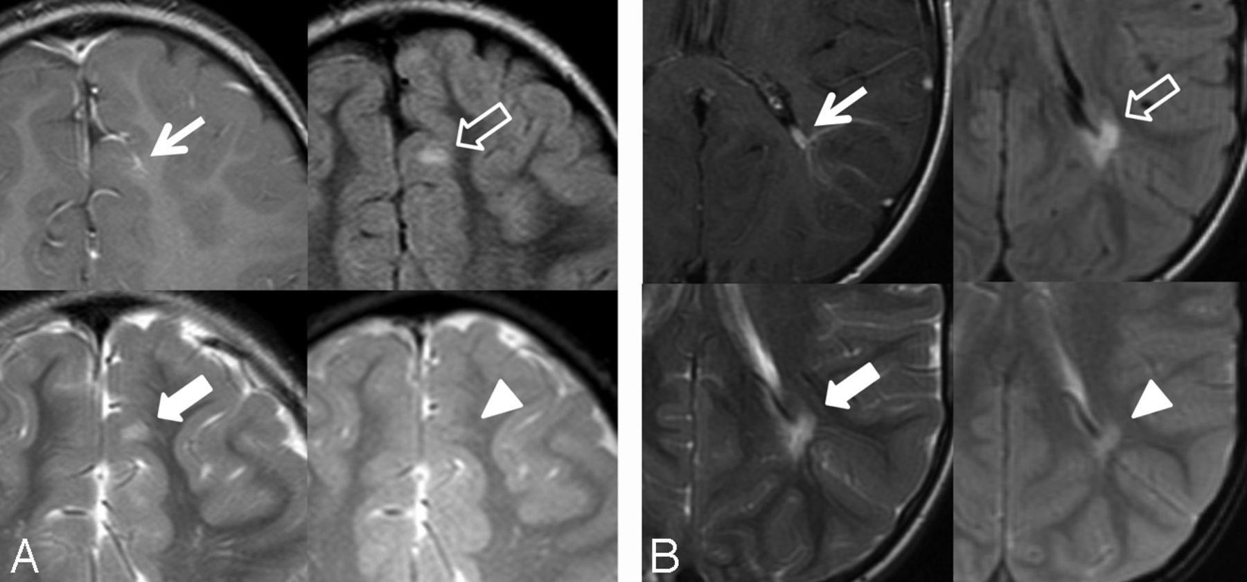
    Two DVAs with associated signal abnormality in a 5-year-old boy (A) and a 7-year-old boy (B). T1WI with contrast (arrow), FLAIR (outlined arrow), T2WI (block arrow), and gradient recalled-echo (arrowhead). A, Left frontal lobe DVA with juxtacortical depth, superficial venous drainage, and associated increased FLAIR and T2 signal abnormality. Note the lack of gradient recalled-echo hypointensity in the same region. B, Left parietal lobe DVA with periventricular depth, deep venous drainage, and associated signal abnormality.

  • Fig 2.
    • Download figure
    • Open in new tab
    • Download powerpoint
    Fig 2.

    DVA with associated signal abnormality (black arrows) and CM (open black arrows) in a 13-month-old boy. A, T1WI with contrast. B, FLAIR. C, T2WI. D, SWI. Right frontal lobe DVA with subcortical depth and bidirectional venous drainage (white arrows).

  • Fig 3.
    • Download figure
    • Open in new tab
    • Download powerpoint
    Fig 3.

    DVA in a 23-month-old boy with associated signal abnormality and parenchymal atrophy. A, T1WI with contrast. B, FLAIR. Left frontal lobe DVA with periventricular depth and deep venous drainage. Note the increased FLAIR and T2 signal abnormality with associated parenchymal atrophy.

  • Fig 4.
    • Download figure
    • Open in new tab
    • Download powerpoint
    Fig 4.

    Percentage signal abnormalities associated with DVAs by age group. n indicates the number of subjects in each age group.

  • Fig 5.
    • Download figure
    • Open in new tab
    • Download powerpoint
    Fig 5.

    FLAIR (A) and T1WI with contrast (B) show signal abnormality in the images of a 23-month-old boy, showing relative increased signal within the drainage territory of the DVA compared with normal contralateral myelinating white matter. Left frontal lobe DVA with periventricular depth and deep venous drainage.

  • Fig 6.
    • Download figure
    • Open in new tab
    • Download powerpoint
    Fig 6.

    DVA with a subtle decreased extent of signal abnormality on follow-up examination. Axial FLAIR images at 2 years (A) and 5 years (B) of age. Note subtle decreased signal intensity in the drainage territory of the DVA along the lateral aspect of the DVA draining vein.

Tables

  • Figures
  • Signal abnormalities related to subject age, sex, CM, and parenchymal atrophy

    Signal AbnormalityNo Signal AbnormalityTotalP′OR (95% CI)aP″
    Total No.26b167c193d
    Demographics
        Age7.3 ± 5.5 [5.1–9.5]11.4 ± 6.6 [10.4–12.4]10.8 ± 6.6 [9.9–11.8].0030.84 (0.76–0.94).001
        Female10 (38.5%)75 (44.9%)85 (44%).5380.94 (0.32–2.83).918
    Associated abnormalities
        Focal atrophy5 (17.8%)3 (1.8%)8 (4.1%)<.00117.1 (2.52–117).004
        CM5 (19.2%)7 (4.2%)12 (6.2%).00319.3 (2.95–126).002
    Location.260.589
        Lobar24 (92.3%)129 (77.2%)153 (79.3%)1.0
        Thalamus/BG0 (0%)6 (3.6%)6 (3.1%)0 (0–∞)
        Cerebellum/BS2 (7.7%)32 (19.2%)34 (17.6%)0.37 (0.05–2.49)
    Depth of draining vein.016.909
        Periventricular8 (30.8%)19 (11.4%)27 (14%)1.0
        Subcortical10 (38.5%)60 (35.9%)70 (36.3%)0.73 (0.18–3.01)
        Juxtacortical8 (30.8%)88 (52.7%)96 (49.7%)0.81 (0.16–4.09)
    Direction of draining vein.001.047
        Superficial9 (34.6%)112 (67.1%)121 (62.7%)1.0
        Deep14 (53.8%)52 (31.1%)66 (34.2%)5.21 (1.22–22.2)
        Both3 (11.6%)3 (1.8%)6 (3.1%)6.85 (0.84–56.0)
    • Note:—BS indicates brain stem; BG, basal ganglia; P′, univariate analysis (t test or χ2 test); P″, multivariate logistic regression; brackets, 95% confidence interval for age.

    • ↵a OR for age is expressed as the likelihood of signal abnormality with each additional year. OR for sex is expressed as the likelihood of signal abnormality for a female subject. ORs for focal atrophy and CM are expressed as the likelihood of signal abnormality in the presence of either associated abnormality. ORs for location, depth, and direction of draining vein are expressed as the likelihood of signal abnormality compared with the first DVA morphologic descriptor (eg, lobar, periventricular, superficial), defined as a baseline OR of 1.0.

    • ↵b % is the percentage of DVAs with a certain characteristic or associated finding divided by the total number of DVAs with signal abnormality.

    • ↵c % is the percentage of DVAs with a certain characteristic or associated finding divided by the total number of DVAs without signal abnormality.

    • ↵d % is the percentage of DVAs with a certain characteristic or associated finding divided by the total number of DVAs.

PreviousNext
Back to top

In this issue

American Journal of Neuroradiology: 35 (8)
American Journal of Neuroradiology
Vol. 35, Issue 8
1 Aug 2014
  • Table of Contents
  • Index by author
  • Complete Issue (PDF)
Advertisement
Print
Download PDF
Email Article

Thank you for your interest in spreading the word on American Journal of Neuroradiology.

NOTE: We only request your email address so that the person you are recommending the page to knows that you wanted them to see it, and that it is not junk mail. We do not capture any email address.

Enter multiple addresses on separate lines or separate them with commas.
Brain Parenchymal Signal Abnormalities Associated with Developmental Venous Anomalies in Children and Young Adults
(Your Name) has sent you a message from American Journal of Neuroradiology
(Your Name) thought you would like to see the American Journal of Neuroradiology web site.
CAPTCHA
This question is for testing whether or not you are a human visitor and to prevent automated spam submissions.
Cite this article
L.L. Linscott, J.L. Leach, B. Zhang, B.V. Jones
Brain Parenchymal Signal Abnormalities Associated with Developmental Venous Anomalies in Children and Young Adults
American Journal of Neuroradiology Aug 2014, 35 (8) 1600-1607; DOI: 10.3174/ajnr.A3960

Citation Manager Formats

  • BibTeX
  • Bookends
  • EasyBib
  • EndNote (tagged)
  • EndNote 8 (xml)
  • Medlars
  • Mendeley
  • Papers
  • RefWorks Tagged
  • Ref Manager
  • RIS
  • Zotero
0 Responses
Respond to this article
Share
Bookmark this article
Brain Parenchymal Signal Abnormalities Associated with Developmental Venous Anomalies in Children and Young Adults
L.L. Linscott, J.L. Leach, B. Zhang, B.V. Jones
American Journal of Neuroradiology Aug 2014, 35 (8) 1600-1607; DOI: 10.3174/ajnr.A3960
del.icio.us logo Twitter logo Facebook logo Mendeley logo
  • Tweet Widget
  • Facebook Like
  • Google Plus One
Purchase

Jump to section

  • Article
    • Abstract
    • ABBREVIATIONS:
    • Materials and Methods
    • Results
    • Discussion
    • Conclusions
    • Footnotes
    • REFERENCES
  • Figures & Data
  • Supplemental
  • Info & Metrics
  • Responses
  • References
  • PDF

Related Articles

  • PubMed
  • Google Scholar

Cited By...

  • Neonatal Developmental Venous Anomalies: Clinicoradiologic Characterization and Follow-Up
  • The Central Vein: FLAIR Signal Abnormalities Associated with Developmental Venous Anomalies in Patients with Multiple Sclerosis
  • Multiple Brain Developmental Venous Anomalies as a Marker for Constitutional Mismatch Repair Deficiency Syndrome
  • Increased Prevalence of Developmental Venous Anomalies in Children with Intracranial Neoplasms
  • Brain Metabolic Abnormalities Associated with Developmental Venous Anomalies
  • Crossref (35)
  • Google Scholar

This article has been cited by the following articles in journals that are participating in Crossref Cited-by Linking.

  • A Review of Clinical and Imaging Findings in Tumefactive Demyelination
    Mariko Nakayama, Shotaro Naganawa, Michelle Ouyang, Karra A. Jones, John Kim, Aristides A. Capizzano, Toshio Moritani
    American Journal of Roentgenology 2021 217 1
  • Prevalence of cerebral cavernous malformations associated with developmental venous anomalies increases with age
    Waleed Brinjikji, Ali El-Rida El-Masri, John T. Wald, Kelly D. Flemming, Giuseppe Lanzino
    Child's Nervous System 2017 33 9
  • Developmental venous anomalies of the brain in children — imaging spectrum and update
    Luke L. Linscott, James L. Leach, Blaise V. Jones, Todd A. Abruzzo
    Pediatric Radiology 2016 46 3
  • Multiple Brain Developmental Venous Anomalies as a Marker for Constitutional Mismatch Repair Deficiency Syndrome
    S.I. Shiran, L. Ben-Sira, R. Elhasid, J. Roth, U. Tabori, M. Yalon, S. Constantini, R. Dvir
    American Journal of Neuroradiology 2018 39 10
  • Increased Prevalence of Developmental Venous Anomalies in Children with Intracranial Neoplasms
    B.V. Jones, L. Linscott, G. Koberlein, T.R. Hummel, J.L. Leach
    American Journal of Neuroradiology 2015 36 9
  • Brain Metabolic Abnormalities Associated with Developmental Venous Anomalies
    M. Larvie, D. Timerman, J.A. Thum
    American Journal of Neuroradiology 2015 36 3
  • Sensitivity of susceptibility-weighted imaging in detecting developmental venous anomalies and associated cavernomas and microhemorrhages in children
    Allen Young, Andrea Poretti, Thangamadhan Bosemani, Reema Goel, Thierry A. G. M. Huisman
    Neuroradiology 2017 59 8
  • Revealing vascular abnormalities and measuring small vessel density in multiple sclerosis lesions using USPIO
    Sagar Buch, Karthikeyan Subramanian, Pavan K. Jella, Yongsheng Chen, Zhen Wu, Kamran Shah, Evanthia Bernitsas, Yulin Ge, E. Mark Haacke
    NeuroImage: Clinical 2021 29
  • Venous pathologies in paediatric neuroradiology: from foetal to adolescent life
    Kshitij Mankad, Asthik Biswas, Maria Camilla Rossi Espagnet, Luke Dixon, Nihaal Reddy, Ai Peng Tan, Ozgur Oztekin, Felice D’Arco, Karuna Shekdar, Prakash Muthusami, Fergus Robertson, Stacy Goergen, Winston Chong
    Neuroradiology 2020 62 1
  • An Overview of Venous Abnormalities Related to the Development of Lesions in Multiple Sclerosis
    E. Mark Haacke, Yulin Ge, Sean K. Sethi, Sagar Buch, Paolo Zamboni
    Frontiers in Neurology 2021 12

More in this TOC Section

  • SyMRI & MR Fingerprinting in Brainstem Myelination
  • Comparison of Image Quality and Radiation Dose in Pediatric Temporal Bone CT Using Photon-Counting Detector CT and Energy-Integrating Detector CT
  • Venous Sinus Stenosis in Mucopolysaccharidosis I
Show more Pediatrics

Similar Articles

Advertisement

Indexed Content

  • Current Issue
  • Accepted Manuscripts
  • Article Preview
  • Past Issues
  • Editorials
  • Editor's Choice
  • Fellows' Journal Club
  • Letters to the Editor
  • Video Articles

Cases

  • Case Collection
  • Archive - Case of the Week
  • Archive - Case of the Month
  • Archive - Classic Case

Special Collections

  • AJNR Awards
  • ASNR Foundation Special Collection
  • Most Impactful AJNR Articles
  • Photon-Counting CT
  • Spinal CSF Leak Articles (Jan 2020-June 2024)

More from AJNR

  • Trainee Corner
  • Imaging Protocols
  • MRI Safety Corner

Multimedia

  • AJNR Podcasts
  • AJNR Scantastics

Resources

  • Turnaround Time
  • Submit a Manuscript
  • Submit a Video Article
  • Submit an eLetter to the Editor/Response
  • Manuscript Submission Guidelines
  • Statistical Tips
  • Fast Publishing of Accepted Manuscripts
  • Graphical Abstract Preparation
  • Imaging Protocol Submission
  • Evidence-Based Medicine Level Guide
  • Publishing Checklists
  • Author Policies
  • Become a Reviewer/Academy of Reviewers
  • News and Updates

About Us

  • About AJNR
  • Editorial Board
  • Editorial Board Alumni
  • Alerts
  • Permissions
  • Not an AJNR Subscriber? Join Now
  • Advertise with Us
  • Librarian Resources
  • Feedback
  • Terms and Conditions
  • AJNR Editorial Board Alumni

American Society of Neuroradiology

  • Not an ASNR Member? Join Now

© 2025 by the American Society of Neuroradiology All rights, including for text and data mining, AI training, and similar technologies, are reserved.
Print ISSN: 0195-6108 Online ISSN: 1936-959X

Powered by HighWire