
Article Figures & Data
Figures
- Fig 1.
Cohort selection.
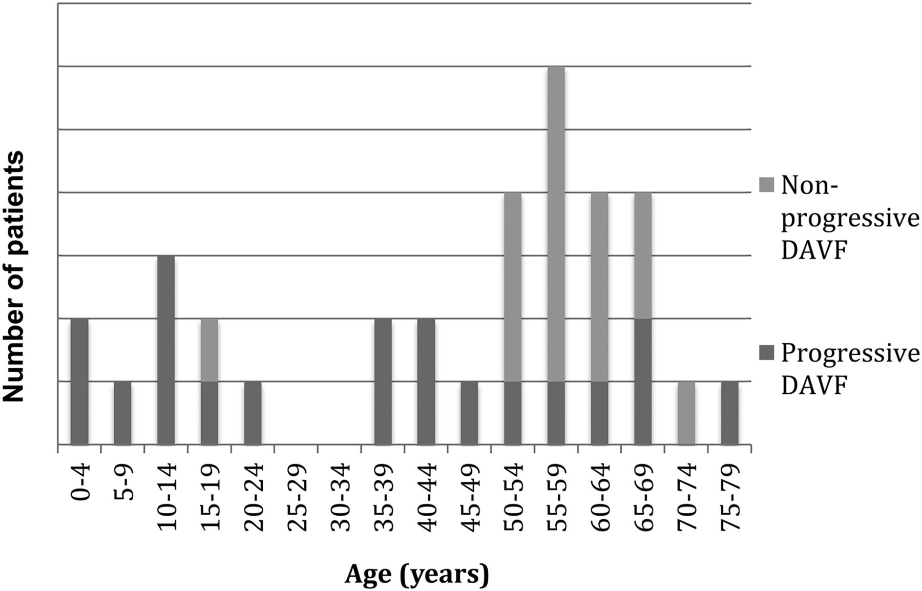
- Fig 2.
Age of diagnosis for patients with progressive-versus-nonprogressive fistulas.
- Fig 3.
Multiple and progressive DAVFs. A 25-year-old female patient with large skull base DAVFs refractory to multiple endovascular treatments. She was initially diagnosed at 18 years of age and died of intracranial hemorrhage 6 years later in the setting of intractable intracranial venous hypertension. CTA images demonstrate an extensive skull base vascular abnormality with dilated cortical veins suggestive of intracranial venous hypertension.
- Fig 4.
Multiple and progressive DAVFs. Same patient as in Fig 3. DSA images before treatment at our institution but 4 years after initial proximal coil embolization of external carotid artery feeders below the skull base at an outside institution demonstrate multiple skull base fistulas associated with venous hypertension, cortical venous reflux, venous sinus dilation, and a jugular bulb outflow stenosis (between white arrows). (Upper row: right vertebral artery lateral, right vertebral artery lateral, left vertebral artery anteroposterior; lower row: left ICA anteroposterior, left external carotid artery anteroposterior, left external carotid artery lateral). A middle meningeal artery to the torcular fistula indicated by the white arrows is shown in greater detail in Fig 5.
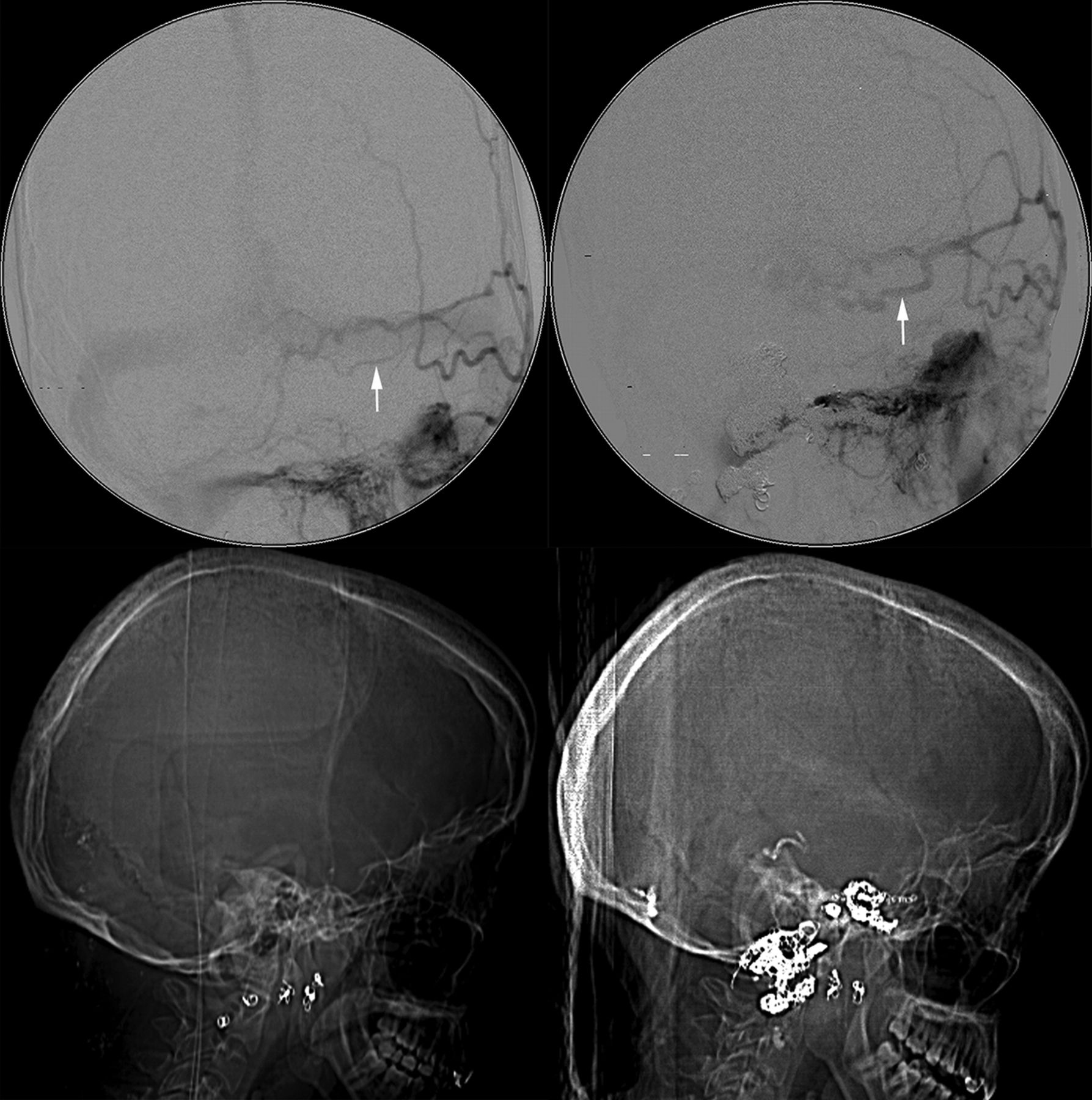
- Fig 5.
Multiple and progressive DAVFs. Same patient as in Figs 3 and 4. DSA demonstrates progression of a left middle meningeal artery to a torcular DAVF with interval enlargement of a feeding artery (arrow in upper left panel versus arrow in upper right panel) during 7 months. Lateral CT scanograms obtained at the same time as DSA provide an overview of treatment with interval deposition of embolic coils and n-BCA glue (lower left versus lower right panel).


Tables
- Table 1:
UCSF intracranial dural arteriovenous fistula cohort with single fistulas versus subset with confirmed enlarging, de novo, multiple, or recurrent fistulas
Enlarging, De Novo, Multiple, or Recurrent Fistulas (n = 34) Remainder of UCSF Intracranial DAVFs (n = 545) P Valuea Mean age at dx ± SD 45 ± 23 y 53 ± 20 y .11b Median age (range) 52.5 y (0.2–77 y) 56 y (0–87 y) Male 35% 49% .16c All Subjects (n = 34) Progressive (n = 19) Nonprogressive (n = 15) P Value Treatments Median treatment procedures (25%, 75%) 5 (3, 9) 3 (2, 4) .0068a Range treatment procedures 2–19 2–7 Any surgical treatment 63% 33% .17b Transarterial embolization 100% 80% .076b Transvenous embolization 74% 67% .72b Ethanol embolization 84% 67% .42b Coil embolization 89% 80% .63b PVA embolization 74% 53% .29b Glue embolization 42% 13% .13b Onyx embolizationc 11% 20% .63b Outcomes at last follow-up Mean follow-up duration 4.6 y 5.1 y 4.0 y .70 Median (range) follow-up duration 2.5 y (56 d to 23 y) 3.9 y (58 d to 17 y) 1.4 y (56 d to 23 y) .37a Death 16% 0% .24b Median outcome (normal = 0, deficits/residual fistula = 1, death = 2) 1 (0, 1) 1 (0, 1) .31a Median last mRS (25%, 75%) 1 (0, 3) 1 (0, 3) .39a Range last mRS 0–6 0–3 Good outcome (last mRS 0–2) 74% 73% 1.0 Median change in mRS (25%, 75%) −0.5 (−1, 0) −1 (−1, 0) .83a Range change mRS −3 to 1 −4 to 2


Jump to section
Related Articles
Cited By...
- Acquired progressive torcular dural arteriovenous fistula after subtotal resection of peritorcular meningioma
- Indications for the Performance of Intracranial Endovascular Neurointerventional Procedures: A Scientific Statement From the American Heart Association
- Pial Artery Supply as an Anatomic Risk Factor for Ischemic Stroke in the Treatment of Intracranial Dural Arteriovenous Fistulas