
Article Figures & Data
Figures
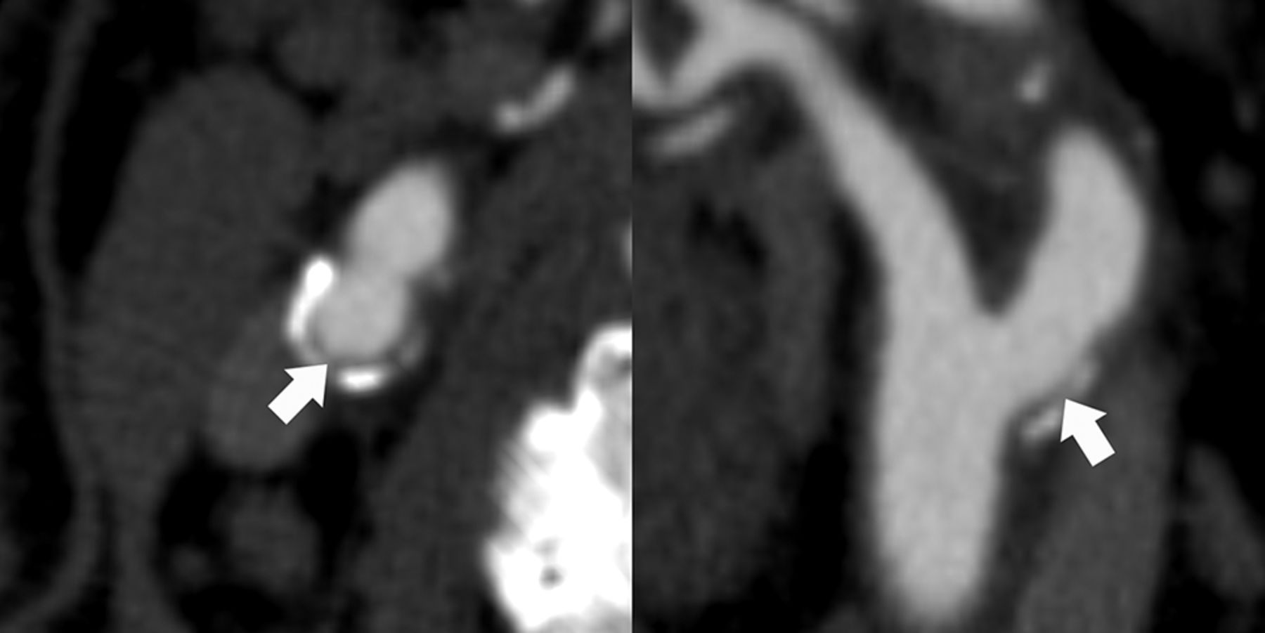
- Fig 1.
Plaque ulceration on MDCTA. Transversal (left) and longitudinal (right) MDCTA images of the carotid bifurcation. A “plaque ulceration,” defined as the extension of contrast material in the atherosclerotic plaque, is visible on both planes (arrows; arrowheads indicate the edges of the plaque ulceration). ECA indicates external carotid artery.
- Fig 2.
Fissured fibrous cap on MDCTA. Transversal (left) and longitudinal (right) MDCTA images of the carotid bifurcation. A “fissured fibrous cap,” defined as an extension of contrast material of <1 mm into the atherosclerotic plaque and an angle of ≥230° with the lumen, is visible only on the transversal plane (arrows).
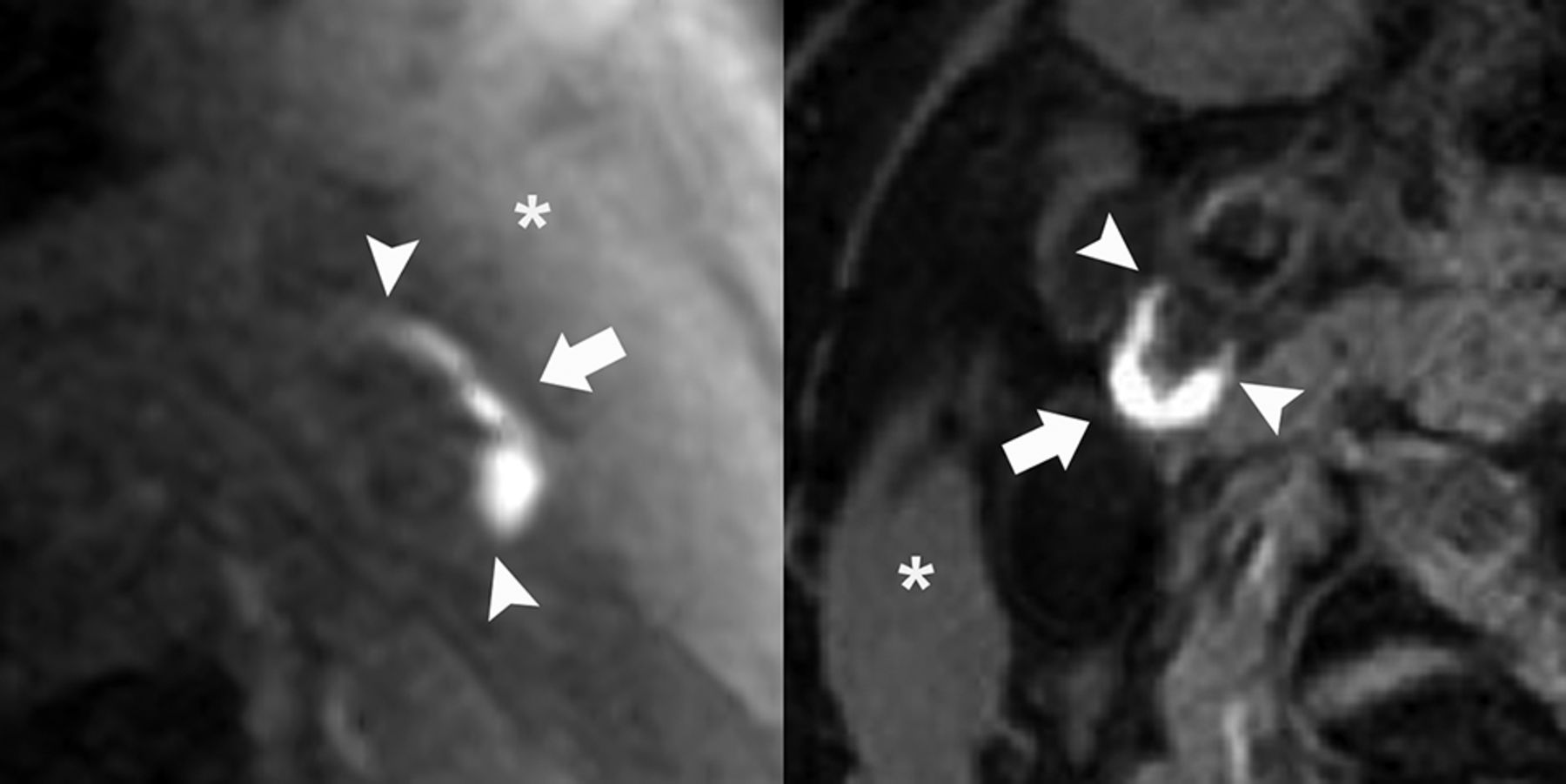
- Fig 3.
Intraplaque hemorrhage on MR imaging. Carotid bifurcations of 2 patients: left, the 3D-T1W fat suppressed spoiled gradient echo sequence (Discovery MR 750; GE Healthcare), and right, the 2D-T1W inversion recovery turbo field echo sequence (Achieva; Philips Healthcare). In both patients, intraplaque hemorrhage is present (arrows; arrowheads indicate the edges of the intraplaque hemorrhage), defined as a hyperintense signal in the atherosclerotic plaque compared with the sternocleidomastoid muscle (asterisk).
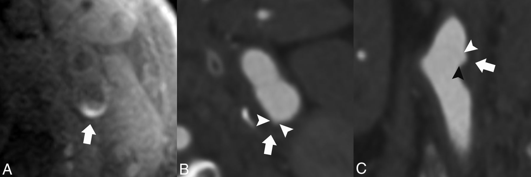
- Fig 4.
Correlated intraplaque hemorrhage on MR imaging and plaque ulceration on MDCTA. An example of MR imaging and MDCTA images of the left carotid bifurcation. On the 3D-T1W fat suppressed echo-spoiled gradient echo MR imaging sequence, intraplaque hemorrhage is visible in the atherosclerotic plaque (A, arrow). MDCTA images show a small ulceration on both the transversal and longitudinal plane at the site of the intraplaque hemorrhage (B and C, arrows; arrowheads indicate the edges of the plaque ulceration).


Tables
Clinical Characteristic Age (yr) 67 ± 9 Male sex 57 (73%) Classification event TIA 30 (38%) Stroke 41 (53%) Amaurosis fugax 7 (9%) Hypercholesterolemia 43 (55%) Hypertension 56 (72%) Diabetes mellitus 19 (24%) Current smoking No 58 (74%) Yes 20 (26%) Body mass index 25.2 (24.3–28.4) Current use Antiplatelet therapy 36 (46%) Oral anticoagulants 0 (0%) Statins 36 (46%) Antihypertensive medication 50 (64%) Antidiabetic medication 13 (17%) History Ischemic stroke or TIA 13 (17%) Ischemic heart disease 16 (21%) Peripheral arterial disease 15 (19%) ↵a Data are mean ± SD, absolute numbers of patients (%), or median (25th–75th percentile).
Vessel Characteristic All Vessels Symptomatic Vessels Contralateral Vessels P Value, Symptomatic versus Contralateral Vessels No. 149 78 71 Plaque ulceration 26 (17%) 21 (27%) 5 (7%) .001b Fissured fibrous cap 13 (9%) 10 (13%) 3 (4%) .06 Calcium volume (mm3) 21.0 (3.6–56.2) 24.2 (6.2–71.8) 17.2 (1.9–53.1) .09 Degree of stenosis (ECST) (%) 51 ± 17 55 ± 17 47 ± 16 .004b Degree of stenosis (NASCET) (%) 8 (0–32) 14 (0–35) 2 (0–26) .03b Intraplaque hemorrhage 38 (26%) 30 (38%) 8 (11%) <.001b - Table 4:
Multivariable OR for the association among clinical characteristics, vessel characteristics, and disrupted plaque surface in all vessels
Characteristic Multivariable (Age, Sex) Multivariablea (Age, Sex, Factors P < .10) OR (95% CI) P Value OR (95% CI) P Value Age 1.05 (1.00–1.10) .07 1.05 (1.00–1.10) .07 Sex 0.42 (0.15–1.12) .08 0.50 (0.17–1.42) .19 Hypertension 1.98 (0.73–5.35) .18 Diabetes mellitus 0.40 (0.14–1.10) .08 0.31 (0.11–0.94) .04b Hypercholesterolemia 1.41 (0.63–3.15) .40 Current smoking 0.48 (0.16–1.46) .20 Intraplaque hemorrhage 3.98 (1.73–9.16) .001b 3.13 (1.25–7.84) .02b Degree of stenosis (ECST, per 10%) 1.42 (1.09–1.84) .009b 1.30 (1.00–1.69) .07 Calcification volume 0.86 (0.69–1.08) .21


Jump to section
Related Articles
Cited By...
- Unifying theory of carotid plaque disruption based on structural phenotypes and forces expressed at the lumen/wall interface
- Proximal Region of Carotid Atherosclerotic Plaque Shows More Intraplaque Hemorrhage: The Plaque at Risk Study
- Carotid Plaque Composition Assessed by CT Predicts Subsequent Cardiovascular Events among Subjects with Carotid Stenosis
- Plaque Composition as a Predictor of Plaque Ulceration in Carotid Artery Atherosclerosis: The Plaque At RISK Study
- Carotid Vessel Wall Imaging on CTA
- Carotid Artery Wall Imaging: Perspective and Guidelines from the ASNR Vessel Wall Imaging Study Group and Expert Consensus Recommendations of the American Society of Neuroradiology
- Risk Factors for Development of Carotid Plaque Components
- Association between Carotid Plaque Features on CTA and Cerebrovascular Ischemia: A Systematic Review and Meta-Analysis
- Molecular Imaging of Vulnerable Coronary Plaque: A Pathophysiologic Perspective
- Imaging Atherosclerosis