Advertisement

Main menu

  • Home
  • Content
    • Current Issue
    • Accepted Manuscripts
    • Article Preview
    • Past Issue Archive
    • Video Articles
    • AJNR Case Collection
    • Case of the Week Archive
    • Case of the Month Archive
    • Classic Case Archive
  • Special Collections
    • AJNR Awards
    • ASNR Foundation Special Collection
    • Most Impactful AJNR Articles
    • Photon-Counting CT
    • Spinal CSF Leak Articles (Jan 2020-June 2024)
  • Multimedia
    • AJNR Podcasts
    • AJNR SCANtastic
    • Trainee Corner
    • MRI Safety Corner
    • Imaging Protocols
  • For Authors
    • Submit a Manuscript
    • Submit a Video Article
    • Submit an eLetter to the Editor/Response
    • Manuscript Submission Guidelines
    • Statistical Tips
    • Fast Publishing of Accepted Manuscripts
    • Graphical Abstract Preparation
    • Imaging Protocol Submission
    • Author Policies
  • About Us
    • About AJNR
    • Editorial Board
    • Editorial Board Alumni
  • More
    • Become a Reviewer/Academy of Reviewers
    • Subscribers
    • Permissions
    • Alerts
    • Feedback
    • Advertisers
    • ASNR Home

User menu

  • Alerts
  • Log in

Search

  • Advanced search
American Journal of Neuroradiology

American Journal of Neuroradiology

ASHNR American Society of Functional Neuroradiology ASHNR American Society of Pediatric Neuroradiology ASSR
  • Alerts
  • Log in

Advanced Search

  • Home
  • Content
    • Current Issue
    • Accepted Manuscripts
    • Article Preview
    • Past Issue Archive
    • Video Articles
    • AJNR Case Collection
    • Case of the Week Archive
    • Case of the Month Archive
    • Classic Case Archive
  • Special Collections
    • AJNR Awards
    • ASNR Foundation Special Collection
    • Most Impactful AJNR Articles
    • Photon-Counting CT
    • Spinal CSF Leak Articles (Jan 2020-June 2024)
  • Multimedia
    • AJNR Podcasts
    • AJNR SCANtastic
    • Trainee Corner
    • MRI Safety Corner
    • Imaging Protocols
  • For Authors
    • Submit a Manuscript
    • Submit a Video Article
    • Submit an eLetter to the Editor/Response
    • Manuscript Submission Guidelines
    • Statistical Tips
    • Fast Publishing of Accepted Manuscripts
    • Graphical Abstract Preparation
    • Imaging Protocol Submission
    • Author Policies
  • About Us
    • About AJNR
    • Editorial Board
    • Editorial Board Alumni
  • More
    • Become a Reviewer/Academy of Reviewers
    • Subscribers
    • Permissions
    • Alerts
    • Feedback
    • Advertisers
    • ASNR Home
  • Follow AJNR on Twitter
  • Visit AJNR on Facebook
  • Follow AJNR on Instagram
  • Join AJNR on LinkedIn
  • RSS Feeds

AJNR is seeking candidates for the AJNR Podcast Editor. Read the position description.

Research ArticleBrain

Usefulness of Subtraction of 3D T2WI-DRIVE from Contrast-Enhanced 3D T1WI: Preoperative Evaluations of the Neurovascular Anatomy of Patients with Neurovascular Compression Syndrome

Y. Masuda, T. Yamamoto, H. Akutsu, M. Shiigai, T. Masumoto, E. Ishikawa, M. Matsuda and A. Matsumura
American Journal of Neuroradiology February 2015, 36 (2) 317-322; DOI: https://doi.org/10.3174/ajnr.A4130
Y. Masuda
aFrom the Departments of Neurosurgery (Y.M., T.Y., H.A., E.I., M.M., A.M.)
  • Find this author on Google Scholar
  • Find this author on PubMed
  • Search for this author on this site
T. Yamamoto
aFrom the Departments of Neurosurgery (Y.M., T.Y., H.A., E.I., M.M., A.M.)
  • Find this author on Google Scholar
  • Find this author on PubMed
  • Search for this author on this site
H. Akutsu
aFrom the Departments of Neurosurgery (Y.M., T.Y., H.A., E.I., M.M., A.M.)
  • Find this author on Google Scholar
  • Find this author on PubMed
  • Search for this author on this site
M. Shiigai
bRadiology (M.S., T.M.), Faculty of Medicine, University of Tsukuba, Tsukuba, Japan.
  • Find this author on Google Scholar
  • Find this author on PubMed
  • Search for this author on this site
T. Masumoto
bRadiology (M.S., T.M.), Faculty of Medicine, University of Tsukuba, Tsukuba, Japan.
  • Find this author on Google Scholar
  • Find this author on PubMed
  • Search for this author on this site
E. Ishikawa
aFrom the Departments of Neurosurgery (Y.M., T.Y., H.A., E.I., M.M., A.M.)
  • Find this author on Google Scholar
  • Find this author on PubMed
  • Search for this author on this site
M. Matsuda
aFrom the Departments of Neurosurgery (Y.M., T.Y., H.A., E.I., M.M., A.M.)
  • Find this author on Google Scholar
  • Find this author on PubMed
  • Search for this author on this site
A. Matsumura
aFrom the Departments of Neurosurgery (Y.M., T.Y., H.A., E.I., M.M., A.M.)
  • Find this author on Google Scholar
  • Find this author on PubMed
  • Search for this author on this site
  • Article
  • Figures & Data
  • Info & Metrics
  • Responses
  • References
  • PDF
Loading

Article Figures & Data

Figures

  • Tables
  • Fig 1.
    • Download figure
    • Open in new tab
    • Download powerpoint
    Fig 1.

    Method of image postprocessing. The first set of 3D T2WI-DRIVE source images (B) is subtracted from the second set of 3D CE T1WI (A) on a pixel-by-pixel basis by using workstations provided by the MR imaging system manufacturer. Then a third set of subtracted source images named sDRICE (C) is produced.

  • Fig 2.
    • Download figure
    • Open in new tab
    • Download powerpoint
    Fig 2.

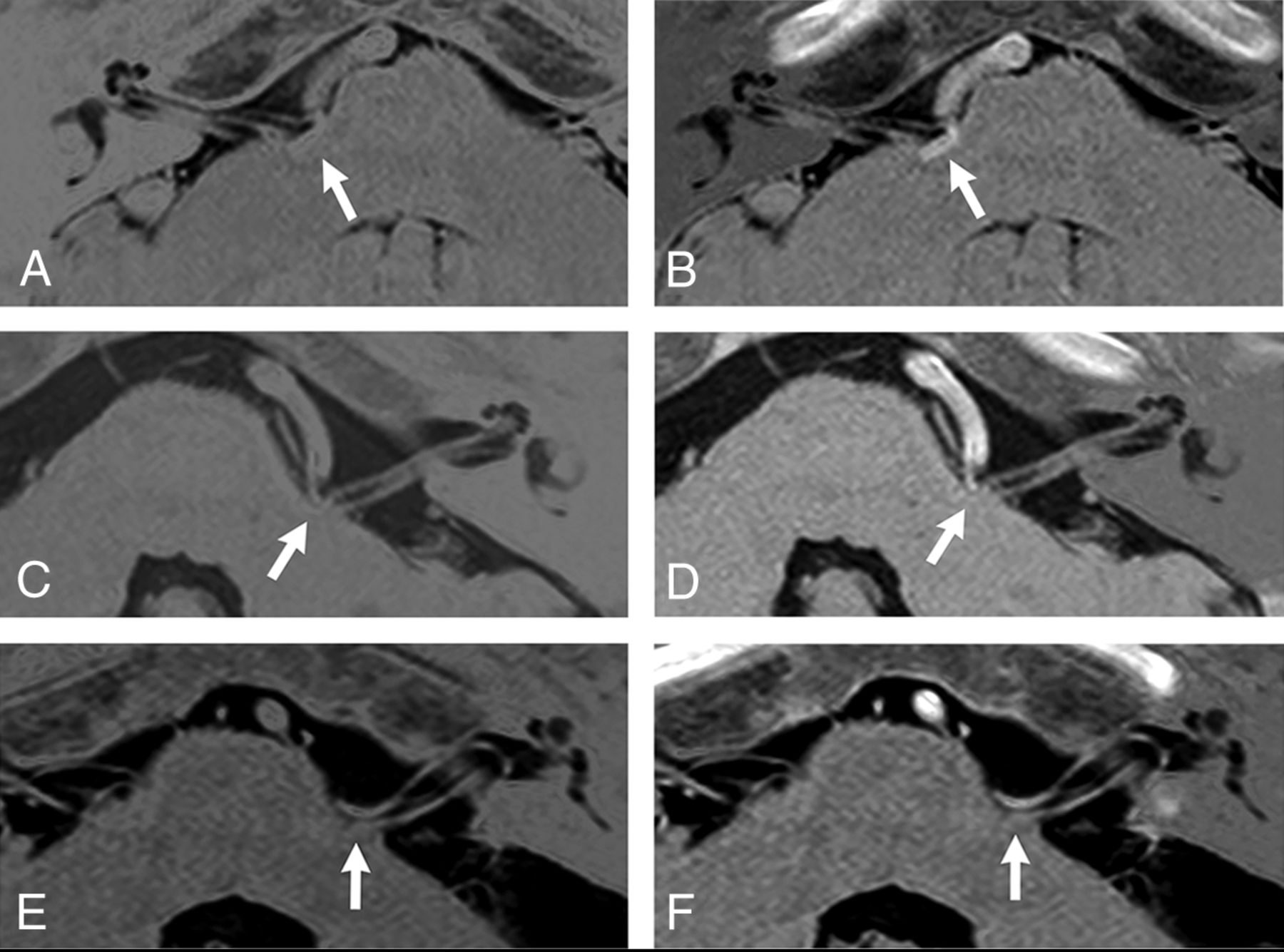
    Comparison between the black/white-reversed 3D T2WI-DRIVE (A, C, E) and sDRICE images (B, D, F) at the NVC (arrow) in patients with HFS. In patients 1 (A and B) and 2 (C and D), the sDRICE images (B and D) clearly show that the offending vessel invaginates into the brain stem and compresses the root exit zone of CN VII, in contrast to the 3D T2WI-DRIVE images (A and C). In patient 6 (E and F), both images clearly show that the posterior cerebral artery compresses CN VII.

  • Fig 3.
    • Download figure
    • Open in new tab
    • Download powerpoint
    Fig 3.

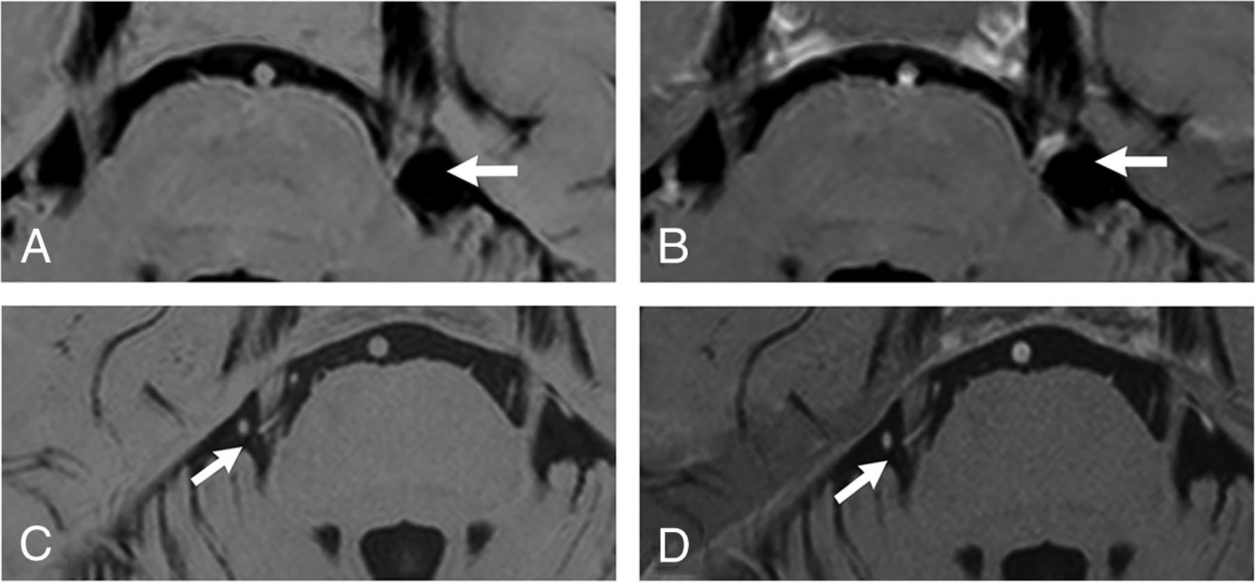
    Comparison between the black/white-reversed 3D T2WI-DRIVE (A and C) and sDRICE images (B and D) at the NVC (arrow) in patients with TN. In patient 7 (A and B), the sDRICE image (B) clearly shows that the offending vessel (the petrosal vein) invaginates into CN V, in contrast to 3D T2WI-DRIVE (A). In patient 10 (C and D), the offending vessel is relatively easily discriminated from CN V even on 3D T2WI-DRIVE (D).

Tables

  • Figures
    • View popup
    Table 1:

    Patient data and outcomes

    Patient No.Age (yr)/SexDiseaseSideOffending VesselResultComplication
    146/FHFSRAICA-PICACureNo
    230/MHFSLVA, AICACureNo
    353/FHFSLPICACureNo
    460/MHFSLVA, AICACureNo
    540/FHFSLPICACureNo
    650/MHFSLAICACureNo
    757/FTNLPetrosal veinCureNo
    861/FTNRSCACureNo
    962/FTNLSCACureNo
    1074/FTNRSCACureNo
    1164/FTNLSCA, AICACureNo
    1233/FTNLVACureNo
    • Note:—AICA indicates anterior inferior cerebellar artery, PICA, posterior inferior cerebellar artery; SCA, superior cerebellar artery; VA, vertebral artery.

    • View popup
    Table 2:

    The lesion conspicuity scoring by the 2 ratersa

    Patient No.DiseaseScores by Rater 1Scores by Rater 2
    3D T2WI-DRIVEsDRICE3D T2WI-DRIVEsDRICE
    1HFS1313
    2HFS1313
    3HFS1213
    4HFS2323
    5HFS2323
    6HFS2322
    7TN1313
    8TN1213
    9TN2323
    10TN3323
    11TN3323
    12TN3333
    • ↵a Score 1, poor, not distinguishable due to no contrast; 2, good; distinguishable by slight contrast; 3, excellent, easily distinguishable by clear contrast.

    • View popup
    Table 3:

    Qualitative and quantitative analyses of 3D T2WI-DRIVE and sDRICE images

    Parameter3D T2WI-DRIVEsDRICEP Value
    Lesion conspicuity on the neurovascular contact pointa
        Rater 1
            Overall (N = 12)2.0 (1–3)3.0 (2–3).006
            HFS (n = 6)1.5 (1–2)3.0 (3–3).023
            TN (n = 6)2.5 (1–3)3.0 (2–3).102
        Rater 2
            Overall (N = 12)2.0 (1–3)3.0 (2–3).004
            HFS (n = 6)1.5 (1–2)3.0 (2–3).038
            TN (n = 6)2.0 (1–3)3.0 (3–3).038
    Contrast (N = 12)b
        Vessel/nerve−1.21 ± 0.720.28 ± 0.15<.001
        Vessel/brain stem−1.08 ± 0.670.17 ± 0.88<.001
    CNR (N = 12)b
        Vessel/nerve−1.80 ± 1.473.34 ± 1.99.007
        Vessel/brain stem−0.97 ± 0.582.25 ± 1.57.003
    • ↵a Scoring data are listed for both raters as rater 1/rater 2 and correspond to the median (with range) across patients. P values were calculated with the Wilcoxon signed rank test.

    • ↵b Contrast and CNR data are means ± SD. The P values were calculated with a paired t test. Contrasta/b = 2(SIa − SIb)/(SIa + SIb), and CNRa/b = (SIa − SIb)/SDa.

PreviousNext
Back to top

In this issue

American Journal of Neuroradiology: 36 (2)
American Journal of Neuroradiology
Vol. 36, Issue 2
1 Feb 2015
  • Table of Contents
  • Index by author
  • Complete Issue (PDF)
Advertisement
Print
Download PDF
Email Article

Thank you for your interest in spreading the word on American Journal of Neuroradiology.

NOTE: We only request your email address so that the person you are recommending the page to knows that you wanted them to see it, and that it is not junk mail. We do not capture any email address.

Enter multiple addresses on separate lines or separate them with commas.
Usefulness of Subtraction of 3D T2WI-DRIVE from Contrast-Enhanced 3D T1WI: Preoperative Evaluations of the Neurovascular Anatomy of Patients with Neurovascular Compression Syndrome
(Your Name) has sent you a message from American Journal of Neuroradiology
(Your Name) thought you would like to see the American Journal of Neuroradiology web site.
CAPTCHA
This question is for testing whether or not you are a human visitor and to prevent automated spam submissions.
Cite this article
Y. Masuda, T. Yamamoto, H. Akutsu, M. Shiigai, T. Masumoto, E. Ishikawa, M. Matsuda, A. Matsumura
Usefulness of Subtraction of 3D T2WI-DRIVE from Contrast-Enhanced 3D T1WI: Preoperative Evaluations of the Neurovascular Anatomy of Patients with Neurovascular Compression Syndrome
American Journal of Neuroradiology Feb 2015, 36 (2) 317-322; DOI: 10.3174/ajnr.A4130

Citation Manager Formats

  • BibTeX
  • Bookends
  • EasyBib
  • EndNote (tagged)
  • EndNote 8 (xml)
  • Medlars
  • Mendeley
  • Papers
  • RefWorks Tagged
  • Ref Manager
  • RIS
  • Zotero
0 Responses
Respond to this article
Share
Bookmark this article
Usefulness of Subtraction of 3D T2WI-DRIVE from Contrast-Enhanced 3D T1WI: Preoperative Evaluations of the Neurovascular Anatomy of Patients with Neurovascular Compression Syndrome
Y. Masuda, T. Yamamoto, H. Akutsu, M. Shiigai, T. Masumoto, E. Ishikawa, M. Matsuda, A. Matsumura
American Journal of Neuroradiology Feb 2015, 36 (2) 317-322; DOI: 10.3174/ajnr.A4130
del.icio.us logo Twitter logo Facebook logo Mendeley logo
  • Tweet Widget
  • Facebook Like
  • Google Plus One
Purchase

Jump to section

  • Article
    • Abstract
    • ABBREVIATIONS:
    • Materials and Methods
    • Results
    • Discussion
    • Conclusions
    • Footnotes
    • REFERENCES
  • Figures & Data
  • Info & Metrics
  • Responses
  • References
  • PDF

Related Articles

  • No related articles found.
  • PubMed
  • Google Scholar

Cited By...

  • No citing articles found.
  • Crossref (9)
  • Google Scholar

This article has been cited by the following articles in journals that are participating in Crossref Cited-by Linking.

  • Reliability of MRI for predicting characteristics of neurovascular conflicts in trigeminal neuralgia: implications for surgical decision making
    Andrei Brînzeu, Landry Drogba, Marc Sindou
    Journal of Neurosurgery 2018
  • A preliminary study of deep learning-based reconstruction specialized for denoising in high-frequency domain: usefulness in high-resolution three-dimensional magnetic resonance cisternography of the cerebellopontine angle
    Hiroyuki Uetani, Takeshi Nakaura, Mika Kitajima, Yuichi Yamashita, Tadashi Hamasaki, Machiko Tateishi, Kosuke Morita, Akira Sasao, Seitaro Oda, Osamu Ikeda, Yasuyuki Yamashita
    Neuroradiology 2021 63 1
  • Acute presentations of intradural lipomas: case reports and a review of the literature
    Luca Massimi, Thailane Maria Feitosa Chaves, François Yves Legninda Sop, Paolo Frassanito, Gianpiero Tamburrini, Massimo Caldarelli
    BMC Neurology 2019 19 1
  • The Use of MRI in Preoperative Decision-Making for Trigeminal Neuralgia: A Single-Center Study
    James Mooney, Nicholas Erickson, Bruce Pittman, Bonita S. Agee, Barton L. Guthrie
    World Neurosurgery 2021 146
  • High-resolution heavily T2-weighted magnetic resonance imaging for evaluation of the pituitary stalk in children with ectopic neurohypophysis
    Imane El Sanharawi, Loukia Tzarouchi, Liesbeth Cardoen, Laetitia Martinerie, Juliane Leger, Jean-Claude Carel, Monique Elmaleh-Berges, Marianne Alison
    Pediatric Radiology 2017 47 5
  • A segmentation-independent volume rendering visualisation method might reduce redundant explorations and post-surgical complications of microvascular decompression
    Bao Wang, Yi Zhang, Yang Ming, Mingxu Ge, Ping Wei, Chuanting Li, Ligang Chen, Xuejun Liu, Shangchen Xu, Yingchao Liu
    European Radiology 2020 30 7
  • The Anterior Inferior Cerebral Artery Variability in the Context of Neurovascular Compression Syndromes: A Narrative Review
    Dawid Kościołek, Mateusz Kobierecki, Mikołaj Tokarski, Konrad Szalbot, Aleksandra Kościołek, Mikołaj Malicki, Sora Wanibuchi, Karol Wiśniewski, Michał Piotrowski, Ernest J. Bobeff, Bartosz M. Szmyd, Dariusz J. Jaskólski
    Biomedicines 2024 12 2
  • Evaluation of Intradural Ecchordosis Physaliphora With Three-Dimensional Fluid-Attenuated Inversion Recovery
    Ryota Kogue, Masayuki Maeda, Maki Umino, Seiya Kishi, Fumine Tanaka, Kazuhiro Tsuchiya, Hajime Sakuma
    Journal of Computer Assisted Tomography 2020 44 5
  • The role of neurovascular conflict in the pathogenesis of classical trigeminal neuralgia and the dynamics of approaches to its visualization
    E. V. Baliazina, O. M. Evusyak, V. A. Baliazin, N. G. Kadyan
    South Russian Journal of Therapeutic Practice 2021 2 1

More in this TOC Section

  • SWI or T2*: Which MRI Sequence to Use in the Detection of Cerebral Microbleeds? The Karolinska Imaging Dementia Study
  • Progression of Microstructural Damage in Spinocerebellar Ataxia Type 2: A Longitudinal DTI Study
  • Statin Therapy Does Not Affect the Radiographic and Clinical Profile of Patients with TIA and Minor Stroke
Show more BRAIN

Similar Articles

Advertisement

Indexed Content

  • Current Issue
  • Accepted Manuscripts
  • Article Preview
  • Past Issues
  • Editorials
  • Editor's Choice
  • Fellows' Journal Club
  • Letters to the Editor
  • Video Articles

Cases

  • Case Collection
  • Archive - Case of the Week
  • Archive - Case of the Month
  • Archive - Classic Case

Special Collections

  • AJNR Awards
  • ASNR Foundation Special Collection
  • Most Impactful AJNR Articles
  • Photon-Counting CT
  • Spinal CSF Leak Articles (Jan 2020-June 2024)

More from AJNR

  • Trainee Corner
  • Imaging Protocols
  • MRI Safety Corner

Multimedia

  • AJNR Podcasts
  • AJNR Scantastics

Resources

  • Turnaround Time
  • Submit a Manuscript
  • Submit a Video Article
  • Submit an eLetter to the Editor/Response
  • Manuscript Submission Guidelines
  • Statistical Tips
  • Fast Publishing of Accepted Manuscripts
  • Graphical Abstract Preparation
  • Imaging Protocol Submission
  • Evidence-Based Medicine Level Guide
  • Publishing Checklists
  • Author Policies
  • Become a Reviewer/Academy of Reviewers
  • News and Updates

About Us

  • About AJNR
  • Editorial Board
  • Editorial Board Alumni
  • Alerts
  • Permissions
  • Not an AJNR Subscriber? Join Now
  • Advertise with Us
  • Librarian Resources
  • Feedback
  • Terms and Conditions
  • AJNR Editorial Board Alumni

American Society of Neuroradiology

  • Not an ASNR Member? Join Now

© 2025 by the American Society of Neuroradiology All rights, including for text and data mining, AI training, and similar technologies, are reserved.
Print ISSN: 0195-6108 Online ISSN: 1936-959X

Powered by HighWire