Advertisement

Main menu

  • Home
  • Content
    • Current Issue
    • Accepted Manuscripts
    • Article Preview
    • Past Issue Archive
    • Video Articles
    • AJNR Case Collection
    • Case of the Week Archive
    • Case of the Month Archive
    • Classic Case Archive
  • Special Collections
    • AJNR Awards
    • ASNR Foundation Special Collection
    • Most Impactful AJNR Articles
    • Photon-Counting CT
    • Spinal CSF Leak Articles (Jan 2020-June 2024)
  • Multimedia
    • AJNR Podcasts
    • AJNR SCANtastic
    • Trainee Corner
    • MRI Safety Corner
    • Imaging Protocols
  • For Authors
    • Submit a Manuscript
    • Submit a Video Article
    • Submit an eLetter to the Editor/Response
    • Manuscript Submission Guidelines
    • Statistical Tips
    • Fast Publishing of Accepted Manuscripts
    • Graphical Abstract Preparation
    • Imaging Protocol Submission
    • Author Policies
  • About Us
    • About AJNR
    • Editorial Board
    • Editorial Board Alumni
  • More
    • Become a Reviewer/Academy of Reviewers
    • Subscribers
    • Permissions
    • Alerts
    • Feedback
    • Advertisers
    • ASNR Home

User menu

  • Alerts
  • Log in

Search

  • Advanced search
American Journal of Neuroradiology

American Journal of Neuroradiology

ASHNR American Society of Functional Neuroradiology ASHNR American Society of Pediatric Neuroradiology ASSR
  • Alerts
  • Log in

Advanced Search

  • Home
  • Content
    • Current Issue
    • Accepted Manuscripts
    • Article Preview
    • Past Issue Archive
    • Video Articles
    • AJNR Case Collection
    • Case of the Week Archive
    • Case of the Month Archive
    • Classic Case Archive
  • Special Collections
    • AJNR Awards
    • ASNR Foundation Special Collection
    • Most Impactful AJNR Articles
    • Photon-Counting CT
    • Spinal CSF Leak Articles (Jan 2020-June 2024)
  • Multimedia
    • AJNR Podcasts
    • AJNR SCANtastic
    • Trainee Corner
    • MRI Safety Corner
    • Imaging Protocols
  • For Authors
    • Submit a Manuscript
    • Submit a Video Article
    • Submit an eLetter to the Editor/Response
    • Manuscript Submission Guidelines
    • Statistical Tips
    • Fast Publishing of Accepted Manuscripts
    • Graphical Abstract Preparation
    • Imaging Protocol Submission
    • Author Policies
  • About Us
    • About AJNR
    • Editorial Board
    • Editorial Board Alumni
  • More
    • Become a Reviewer/Academy of Reviewers
    • Subscribers
    • Permissions
    • Alerts
    • Feedback
    • Advertisers
    • ASNR Home
  • Follow AJNR on Twitter
  • Visit AJNR on Facebook
  • Follow AJNR on Instagram
  • Join AJNR on LinkedIn
  • RSS Feeds

AJNR is seeking candidates for the AJNR Podcast Editor. Read the position description.

Research ArticlePediatrics

Comparing 3T T1-Weighted Sequences in Identifying Hyperintense Punctate Lesions in Preterm Neonates

D. Tortora, V. Panara, P.A. Mattei, A. Tartaro, R. Salomone, S. Domizio, A.R. Cotroneo and M. Caulo
American Journal of Neuroradiology March 2015, 36 (3) 581-586; DOI: https://doi.org/10.3174/ajnr.A4144
D. Tortora
aFrom the Radiology Unit (D.T., V.P., A.R.C., M.C.)
dITAB-Institute of Advanced Biomedical Technologies (D.T., V.P., A.T., M.C.)
  • Find this author on Google Scholar
  • Find this author on PubMed
  • Search for this author on this site
V. Panara
aFrom the Radiology Unit (D.T., V.P., A.R.C., M.C.)
dITAB-Institute of Advanced Biomedical Technologies (D.T., V.P., A.T., M.C.)
  • Find this author on Google Scholar
  • Find this author on PubMed
  • Search for this author on this site
P.A. Mattei
bOphthalmology Clinic (P.A.M.)
  • Find this author on Google Scholar
  • Find this author on PubMed
  • Search for this author on this site
A. Tartaro
aFrom the Radiology Unit (D.T., V.P., A.R.C., M.C.)
dITAB-Institute of Advanced Biomedical Technologies (D.T., V.P., A.T., M.C.)
eDepartment of Neuroscience and Imaging (A.T., A.R.C., M.C.), University “G. d'Annunzio” of Chieti, Chieti, Italy.
  • Find this author on Google Scholar
  • Find this author on PubMed
  • Search for this author on this site
R. Salomone
cNeonatology and Neonatal Intensive Care Unit (R.S., S.D.), University Hospital of Chieti, Chieti, Italy
  • Find this author on Google Scholar
  • Find this author on PubMed
  • Search for this author on this site
S. Domizio
cNeonatology and Neonatal Intensive Care Unit (R.S., S.D.), University Hospital of Chieti, Chieti, Italy
  • Find this author on Google Scholar
  • Find this author on PubMed
  • Search for this author on this site
A.R. Cotroneo
aFrom the Radiology Unit (D.T., V.P., A.R.C., M.C.)
eDepartment of Neuroscience and Imaging (A.T., A.R.C., M.C.), University “G. d'Annunzio” of Chieti, Chieti, Italy.
  • Find this author on Google Scholar
  • Find this author on PubMed
  • Search for this author on this site
M. Caulo
aFrom the Radiology Unit (D.T., V.P., A.R.C., M.C.)
dITAB-Institute of Advanced Biomedical Technologies (D.T., V.P., A.T., M.C.)
eDepartment of Neuroscience and Imaging (A.T., A.R.C., M.C.), University “G. d'Annunzio” of Chieti, Chieti, Italy.
  • Find this author on Google Scholar
  • Find this author on PubMed
  • Search for this author on this site
  • Article
  • Figures & Data
  • Info & Metrics
  • Responses
  • References
  • PDF
Loading

Article Figures & Data

Figures

  • Tables
  • Fig 1.
    • Download figure
    • Open in new tab
    • Download powerpoint
    Fig 1.

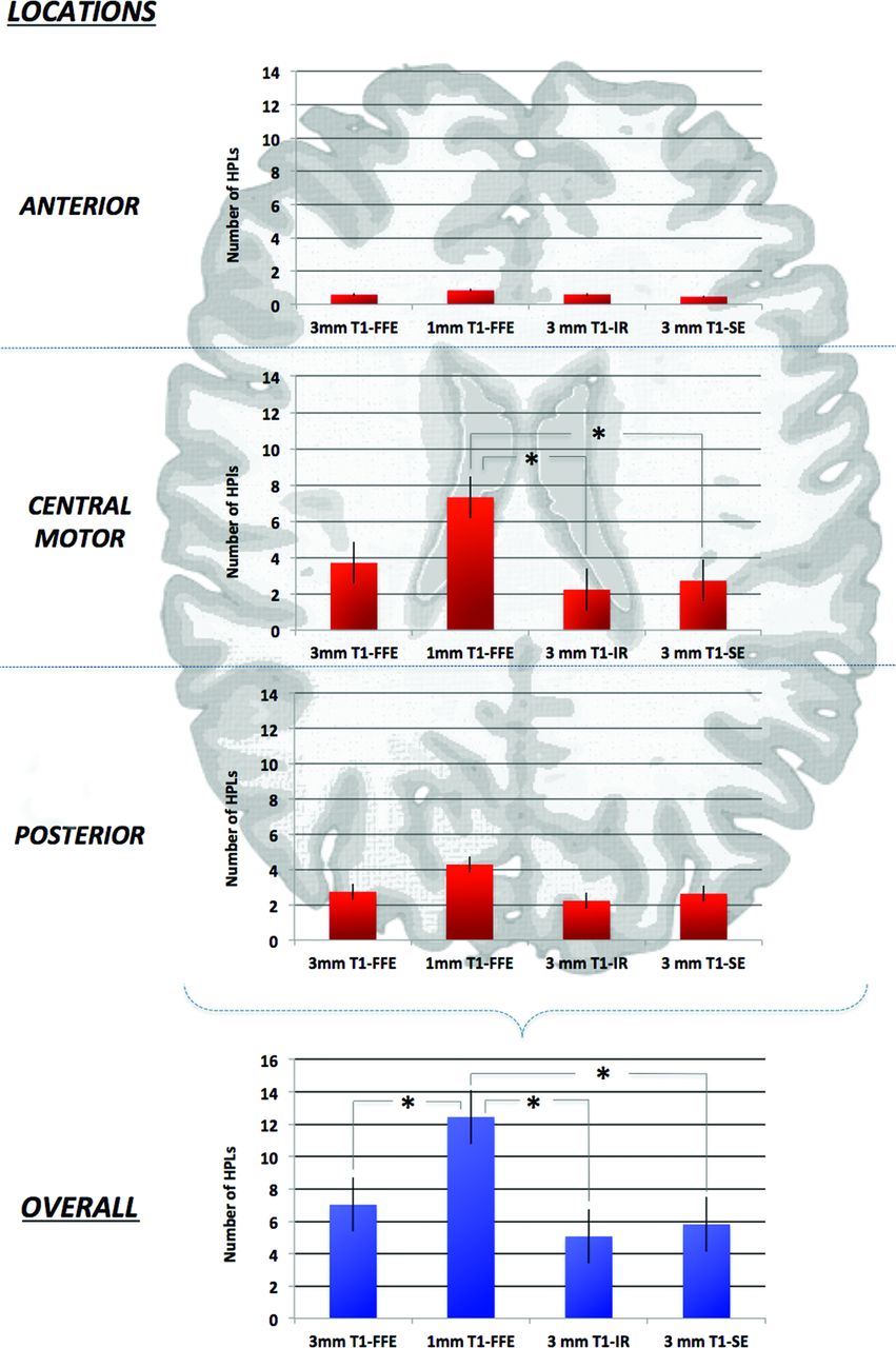
    Schematic representation of a typical neonatal brain. Only the central (motor) location on the 1-mm reformatted T1 3D-FFE sequence scored a significantly greater number of HPLs compared with IR and SE sequences. The 1-mm axial reformatted T1 3D-FFE sequence identified a significant overall greater number of HPLs than other 3-mm sequences (P < .01).

  • Fig 2.
    • Download figure
    • Open in new tab
    • Download powerpoint
    Fig 2.

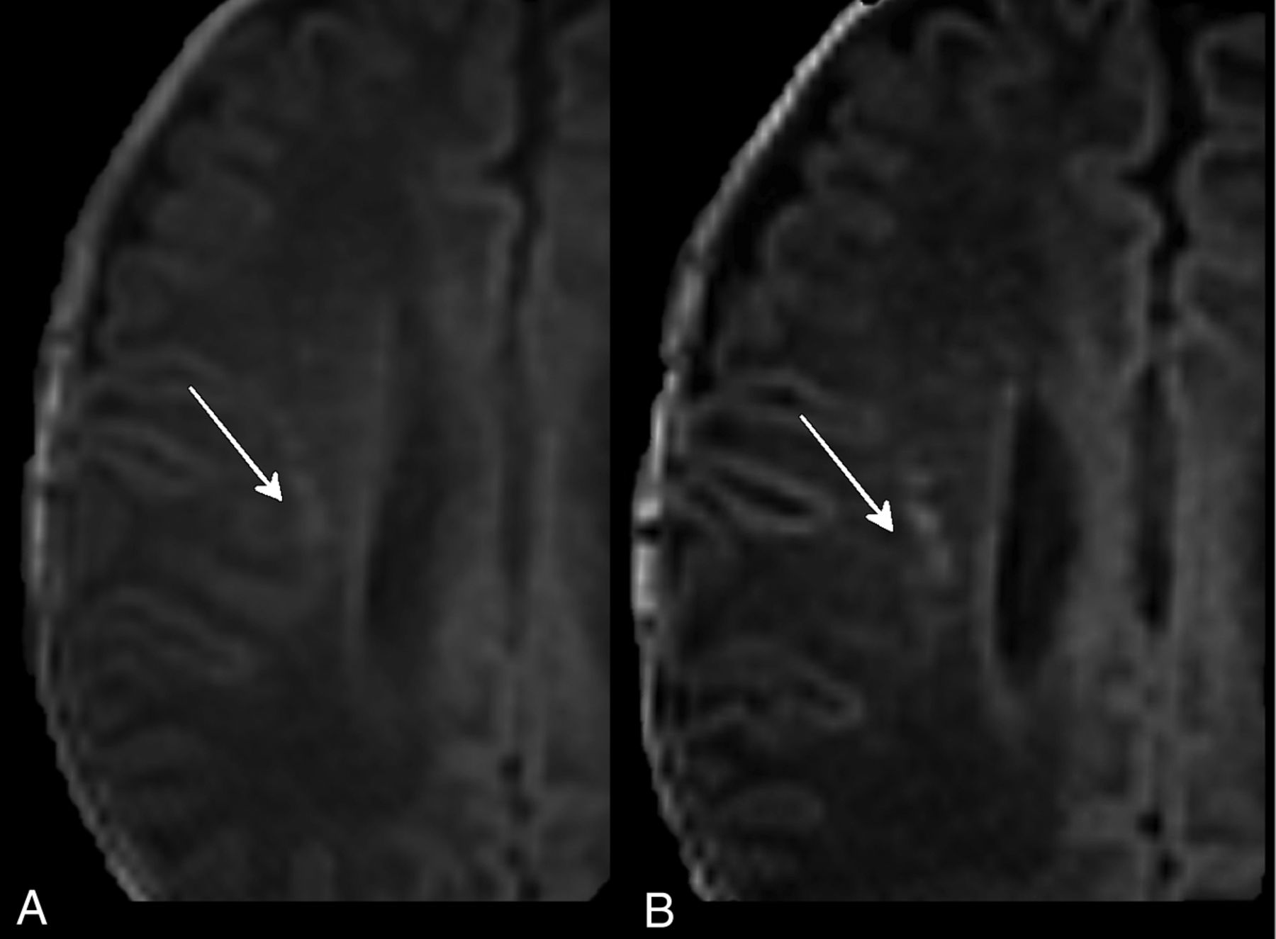
    Three-millimeter (A) and 1-mm (B) axial reformatted T1 3D-FFE sequences of PNs affected by PVL. The thinner sections (B) separated lesions that were seen as confluent with the thicker sections (A).

Tables

  • Figures
    • View popup
    Table 1:

    MRI sequence parameters

    SequenceSection Thickness (mm)Intersection (mm)TR (ms)TE (ms)TI (ms)FASAR (W/Kg)dB
    Axial T1 SE30.566311––<0.2−6.5
    Axial T1 IR30.5493815400–<0.17.2
    T1 3D-FFE1 and 309.24.3–10°0.05.4
    Axial T2-TSE30.5300080––<0.26.5
    Axial DWI41238065––<0.19.5
    • Note:—SAR indicates specific absorption rate; FA, flip angle.

    • View popup
    Table 2:

    HPLs identified in consensus reading

    PatientSexOutcomeOverall
    3-mm T1 FFE1-mm T1 FFE3-mm T1 IR3-mm T1 SE
    1F1470NA
    2F0370NA
    3F112206NA
    4F0386NA
    5M181254
    6F11635816
    7M091267
    8M117191011
    9M1122599
    10F19111110
    11M03524
    12FNA51474
    13M1691111
    14M1213099
    15M115291715
    16M1613814
    17MNA49NANA
    18F081100
    19M06912
    20F161256
    21M04900
    22M14834
    23M12422
    24F141031
    25M11752
    26M11515913
    27M051033
    28M02500
    29M12800
    30F11500
    31F15864
    Total218386152151
    • Note:—NA indicates not available.

    • View popup
    Table 3:

    Interreader agreement and pulse sequence performancesa

    Pulse SequencesAgreement of Reader 1 and Reference StandardAgreement of Reader 2 and Reference StandardInterreader Agreement
    3-mm T1 FFE0.8930.8220.844
    1-mm T1 FFE0.9880.9920.986
    3-mm T1 IR0.7760.6310.653
    3-mm T1 SE0.7930.7970.820
    • ↵a Data are intraclass correlation coefficients.

PreviousNext
Back to top

In this issue

American Journal of Neuroradiology: 36 (3)
American Journal of Neuroradiology
Vol. 36, Issue 3
1 Mar 2015
  • Table of Contents
  • Index by author
  • Complete Issue (PDF)
Advertisement
Print
Download PDF
Email Article

Thank you for your interest in spreading the word on American Journal of Neuroradiology.

NOTE: We only request your email address so that the person you are recommending the page to knows that you wanted them to see it, and that it is not junk mail. We do not capture any email address.

Enter multiple addresses on separate lines or separate them with commas.
Comparing 3T T1-Weighted Sequences in Identifying Hyperintense Punctate Lesions in Preterm Neonates
(Your Name) has sent you a message from American Journal of Neuroradiology
(Your Name) thought you would like to see the American Journal of Neuroradiology web site.
CAPTCHA
This question is for testing whether or not you are a human visitor and to prevent automated spam submissions.
Cite this article
D. Tortora, V. Panara, P.A. Mattei, A. Tartaro, R. Salomone, S. Domizio, A.R. Cotroneo, M. Caulo
Comparing 3T T1-Weighted Sequences in Identifying Hyperintense Punctate Lesions in Preterm Neonates
American Journal of Neuroradiology Mar 2015, 36 (3) 581-586; DOI: 10.3174/ajnr.A4144

Citation Manager Formats

  • BibTeX
  • Bookends
  • EasyBib
  • EndNote (tagged)
  • EndNote 8 (xml)
  • Medlars
  • Mendeley
  • Papers
  • RefWorks Tagged
  • Ref Manager
  • RIS
  • Zotero
0 Responses
Respond to this article
Share
Bookmark this article
Comparing 3T T1-Weighted Sequences in Identifying Hyperintense Punctate Lesions in Preterm Neonates
D. Tortora, V. Panara, P.A. Mattei, A. Tartaro, R. Salomone, S. Domizio, A.R. Cotroneo, M. Caulo
American Journal of Neuroradiology Mar 2015, 36 (3) 581-586; DOI: 10.3174/ajnr.A4144
del.icio.us logo Twitter logo Facebook logo Mendeley logo
  • Tweet Widget
  • Facebook Like
  • Google Plus One
Purchase

Jump to section

  • Article
    • Abstract
    • ABBREVIATIONS:
    • Materials and Methods
    • Results
    • Discussion
    • Conclusions
    • Footnotes
    • REFERENCES
  • Figures & Data
  • Info & Metrics
  • Responses
  • References
  • PDF

Related Articles

  • No related articles found.
  • PubMed
  • Google Scholar

Cited By...

  • Prediction of adverse motor outcome for neonates with punctate white matter lesions by MRI images using radiomics strategy: protocol for a prospective cohort multicentre study
  • Differences in subependymal vein anatomy may predispose preterm infants to GMH-IVH
  • Characterization of Extensive Microstructural Variations Associated with Punctate White Matter Lesions in Preterm Neonates
  • Quiet T1-Weighted Pointwise Encoding Time Reduction with Radial Acquisition for Assessing Myelination in the Pediatric Brain
  • Crossref (17)
  • Google Scholar

This article has been cited by the following articles in journals that are participating in Crossref Cited-by Linking.

  • Intracranial hemorrhage in neonates: A review of etiologies, patterns and predicted clinical outcomes
    Ai Peng Tan, Patricia Svrckova, Frances Cowan, Wui Khean Chong, Kshitij Mankad
    European Journal of Paediatric Neurology 2018 22 4
  • Early Pain Exposure Influences Functional Brain Connectivity in Very Preterm Neonates
    Domenico Tortora, Mariasavina Severino, Carlo Di Biase, Maryia Malova, Alessandro Parodi, Diego Minghetti, Cristina Traggiai, Sara Uccella, Luca Boeri, Giovanni Morana, Andrea Rossi, Luca Antonio Ramenghi
    Frontiers in Neuroscience 2019 13
  • Prematurity and brain perfusion: Arterial spin labeling MRI
    Domenico Tortora, Peter Angelo Mattei, Riccardo Navarra, Valentina Panara, Rita Salomone, Andrea Rossi, John A. Detre, Massimo Caulo
    NeuroImage: Clinical 2017 15
  • Differences in subependymal vein anatomy may predispose preterm infants to GMH–IVH
    Domenico Tortora, Mariasavina Severino, Mariya Malova, Alessandro Parodi, Giovanni Morana, Jan Sedlacik, Paul Govaert, Joseph J Volpe, Andrea Rossi, Luca Antonio Ramenghi
    Archives of Disease in Childhood - Fetal and Neonatal Edition 2018 103 1
  • Quiet T1-Weighted Pointwise Encoding Time Reduction with Radial Acquisition for Assessing Myelination in the Pediatric Brain
    N. Aida, T. Niwa, Y. Fujii, K. Nozawa, M. Enokizono, K. Murata, T. Obata
    American Journal of Neuroradiology 2016 37 8
  • Characterization of Extensive Microstructural Variations Associated with Punctate White Matter Lesions in Preterm Neonates
    X. Li, J. Gao, M. Wang, J. Zheng, Y. Li, E.S. Hui, M. Wan, J. Yang
    American Journal of Neuroradiology 2017 38 6
  • A multi-view pyramid network for skull stripping on neonatal T1-weighted MRI
    Yan Gao, Jie Li, Haojun Xu, Miaomiao Wang, Congcong Liu, Yannan Cheng, Mengxuan Li, Jian Yang, Xianjun Li
    Magnetic Resonance Imaging 2019 63
  • Nosological Differences in the Nature of Punctate White Matter Lesions in Preterm Infants
    Mariya Malova, Elena Morelli, Valentina Cardiello, Domenico Tortora, Mariasavina Severino, Maria Grazia Calevo, Alessandro Parodi, Laura Costanza De Angelis, Diego Minghetti, Andrea Rossi, Luca Antonio Ramenghi
    Frontiers in Neurology 2021 12
  • Perinatal MRI diffusivity is related to early assessment of motor performance in preterm neonates
    Riccardo Navarra, Carlo Sestieri, Emanuela Conte, Rita Salomone, Peter A Mattei, Gian L Romani, Sergio Domizio, Massimo Caulo
    The Neuroradiology Journal 2016 29 2
  • Medical Image Computing and Computer Assisted Intervention – MICCAI 2019
    Yalong Liu, Jie Li, Ying Wang, Miaomiao Wang, Xianjun Li, Zhicheng Jiao, Jian Yang, Xingbo Gao
    2019 11766

More in this TOC Section

  • SyMRI & MR Fingerprinting in Brainstem Myelination
  • Comparison of Image Quality and Radiation Dose in Pediatric Temporal Bone CT Using Photon-Counting Detector CT and Energy-Integrating Detector CT
  • Dandy-Walker Phenotype with Brainstem Involvement
Show more Pediatrics

Similar Articles

Advertisement

Indexed Content

  • Current Issue
  • Accepted Manuscripts
  • Article Preview
  • Past Issues
  • Editorials
  • Editor's Choice
  • Fellows' Journal Club
  • Letters to the Editor
  • Video Articles

Cases

  • Case Collection
  • Archive - Case of the Week
  • Archive - Case of the Month
  • Archive - Classic Case

Special Collections

  • AJNR Awards
  • ASNR Foundation Special Collection
  • Most Impactful AJNR Articles
  • Photon-Counting CT
  • Spinal CSF Leak Articles (Jan 2020-June 2024)

More from AJNR

  • Trainee Corner
  • Imaging Protocols
  • MRI Safety Corner

Multimedia

  • AJNR Podcasts
  • AJNR Scantastics

Resources

  • Turnaround Time
  • Submit a Manuscript
  • Submit a Video Article
  • Submit an eLetter to the Editor/Response
  • Manuscript Submission Guidelines
  • Statistical Tips
  • Fast Publishing of Accepted Manuscripts
  • Graphical Abstract Preparation
  • Imaging Protocol Submission
  • Evidence-Based Medicine Level Guide
  • Publishing Checklists
  • Author Policies
  • Become a Reviewer/Academy of Reviewers
  • News and Updates

About Us

  • About AJNR
  • Editorial Board
  • Editorial Board Alumni
  • Alerts
  • Permissions
  • Not an AJNR Subscriber? Join Now
  • Advertise with Us
  • Librarian Resources
  • Feedback
  • Terms and Conditions
  • AJNR Editorial Board Alumni

American Society of Neuroradiology

  • Not an ASNR Member? Join Now

© 2025 by the American Society of Neuroradiology All rights, including for text and data mining, AI training, and similar technologies, are reserved.
Print ISSN: 0195-6108 Online ISSN: 1936-959X

Powered by HighWire