
Article Figures & Data
Figures
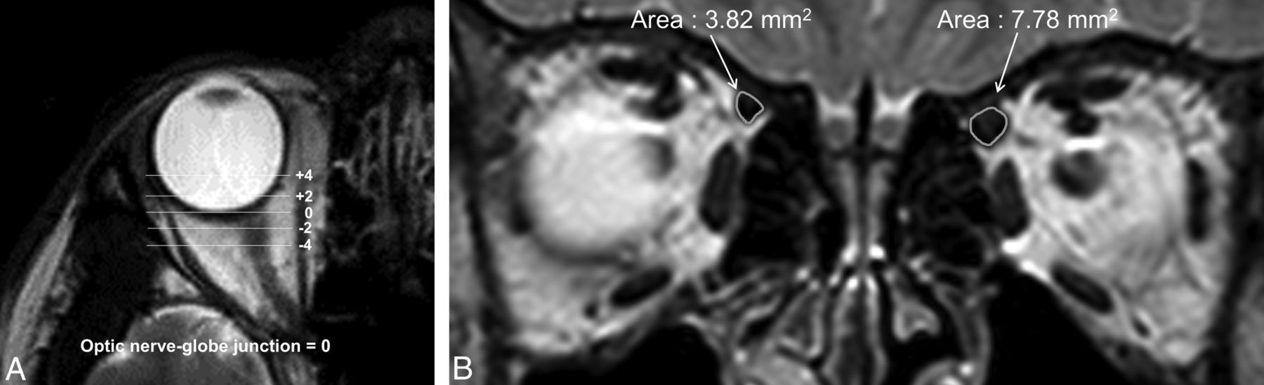
- Fig 1.
MR imaging in a patient with right superior oblique muscle palsy. A, The optic nerve–globe junction was defined as the standard plane (plane “0”), and SO areas were measured in the coronal sections of 5 contiguous planes, including the standard plane and planes that were 2 and 4 mm anterior or posterior to the standard plane. B, T2-weighted coronal image of the orbit and SO. The right SO is hypoplastic compared with the left. The area surrounded by the curvilinear line was measured in 5 different planes, by using DTU-710 (Wacom) and PACS software, which provides automatic measurements for area. The volume of the SO was defined as the sum of SO areas at the 5 planes multiplied by 2 mm.
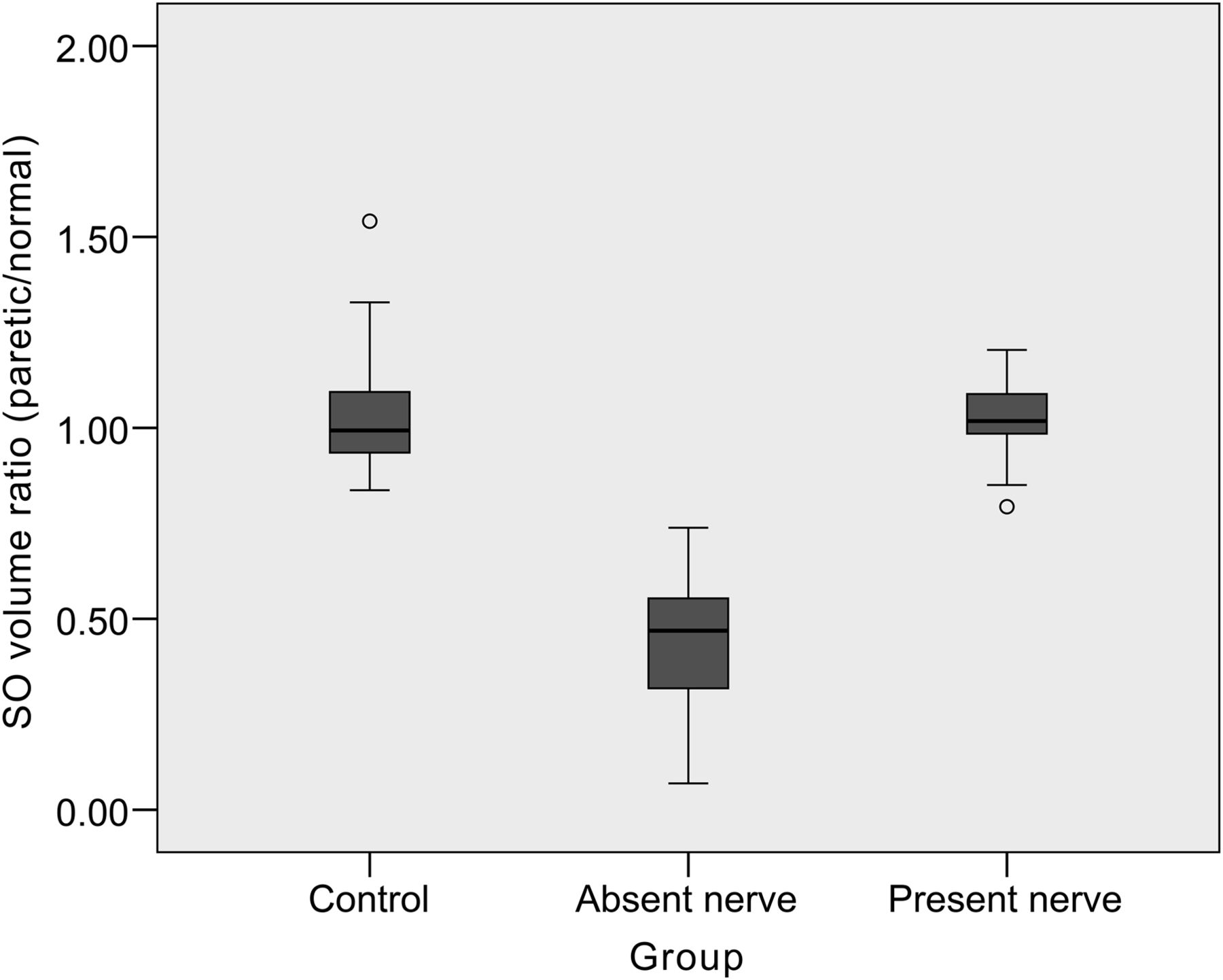
- Fig 2.
Boxplots of the superior oblique muscle volume ratio of the paretic-to-normal side in patients with congenital superior oblique palsy without (absent group) and with (present group) an ipsilateral trochlear nerve compared with controls. The SO volume ratio was significantly smaller in the absent group (P < .001) compared with controls. There was no significant difference of the SO volume ratio between controls and the present group.
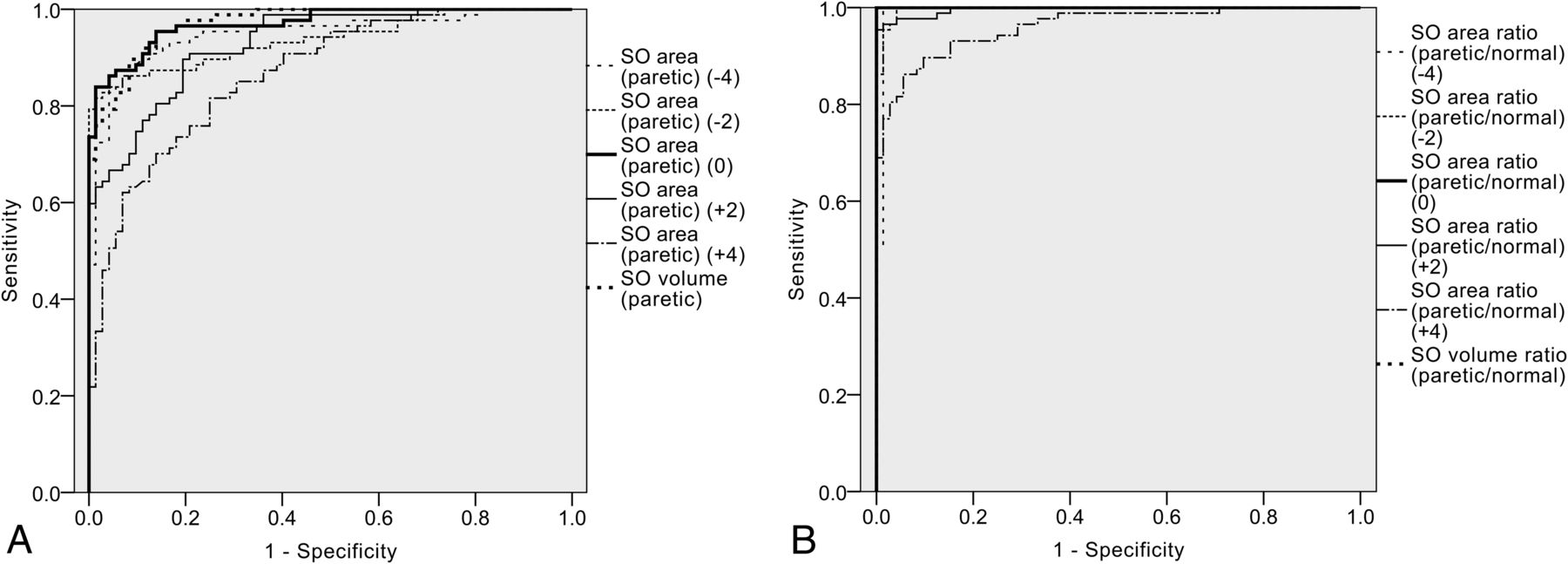
- Fig 3.
Receiver operating characteristic curves of paretic side SO areas, SO volume, paretic/normal side ratios of the SO area and SO volume for predicting trochlear nerve absence. The area under the receiver operating characteristic curves was largest for the paretic/normal side ratios of SO volume and SO area at the optic nerve–globe junction, with an AUC value of >0.950. AUCs of the paretic side SO areas were much smaller at the anterior planes (+4, +2) compared with the standard plane (0) or posterior planes (−2, −4).

Tables
Patient characteristics and the superior oblique muscle area and volume measured in controls and patients with congenital superior oblique palsy without (absent group) and with (present group) an ipsilateral trochlear nerve
Control (n = 34) Absent Group (n = 87) Present Group (n = 38) P Value Age at time of imaging (yr) 16.1 ± 20.7 (1–69) 15.2 ± 17.9 (1–64) 11.5 ± 13.0 (1–58) .896a Age of onset (yr) 10.8 ± 16.9 (0–63) 7.7 ± 13.4 (0–58) .317b Interval between onset and imaging (yr) 3.8 ± 6.4 (0–44) 3.8 ± 4.8 (0–23) .967b Male sex 16 (47.0%) 55 (63.2%) 21 (55.3%) .283c Paretic side Right 48 (55.2%) 17 (44.7%) .085c Left 39 (44.8%) 21 (55.3%) SO area “0” (P) 4.58 ± 1.05 (2.68–6.89) 2.12 ± 0.96 (0.34–4.59) 5.01 ± 1.02 (3.39–7.78) <.001a SO area “0” (N) 4.43 ± 1.23 (2.41–8.53) 5.34 ± 1.31 (2.17–10.45) 4.87 ± 1.11 (3.11–8.17) .001a SO area “0” ratio (P/N)d 1.06 ± 0.18 (0.78–1.42) 0.40 ± 0.15 (0.05–0.75) 1.04 ± 0.10 (0.86–1.21) <.001a SO volume (P)e 41.57 ± 8.15 (27.03–61.17) 21.53 ± 8.69 (3.92–40.12) 46.99 ± 9.50 (28.52–75.99) <.001a SO volume (N)f 40.94 ± 9.87 (24.16–73.10) 49.31 ± 11.15 (27.93–94.04) 46.76 ± 9.60 (30.95–76.00) .001a SO volume ratio (P/N)d 1.03 ± 0.15 (0.84–1.54) 0.44 ± 0.15 (0.07–0.74) 1.03 ± 0.10 (0.79–1.20) <.001a Note:—P indicates paretic side; N, normal side; “0”, measured at the optic nerve-globe junction (standard plane).
↵a P value by ANOVA.
↵b P value by independent t test.
↵c P value by Pearson χ2.
↵d Left-to-right ratio for controls and paretic side to normal side ratio for patients with congenital SOP in the absent and present group.
↵e Left side for controls.
↵f Right side for controls.


Jump to section
Related Articles
Cited By...
- High-Resolution 7T MR Imaging of the Trochlear Nerve
- Quantitative analysis of structure-function relationship between ocular motility and superior oblique muscle hypoplasia in unilateral superior oblique palsy
- Trochlear Groove and Trochlear Cistern: Useful Anatomic Landmarks for Identifying the Tentorial Segment of Cranial Nerve IV on MRI