Advertisement

Main menu

  • Home
  • Content
    • Current Issue
    • Publication Preview--Ahead of Print
    • Past Issue Archive
    • Case of the Week Archive
    • Classic Case Archive
    • Case of the Month Archive
    • COVID-19 Content and Resources
  • For Authors
    • Author Policies
    • Manuscript Submission Guidelines
  • About Us
    • About AJNR
    • American Society of Neuroradiology
  • Submit a Manuscript
  • Podcasts
    • Podcasts
    • Subscribe on iTunes
  • More
    • Subscribers
    • Permissions
    • Advertisers
    • Alerts
    • Feedback

User menu

  • Subscribe
  • Alerts
  • Log in

Search

  • Advanced search
American Journal of Neuroradiology

American Journal of Neuroradiology

  • Subscribe
  • Alerts
  • Log in

Advanced Search

  • Home
  • Content
    • Current Issue
    • Publication Preview--Ahead of Print
    • Past Issue Archive
    • Case of the Week Archive
    • Classic Case Archive
    • Case of the Month Archive
    • COVID-19 Content and Resources
  • For Authors
    • Author Policies
    • Manuscript Submission Guidelines
  • About Us
    • About AJNR
    • American Society of Neuroradiology
  • Submit a Manuscript
  • Podcasts
    • Podcasts
    • Subscribe on iTunes
  • More
    • Subscribers
    • Permissions
    • Advertisers
    • Alerts
    • Feedback
  • Follow AJNR on Twitter
  • Visit AJNR on Facebook
  • Follow AJNR on Instagram
  • Join AJNR on LinkedIn
  • RSS Feeds
LetterLETTER

Panhypopituitarism with Ectopic Posterior Pituitary Lobe, Heterotopia, Polymicrogyria, Corpus Callosum Dysgenesis, and Optic Chiasm/Nerve Hypoplasia: Is That an Undefined Neuronal Migration Syndrome?

N.C. Ören, E. Cagıltay, F. Akay and S. Toyran
American Journal of Neuroradiology May 2015, 36 (5) E33-E35; DOI: https://doi.org/10.3174/ajnr.A4305
N.C. Ören
aDepartment of Radiology
  • Find this author on Google Scholar
  • Find this author on PubMed
  • Search for this author on this site
E. Cagıltay
bDepartment of Endocrinology
  • Find this author on Google Scholar
  • Find this author on PubMed
  • Search for this author on this site
F. Akay
cDepartment of Ophthalmology Izmir Military Hospital, Izmir, Turkey
  • Find this author on Google Scholar
  • Find this author on PubMed
  • Search for this author on this site
S. Toyran
cDepartment of Ophthalmology Izmir Military Hospital, Izmir, Turkey
  • Find this author on Google Scholar
  • Find this author on PubMed
  • Search for this author on this site
  • Article
  • Figures & Data
  • Info & Metrics
  • References
  • PDF
Loading

We read with interest the article by Mitchell et al1 of 4 cases of ectopic posterior pituitary lobe and periventricular heterotopia and the letter about this study from Bergson et al.2 The authors suggested that an underlying genetic abnormality or a common mechanism or both were the most likely explanation for the findings. Bergson et al also suggested that it might be a complex form of a septo-optic dysplasia. We report a case that shares similar findings with those1,2 but is also associated with corpus callosum dysgenesis, and we discuss the common underlying mechanism.

A 19-year-old man with a history of panhypopituitarism and intellectual and motor disability came to our department for MR imaging. He did not have a history of seizures except from a febrile convulsion episode at 3 years of age. The ophthalmologic examination was positive for a pale left optic disc. MR imaging revealed an ectopic posterior pituitary lobe, a hypoplastic pituitary gland, corpus callosum dysgenesis, and a hypoplastic optic chiasm on the T1-weighted sagittal and coronal images (Fig 1A–C). Absence or a marked hypoplasia of the pituitary stalk was detected incidentally on coronal images. Coronal 3D T1-weighted, axial T2-weighted, and FLAIR images showed right frontal cortical dysplasia (defined as polymicrogyria) and a strip of ectopic gray matter (defined as subcortical heterotopia) extending from the head of the right caudate nuclei to the orbitofrontal lobe. In addition, subependymal nodular heterotopia on the frontal, temporal, and occipital horns of the lateral ventricles bilaterally and a hypoplastic left optic nerve with a hypoplastic chiasm were detected on 3D T1-weighted coronal images also (Fig 2A–D).

Fig 1.
  • Download figure
  • Open in new tab
  • Download powerpoint
Fig 1.

A, Sagittal T1-weighted image shows a hyperintensity corresponding to an ectopic posterior pituitary lobe located on the floor of the third ventricle (white arrow) with corpus callosum dysgenesis (star). The pituitary gland is small, and the stalk is not visualized incidentally. B, Coronal T1-weighted image shows hyperintensity corresponding to an ectopic posterior pituitary lobe that is located on the floor of the third ventricle (white arrow). C, Coronal T1-weighted image shows hypoplasia of the left optic nerve (arrowhead).

Fig 2.
  • Download figure
  • Open in new tab
  • Download powerpoint
Fig 2.

A and B, Axial T2-weighted and FLAIR images show cortical dysplasia (polymicrogyria) in the left orbitofrontal lobe (thin arrow). C, Coronal 3D T1-weighted image shows left subcortical heterotopia (thick arrow). D, Coronal 3D T1-weighted image shows subependymal nodular heterotopia near the lateral ventricle (arrowheads).

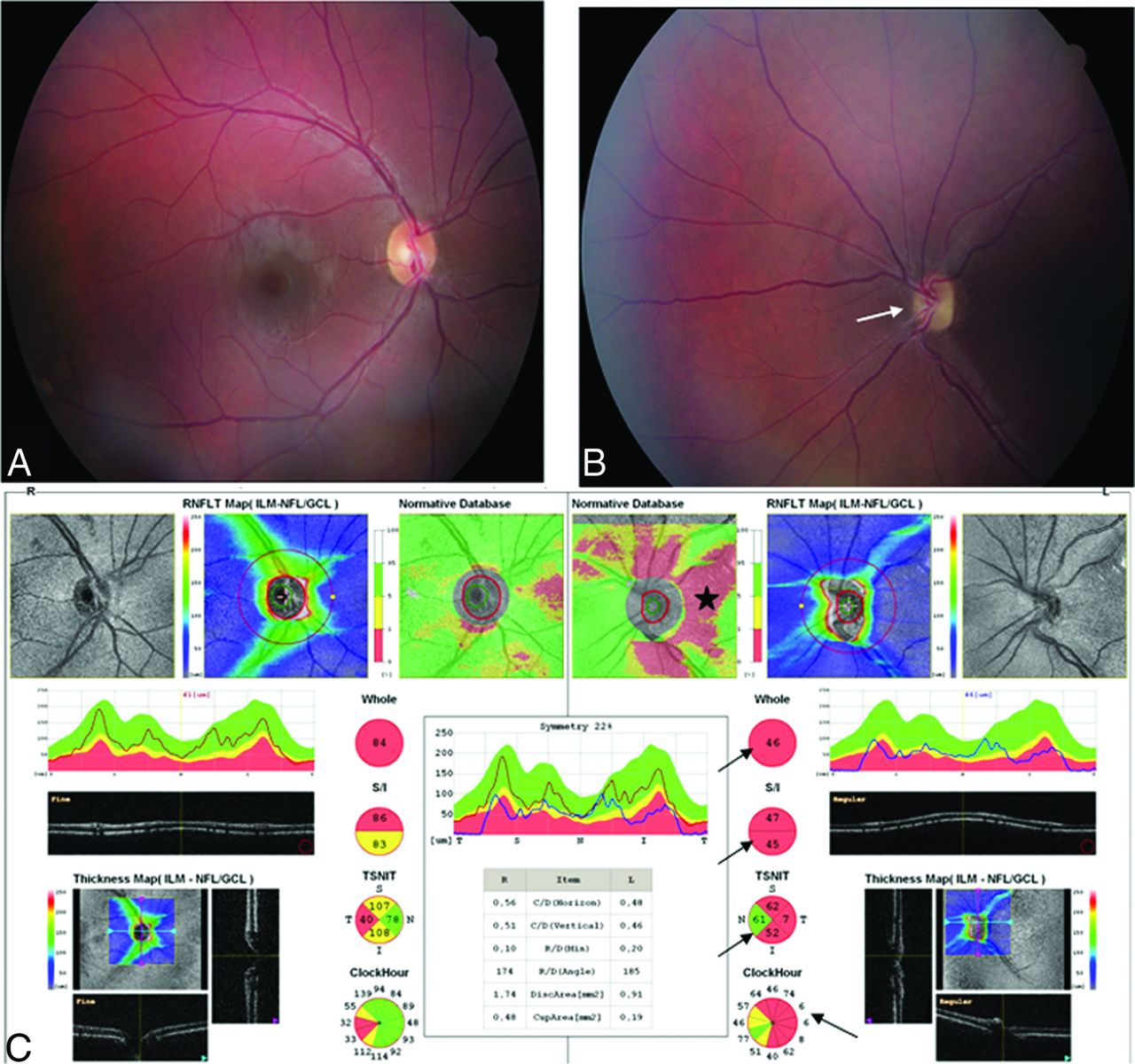
Neuronal migration is essential for brain development and normal functioning of the nervous system. Neuronal migration disorders (NMD) are a heterogeneous group of disorders that share similar underlying mechanisms such as an error in neuronal migration during neurogenesis. Lissencephaly (smooth brain), heterotopia, polymicrogyria, and schizencephaly are the most common NMD. Subcortical and subependymal nodular heterotopias with polymicrogyria were the examples of NMD in our case as well as in the cases reported by the authors.1,2 These cases were different from septo-optic dysplasia because of the presence of a septum pellucidum. Another interesting point in our case is corpus callosum dysgenesis, which was detected in another similar case reported previously.3 The corpus callosum is the largest commissure connecting the cerebral hemispheres. Some molecules are secreted by midline glial populations that are involved in attracting and repelling axons so that they cross the midline and form the corpus callosum.4 An abnormal migrational mechanism is also supported by the associations of corpus callosum developmental abnormalities with NMD, such as classic lissencephaly, cobblestone lissencephalies, polymicrogyria, schizencephaly, and heterotopia.4 During the patient's eye examination, retinal thickness was assessed by spectral domain optical coherence tomography. The ganglion cell and inner nuclear layers were thinner than those in the normal population. This size leads to a thinner retinal nerve fiber layer, which follows the optic nerve toward the optical chiasm (Fig 3A–C). This finding was identified as more evidence of the defective migrational mechanism in the physiopathology for this complex condition.

Fig 3.
  • Download figure
  • Open in new tab
  • Download powerpoint
Fig 3.

Fundus photography of right (A) and left (B) eyes. The optic disc is hypoplastic in the left eye (white arrow). C, Spectral domain optical coherence tomography analysis reveals thinning of the bilateral retinal nerve fiber layer, more on the left side than the right (red indicates the decrease of the ganglion cells in the retinal nerve fiber layer, star and black arrows).

In conclusion, we agree with the authors and support the hypothesis that these cases may occur with a common mechanism that may be related to an error during neuronal migration in early embryogenesis. These similar clinical and radiologic findings may also indicate an undefined neuronal migration syndrome.

REFERENCES

  1. 1.↵
    1. Mitchell LA,
    2. Thomas PQ,
    3. Zacharin MR, et al
    . Ectopic posterior pituitary lobe and periventricular heterotopia: cerebral malformations with the same underlying mechanism? AJNR Am J Neuroradiol 2002;23:1475–81
    Abstract/FREE Full Text
  2. 2.↵
    1. Bergson JC,
    2. Garg VK,
    3. Chang J
    . Ectopic posterior pituitary lobe and cortical dysplasia. AJNR Am J Neuroradiol 2007;28:198–99
    FREE Full Text
  3. 3.↵
    1. Arslan H,
    2. Saylık M,
    3. Akdeniz H
    . MRI findings of coexistence of ectopic neurohypophysis, corpus callosum dysgenesis, and periventricular neuronal heterotopia. J Clin Imaging Sci 2014;4:22
    CrossRefPubMed
  4. 4.↵
    1. Kułak P,
    2. Gościk E,
    3. Śmigielska-Kuzia J
    . Abnormalities of the corpus callosum: magnetic resonance imaging analysis in children. Prog Health Sci 2012;2:57–63
  • © 2015 by American Journal of Neuroradiology
PreviousNext
Back to top

In this issue

American Journal of Neuroradiology: 36 (5)
American Journal of Neuroradiology
Vol. 36, Issue 5
1 May 2015
  • Table of Contents
  • Index by author
  • Complete Issue (PDF)
Advertisement
Print
Download PDF
Email Article

Thank you for your interest in spreading the word on American Journal of Neuroradiology.

NOTE: We only request your email address so that the person you are recommending the page to knows that you wanted them to see it, and that it is not junk mail. We do not capture any email address.

Enter multiple addresses on separate lines or separate them with commas.
Panhypopituitarism with Ectopic Posterior Pituitary Lobe, Heterotopia, Polymicrogyria, Corpus Callosum Dysgenesis, and Optic Chiasm/Nerve Hypoplasia: Is That an Undefined Neuronal Migration Syndrome?
(Your Name) has sent you a message from American Journal of Neuroradiology
(Your Name) thought you would like to see the American Journal of Neuroradiology web site.
CAPTCHA
This question is for testing whether or not you are a human visitor and to prevent automated spam submissions.
Citation Tools
Panhypopituitarism with Ectopic Posterior Pituitary Lobe, Heterotopia, Polymicrogyria, Corpus Callosum Dysgenesis, and Optic Chiasm/Nerve Hypoplasia: Is That an Undefined Neuronal Migration Syndrome?
N.C. Ören, E. Cagıltay, F. Akay, S. Toyran
American Journal of Neuroradiology May 2015, 36 (5) E33-E35; DOI: 10.3174/ajnr.A4305

Citation Manager Formats

  • BibTeX
  • Bookends
  • EasyBib
  • EndNote (tagged)
  • EndNote 8 (xml)
  • Medlars
  • Mendeley
  • Papers
  • RefWorks Tagged
  • Ref Manager
  • RIS
  • Zotero
Share
Panhypopituitarism with Ectopic Posterior Pituitary Lobe, Heterotopia, Polymicrogyria, Corpus Callosum Dysgenesis, and Optic Chiasm/Nerve Hypoplasia: Is That an Undefined Neuronal Migration Syndrome?
N.C. Ören, E. Cagıltay, F. Akay, S. Toyran
American Journal of Neuroradiology May 2015, 36 (5) E33-E35; DOI: 10.3174/ajnr.A4305
del.icio.us logo Digg logo Reddit logo Twitter logo CiteULike logo Facebook logo Google logo Mendeley logo
  • Tweet Widget
  • Facebook Like
  • Google Plus One
Purchase

Jump to section

  • Article
    • REFERENCES
  • Figures & Data
  • Info & Metrics
  • References
  • PDF

Related Articles

  • No related articles found.
  • PubMed
  • Google Scholar

Cited By...

  • No citing articles found.
  • Crossref (3)
  • Google Scholar

This article has been cited by the following articles in journals that are participating in Crossref Cited-by Linking.

  • Imaging of the Sella and Parasellar Region
    John L. Go, Anandh G. Rajamohan
    Radiologic Clinics of North America 2017 55 1
  • Diagnostic Imaging: Brain
    2016
  • Imaging in Neurology
    2016

More in this TOC Section

  • Reply:
  • Regarding “Altered Blood Flow in the Ophthalmic and Internal Carotid Arteries in Patients with Age-Related Macular Degeneration Measured Using Noncontrast MR Angiography at 7T”
  • Fair Performance of CT in Diagnosing Unilateral Vocal Fold Paralysis
Show more LETTERS

Similar Articles

Advertisement

News and Updates

  • Lucien Levy Best Research Article Award
  • Thanks to our 2022 Distinguished Reviewers

Resources

  • Evidence-Based Medicine Level Guide
  • AJNR Podcast Archive
  • Librarian Resources
  • Terms and Conditions

Opportunities

  • Get Peer Review Credit from Publons

American Society of Neuroradiology

  • Neurographics
  • ASNR Annual Meeting
  • Fellowship Portal

© 2023 by the American Society of Neuroradiology | Print ISSN: 0195-6108 Online ISSN: 1936-959X

Powered by HighWire