
Article Figures & Data
Figures
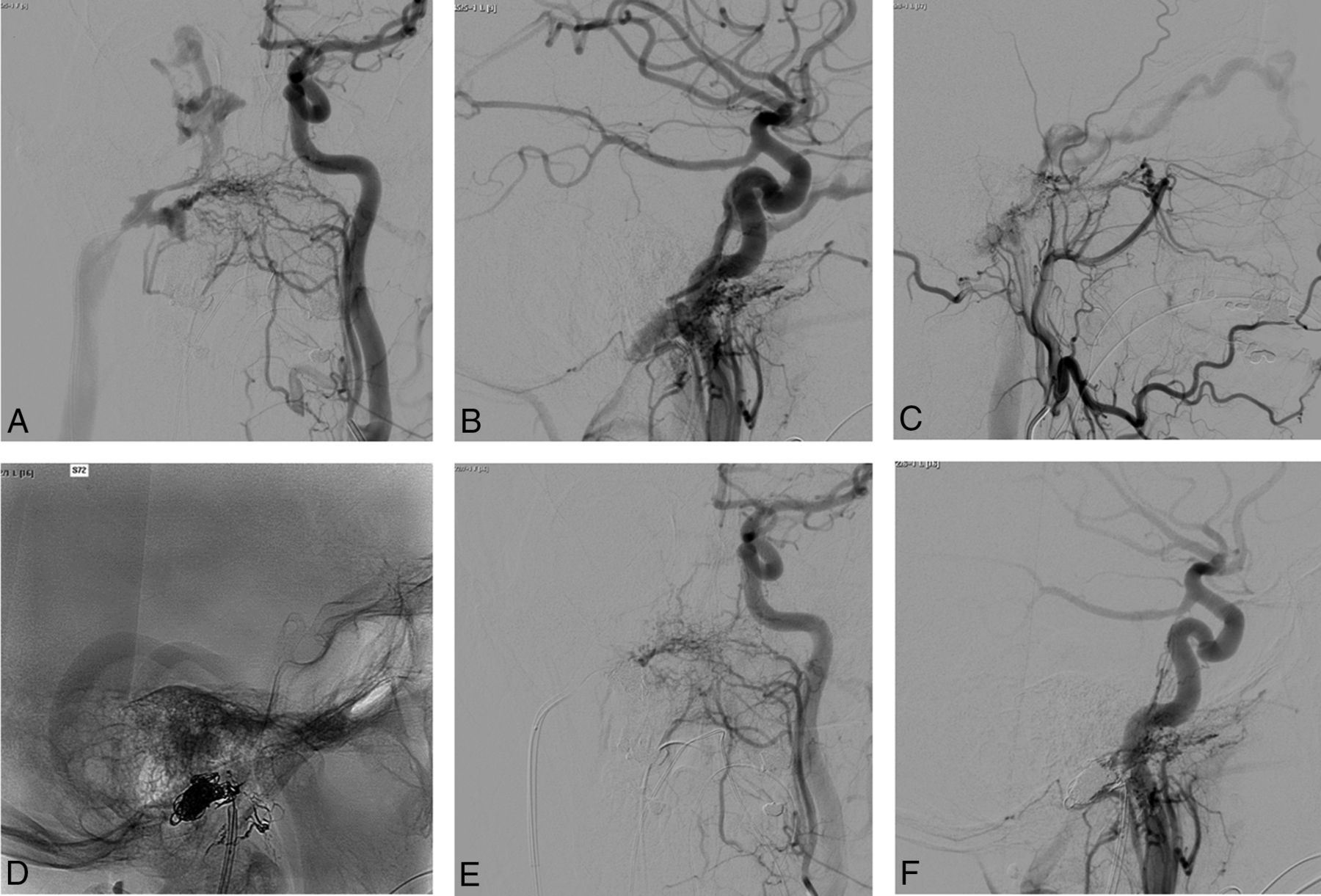
- Fig 1.
Pretherapeutic left common carotid artery DSA in anteroposterior (A) and lateral (B) projections and external carotid artery DSA in a lateral projection (C) highlighting a right jugular foramen dAVF with venous reflux into the right inferior petrosal sinus, the right cavernous sinus, and the right superior ophthalmic vein in a patient presenting with right chemosis and exophthalmia. D, Note the cast of Onyx (Covidien, Irvine, California) after an arterial embolization. Posttherapeutic left common carotid injections in anteroposterior (E) and lateral (F) projections.
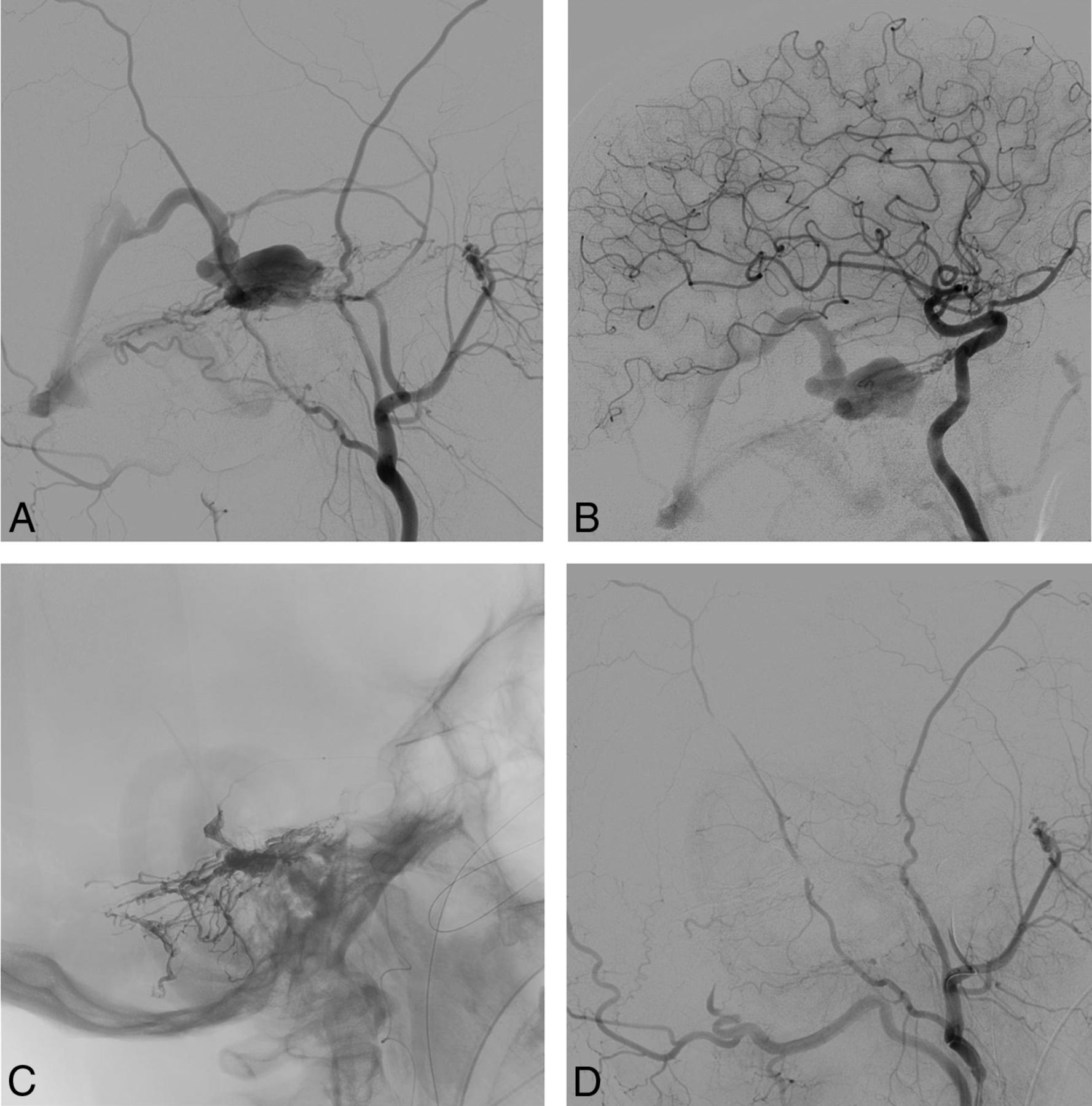
- Fig 2.
Lateral (A) and anteroposterior (B) projections of a pretherapeutic left external carotid artery DSA showing a complex fistula of the posterior third of the superior sagittal sinus in a patient with signs of intracranial hypertension. Lateral projection (C) of the left vertebral artery injection in the same patient shows multiple fistulous points on the transverse and sigmoid sinuses. D, A lateral skull x-ray with the important cast of Onyx used to treat the fistulas. Posttherapeutic lateral (E) and anteroposterior (F) projections of the left external carotid artery injection.
- Fig 3.
Pretherapeutic lateral projections of the right external (A) and internal (B) carotid artery DSA highlighting a tentorial dAVF with venous ectasia of the third portion of the basal vein in a patient with a trochlear nerve deficit. C, Note the cast of Onyx (Covidien, Irvine, California) after injection through the middle meningeal artery branches. D, Posttherapeutic right external carotid artery DSA in a lateral projection without a residual fistula.

Tables
Variable Patients (n = 13) Age (yr) (median) (range) 50.3 (15–72) Men 9 (69.2%) Clinical signs Pulsatile tinnitus 5 (38.5%) Chemosis 8 (61.5%) Exophthalmia 5 (38.5%) Loss of visual acuity 5 (38.5%) Ocular hypertension 1 (7.7.%) Oculomotor palsy 4 (30.8%) Third CN palsy 2 (15.4%) Fourth CN palsy 2 (15.4%) Sixth CN palsy 3 (23.1%) Papillary edema 9 (69.2%) Time between first sign and diagnosis (mo) 10 (1–36) mRS score before treatment 1 9 (69.2%) 2 4 (30.8%) Note:—CN indicates cranial nerve.
Variable No. (%) Total No. of embolization sessions 23 Embolization per patient (mean) (range) 1.8 (1–3) Venous approach 14 (60.8%) Arterial approach 8 (34.8%) Combined approach 1 (4.3%) Overall success rate 12 (52.2%) Incomplete closed fistula 4 (15.4%) Impossible to catheterize 2 (7.7%) Embolic agent used Onyx 12 (52.2%) Coils + Onyx 5 (21.7) Glubran Tiss 4 (17.4%) Coils 2 (8.8%) Success rate By patient 11/14 (78.5%) Associated microsurgery 1/14 (7.2%) Complications Permanent 2 (8.7%) Death 1 (4.3%) Follow-up Mean (range) (mo) 10.1 (1–48) Last mRS 0 3 (23.1%) 1 6 (46.2%) 2 2 (15.4%) 3 1 (7.2%) 6 1 (7.2%) Ophthalmologic follow-up Normal findings 8 (61.5%) Central scotoma 1 (7.2%) Third and sixth nerve palsy 1 (7.2%) Ocular hypertonia 1 (7.2%) Persistent papillary edema 1 (7.2%) Lost to follow-up 1 (7.2%)


Jump to section
Related Articles
Cited By...
- No citing articles found.