
Article Figures & Data
Figures
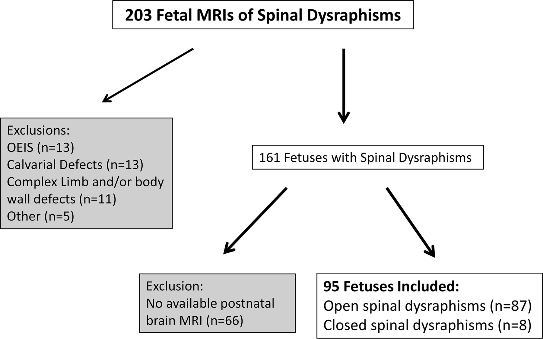
- Fig 1.
Breakdown of the fetuses included in our study. OEIS indicates omphalocele-exstrophy-imperforate anus-spinal defects.
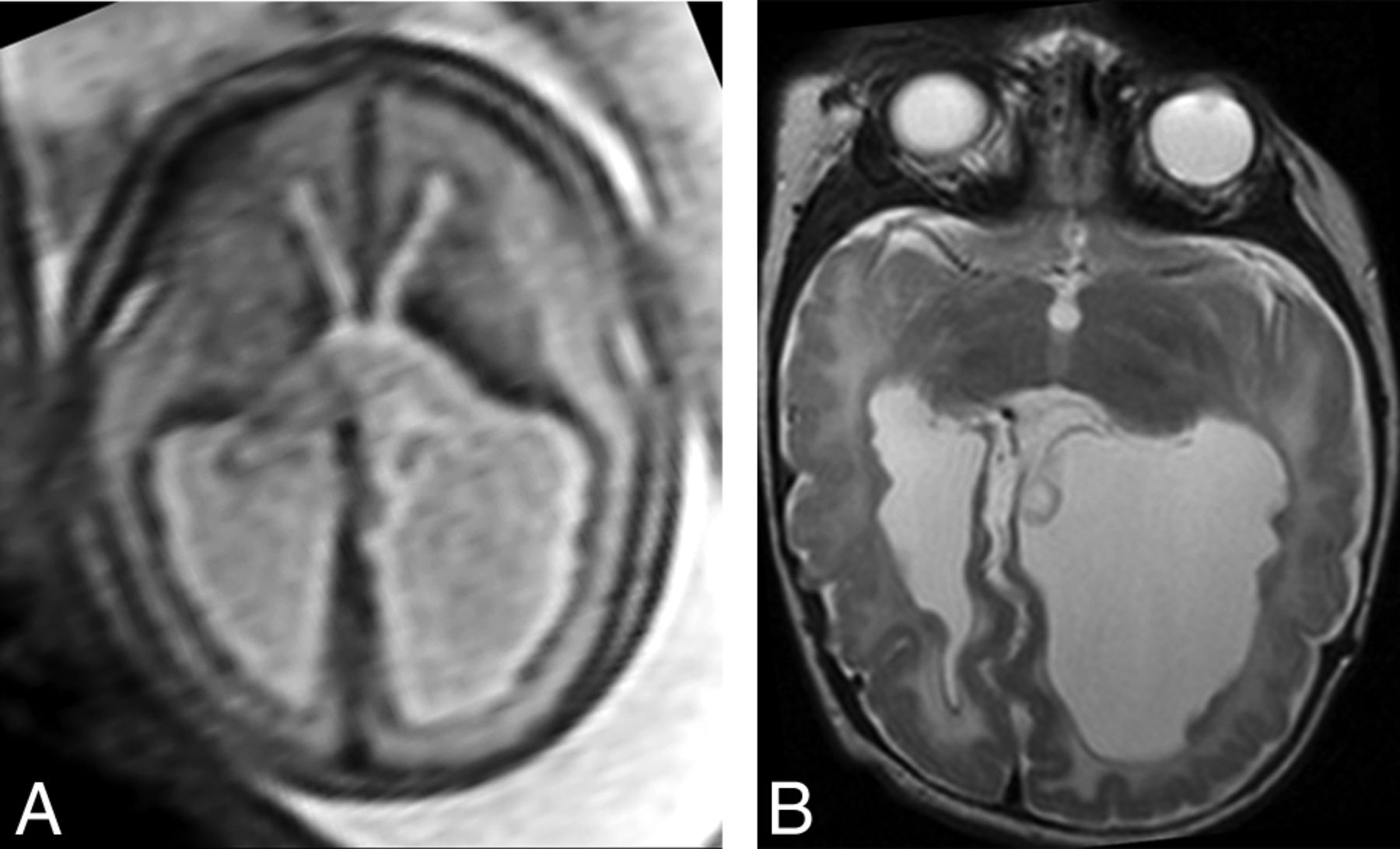
- Fig 2.
Example of SEH prospectively diagnosed on fetal MR imaging (true-positive finding). Axial T2 single-shot fast spin-echo imaging of the fetal brain at 25 weeks' GA (A) demonstrates multiple nodules along the ependymal surfaces of the lateral ventricles suspicious for SEH. Postnatal brain MR imaging at 5 weeks of age (B) confirms the presence of bilateral SEH on axial T2 FSE.
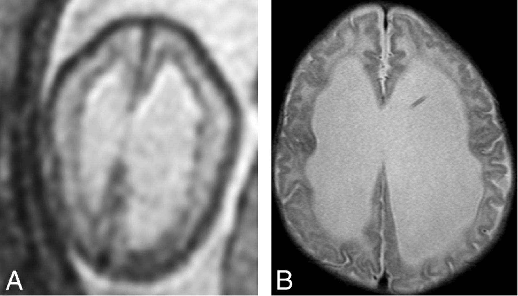
- Fig 3.
An example of subependymal nodularity giving the false appearance of SEH (false-positive finding). Axial T2 single-shot fast spin-echo image from fetal MR imaging at 24 weeks' GA (A) demonstrates nodularity along the ependymal surfaces of the lateral ventricles, giving the appearance of SEH. However, postnatal MR imaging at 8 weeks of age (B) does not demonstrate any SEH.
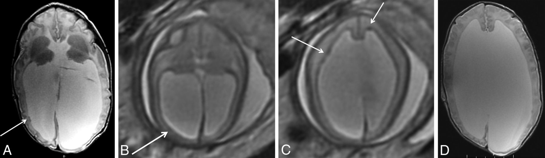
- Fig 4.
Example of a false-negative finding in a patient with SEH. Postnatal brain MR imaging at 15 days of age (A) demonstrates 2 small SEH along the right lateral ventricle (arrow) on axial T2 FSE imaging. In retrospect, there is some subtle asymmetric irregularity of the subependyma (arrow) on fetal MR imaging at 24 weeks' GA (B) on axial T2 single-shot fast spin-echo imaging. However, other areas of nodularity on fetal MR imaging in this patient (C, arrows) do not correspond to SEH postnatally (D).


Jump to section
Related Articles
Cited By...
- Early Diagnosis of Tuberous Sclerosis Complex: Prenatal Diagnosis
- Fetal Intraventricular Hemorrhage in Open Neural Tube Defects: Prenatal Imaging Evaluation and Perinatal Outcomes
- Reliability of MR Imaging-Based Posterior Fossa and Brain Stem Measurements in Open Spinal Dysraphism in the Era of Fetal Surgery