Advertisement

Main menu

  • Home
  • Content
    • Current Issue
    • Accepted Manuscripts
    • Article Preview
    • Past Issue Archive
    • Video Articles
    • AJNR Case Collection
    • Case of the Week Archive
    • Case of the Month Archive
    • Classic Case Archive
  • Special Collections
    • AJNR Awards
    • ASNR Foundation Special Collection
    • Most Impactful AJNR Articles
    • Photon-Counting CT
    • Spinal CSF Leak Articles (Jan 2020-June 2024)
  • Multimedia
    • AJNR Podcasts
    • AJNR SCANtastic
    • Trainee Corner
    • MRI Safety Corner
    • Imaging Protocols
  • For Authors
    • Submit a Manuscript
    • Submit a Video Article
    • Submit an eLetter to the Editor/Response
    • Manuscript Submission Guidelines
    • Statistical Tips
    • Fast Publishing of Accepted Manuscripts
    • Graphical Abstract Preparation
    • Imaging Protocol Submission
    • Author Policies
  • About Us
    • About AJNR
    • Editorial Board
    • Editorial Board Alumni
  • More
    • Become a Reviewer/Academy of Reviewers
    • Subscribers
    • Permissions
    • Alerts
    • Feedback
    • Advertisers
    • ASNR Home

User menu

  • Alerts
  • Log in

Search

  • Advanced search
American Journal of Neuroradiology

American Journal of Neuroradiology

ASHNR American Society of Functional Neuroradiology ASHNR American Society of Pediatric Neuroradiology ASSR
  • Alerts
  • Log in

Advanced Search

  • Home
  • Content
    • Current Issue
    • Accepted Manuscripts
    • Article Preview
    • Past Issue Archive
    • Video Articles
    • AJNR Case Collection
    • Case of the Week Archive
    • Case of the Month Archive
    • Classic Case Archive
  • Special Collections
    • AJNR Awards
    • ASNR Foundation Special Collection
    • Most Impactful AJNR Articles
    • Photon-Counting CT
    • Spinal CSF Leak Articles (Jan 2020-June 2024)
  • Multimedia
    • AJNR Podcasts
    • AJNR SCANtastic
    • Trainee Corner
    • MRI Safety Corner
    • Imaging Protocols
  • For Authors
    • Submit a Manuscript
    • Submit a Video Article
    • Submit an eLetter to the Editor/Response
    • Manuscript Submission Guidelines
    • Statistical Tips
    • Fast Publishing of Accepted Manuscripts
    • Graphical Abstract Preparation
    • Imaging Protocol Submission
    • Author Policies
  • About Us
    • About AJNR
    • Editorial Board
    • Editorial Board Alumni
  • More
    • Become a Reviewer/Academy of Reviewers
    • Subscribers
    • Permissions
    • Alerts
    • Feedback
    • Advertisers
    • ASNR Home
  • Follow AJNR on Twitter
  • Visit AJNR on Facebook
  • Follow AJNR on Instagram
  • Join AJNR on LinkedIn
  • RSS Feeds

AJNR is seeking candidates for the AJNR Podcast Editor. Read the position description.

Research ArticleHEAD & NECK

Do Radiologists Report the TNM Staging in Radiology Reports for Head and Neck Cancers? A National Survey Study

B. Ko, U. Parvathaneni, P.A. Hudgins and Y. Anzai
American Journal of Neuroradiology August 2016, 37 (8) 1504-1509; DOI: https://doi.org/10.3174/ajnr.A4742
B. Ko
aFrom the Departments of Radiation Oncology (B.K., U.P.)
  • Find this author on Google Scholar
  • Find this author on PubMed
  • Search for this author on this site
  • ORCID record for B. Ko
U. Parvathaneni
aFrom the Departments of Radiation Oncology (B.K., U.P.)
  • Find this author on Google Scholar
  • Find this author on PubMed
  • Search for this author on this site
  • ORCID record for U. Parvathaneni
P.A. Hudgins
cDepartment of Radiology and Imaging Sciences (P.A.H.), Emory University, Atlanta, Georgia
  • Find this author on Google Scholar
  • Find this author on PubMed
  • Search for this author on this site
  • ORCID record for P.A. Hudgins
Y. Anzai
bRadiology (Y.A.), University of Washington, Seattle, Washington
dDepartment of Radiology and Imaging Sciences (Y.A.), University of Utah Health Care, Salt Lake City, Utah.
  • Find this author on Google Scholar
  • Find this author on PubMed
  • Search for this author on this site
  • ORCID record for Y. Anzai
  • Article
  • Figures & Data
  • Info & Metrics
  • Responses
  • References
  • PDF
Loading

Article Figures & Data

Figures

  • Tables
  • Fig 1.
    • Download figure
    • Open in new tab
    • Download powerpoint
    Fig 1.

    Survey questions.

  • Fig 2.
    • Download figure
    • Open in new tab
    • Download powerpoint
    Fig 2.

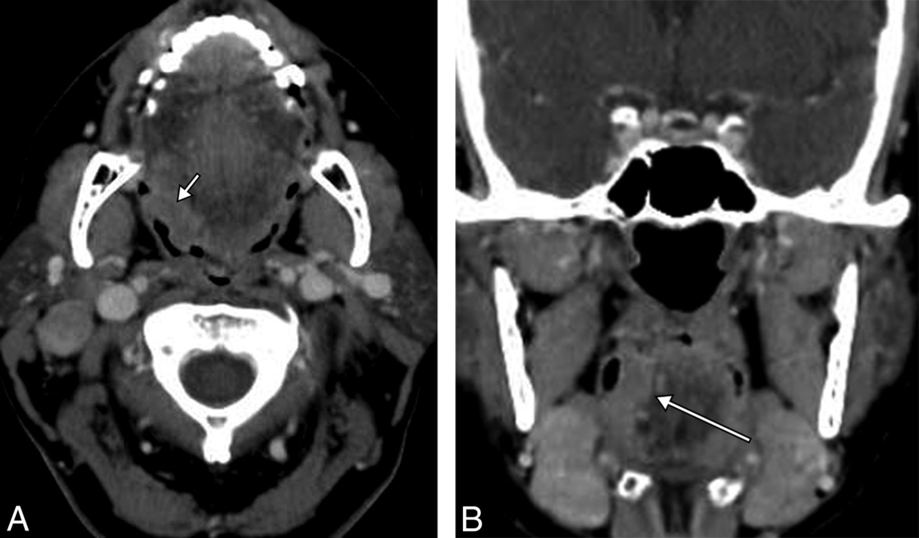
    Contrast-enhanced axial (A) and coronal (B) CT images of a patient with posterior pharyngeal wall cancer. The transaxial dimension is 2.5 cm (short arrow), though the craniocaudal dimension exceeds 4 cm (long arrow). Therefore, based on size alone, the stage is T3. Addition of concurrent chemotherapy to radiation therapy would be appropriate in a T3, but not a T2 lesion. Additional factors that would upstage this tumor, as described in American Joint Committee on Cancer, 7th edition (https://cancerstaging.org/Pages/default.aspx) are extension to the larynx, involvement of extrinsic tongue muscles, medial pterygoid muscle involvement, or hard palate or mandible invasion. These are all imaging-based characteristics.

  • Fig 3.
    • Download figure
    • Open in new tab
    • Download powerpoint
    Fig 3.

    Contrast-enhanced axial (A) and coronal (B) CT images of a patient with squamous cell carcinoma of the right base of the tongue. Although the transaxial dimension is 1.8 cm (short arrow), the craniocaudal dimension is 3.5 cm, technically at least a T2 lesion, if only the size is considered. These measurements can only be accurately acquired on imaging. Note that the largest craniocaudal dimension (long arrow) is submucosal, and on the basis of physical examination alone, this tumor could be grossly understaged.

  • Fig 4.
    • Download figure
    • Open in new tab
    • Download powerpoint
    Fig 4.

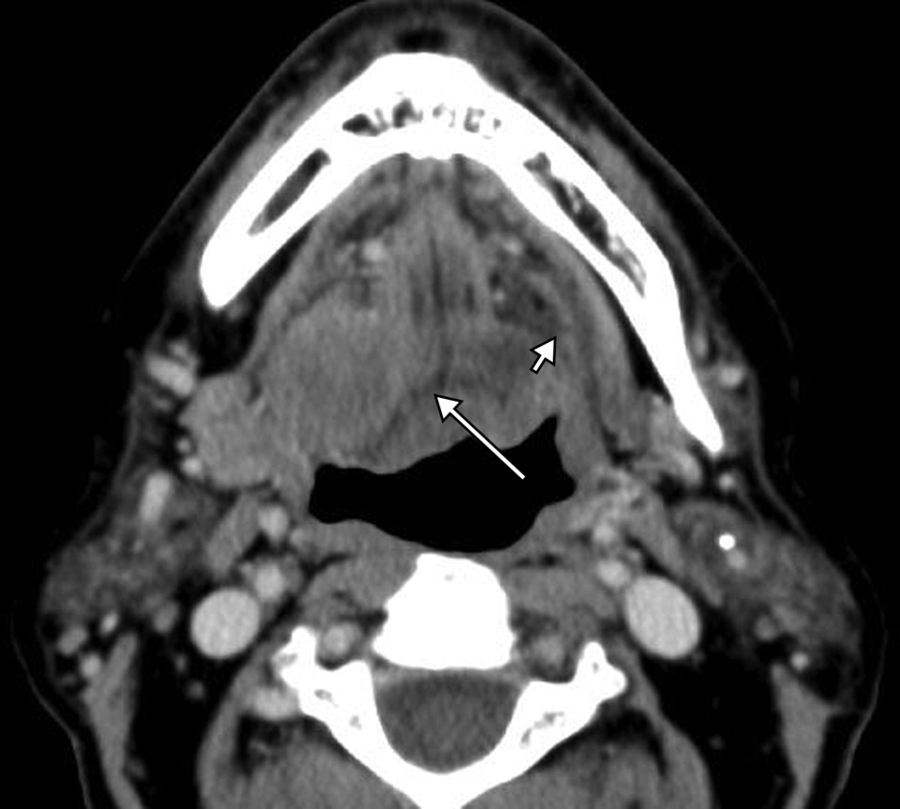
    Axial contrast-enhanced CT at the floor of mouth level. Clinically, this tumor was staged as T2 right oral cavity squamous cell carcinoma because there was mucosal ulceration and a >2-cm palpable mass. However, there is invasion of the lateral and posterior genioglossus muscle (long arrow). Note a normal hyoglossus muscle on the left (short arrow); tumor has completely replaced right hyoglossus muscle. This is upstaged to T4a on the basis of CT findings, changing the prognosis and treatment.

Tables

  • Figures
    • View popup
    Table 1:

    Survey responder demographics

    VariableNo. of ResponsesFrequency
    Nature of Practice229
        Academic14462.8%
        Private5423.8%
        Both3113.5%
    Years of Experience229
        <2 years167.0%
        2–5 years3917.0%
        5–8 years2912.7%
        >8 years14563.3%
    Subspecialty229
        Head and neck radiology16572.1%
        Other subspecialization of radiology6427.9%
    • View popup
    Table 2:

    Perceived barriers to incorporating staging in radiology reportsa

    AnswerNo. of Responses (n = 227)Frequency
    Afraid of inaccuracy13459.0%
    Unable to remember staging classification13258.2%
    Time-consuming10646.7%
    Not required8135.7%
    Other7432.6%
    No reimbursement4118.1%
    • ↵a The sum of responses exceeds the total number of responses (n = 227) because participants were able to choose multiple answers for this particular question.

    • View popup
    Table 3:

    Reasons for assigning TN staging in radiology reportsa

    AnswerNo. of Responses (n = 115)Frequency
    Believe in added value required from surgery or oncology colleagues6052.2%
    Help the treatment decision5043.5%
    Not applicable4034.8%
    Educational value3833.0%
    Other1714.8%
    • ↵a The sum of responses exceeds the total number of responses (n = 115) because participants were able to choose multiple answers for this particular question.

    • View popup
    Table 4:

    Perceived importance of incorporating TN staging in radiology reports

    AnswerNo. of Responses (n = 229)Frequency
    Very important4419.2%
    Somewhat important6829.7%
    Neutral8034.9%
    Not very important187.9%
    Not important198.3%
PreviousNext
Back to top

In this issue

American Journal of Neuroradiology: 37 (8)
American Journal of Neuroradiology
Vol. 37, Issue 8
1 Aug 2016
  • Table of Contents
  • Index by author
  • Complete Issue (PDF)
Advertisement
Print
Download PDF
Email Article

Thank you for your interest in spreading the word on American Journal of Neuroradiology.

NOTE: We only request your email address so that the person you are recommending the page to knows that you wanted them to see it, and that it is not junk mail. We do not capture any email address.

Enter multiple addresses on separate lines or separate them with commas.
Do Radiologists Report the TNM Staging in Radiology Reports for Head and Neck Cancers? A National Survey Study
(Your Name) has sent you a message from American Journal of Neuroradiology
(Your Name) thought you would like to see the American Journal of Neuroradiology web site.
CAPTCHA
This question is for testing whether or not you are a human visitor and to prevent automated spam submissions.
Cite this article
B. Ko, U. Parvathaneni, P.A. Hudgins, Y. Anzai
Do Radiologists Report the TNM Staging in Radiology Reports for Head and Neck Cancers? A National Survey Study
American Journal of Neuroradiology Aug 2016, 37 (8) 1504-1509; DOI: 10.3174/ajnr.A4742

Citation Manager Formats

  • BibTeX
  • Bookends
  • EasyBib
  • EndNote (tagged)
  • EndNote 8 (xml)
  • Medlars
  • Mendeley
  • Papers
  • RefWorks Tagged
  • Ref Manager
  • RIS
  • Zotero
0 Responses
Respond to this article
Share
Bookmark this article
Do Radiologists Report the TNM Staging in Radiology Reports for Head and Neck Cancers? A National Survey Study
B. Ko, U. Parvathaneni, P.A. Hudgins, Y. Anzai
American Journal of Neuroradiology Aug 2016, 37 (8) 1504-1509; DOI: 10.3174/ajnr.A4742
del.icio.us logo Twitter logo Facebook logo Mendeley logo
  • Tweet Widget
  • Facebook Like
  • Google Plus One
Purchase

Jump to section

  • Article
    • Abstract
    • ABBREVIATIONS:
    • Materials and Methods
    • Results
    • Discussion
    • Conclusions
    • References
  • Figures & Data
  • Info & Metrics
  • Responses
  • References
  • PDF

Related Articles

  • No related articles found.
  • PubMed
  • Google Scholar

Cited By...

  • Survey of Head and Neck Practice
  • Crossref (9)
  • Google Scholar

This article has been cited by the following articles in journals that are participating in Crossref Cited-by Linking.

  • Magnetic resonance imaging based radiomics signature for the preoperative discrimination of stage I-II and III-IV head and neck squamous cell carcinoma
    Jiliang Ren, Jie Tian, Ying Yuan, Di Dong, Xiaoxia Li, Yiqian Shi, Xiaofeng Tao
    European Journal of Radiology 2018 106
  • One-slice CT image based kernelized radiomics model for the prediction of low/mid-grade and high-grade HNSCC
    Junyong Ye, Jin Luo, Shengsheng Xu, Wenli Wu
    Computerized Medical Imaging and Graphics 2020 80
  • Common Data Elements in Head and Neck Radiology Reporting
    Anandh G. Rajamohan, Vishal Patel, Nasim Sheikh-Bahaei, Chia-Shang J. Liu, John L. Go, Paul E. Kim, Wende Gibbs, Jay Acharya
    Neuroimaging Clinics of North America 2020 30 3
  • Exploring Multilingual Large Language Models for Enhanced TNM Classification of Radiology Report in Lung Cancer Staging
    Hidetoshi Matsuo, Mizuho Nishio, Takaaki Matsunaga, Koji Fujimoto, Takamichi Murakami
    Cancers 2024 16 21
  • Pathological tumor volume predicts survival outcomes in oral squamous cell carcinoma
    Nobuaki Mukoyama, Hidenori Suzuki, Nobuhiro Hanai, Michihiko Sone, Yasuhisa Hasegawa
    Oncology Letters 2018
  • An international survey of diffusion and perfusion magnetic resonance imaging implementation in the head and neck
    Steve Connor, Alexander Christoforou, Philip Touska, Soraya Robinson, Nancy J. Fischbein, Pim de Graaf, Anne R. J. Péporté, Jussi Hirvonen, Darka Hadnadjev Šimonji, Gloria J. Guzmán Pérez-Carrillo, Xin (Cynthia) Wu, Christine Glastonbury, Kristine M. Mosier, Ashok Srinivasan
    European Radiology 2025
  • Bildgebung zur Operationsplanung
    F. Bootz, S. Greschus
    HNO 2017 65 6
  • Communication in radiology: evaluation of terminology and TNM descriptor use at a cancer center
    Thiago Pereira Fernandes da Silva, Gustavo Gomes Mendes, Valdair Francisco Muglia, Rubens Chojniak, Paula Nicole Vieira Pinto Barbosa
    Radiologia Brasileira 2022 55 6
  • Survey of Head and Neck Practice
    D.M. Yousem
    American Journal of Neuroradiology 2017 38 1

More in this TOC Section

  • Chondrosarcoma vs Synovial Chondromatosis: Imaging
  • WHO Classification Update: Nasal&Skull Base Tumors
  • Peritumoral Signal in Vestibular Schwannomas
Show more HEAD & NECK

Similar Articles

Advertisement

Indexed Content

  • Current Issue
  • Accepted Manuscripts
  • Article Preview
  • Past Issues
  • Editorials
  • Editor's Choice
  • Fellows' Journal Club
  • Letters to the Editor
  • Video Articles

Cases

  • Case Collection
  • Archive - Case of the Week
  • Archive - Case of the Month
  • Archive - Classic Case

Special Collections

  • AJNR Awards
  • ASNR Foundation Special Collection
  • Most Impactful AJNR Articles
  • Photon-Counting CT
  • Spinal CSF Leak Articles (Jan 2020-June 2024)

More from AJNR

  • Trainee Corner
  • Imaging Protocols
  • MRI Safety Corner

Multimedia

  • AJNR Podcasts
  • AJNR Scantastics

Resources

  • Turnaround Time
  • Submit a Manuscript
  • Submit a Video Article
  • Submit an eLetter to the Editor/Response
  • Manuscript Submission Guidelines
  • Statistical Tips
  • Fast Publishing of Accepted Manuscripts
  • Graphical Abstract Preparation
  • Imaging Protocol Submission
  • Evidence-Based Medicine Level Guide
  • Publishing Checklists
  • Author Policies
  • Become a Reviewer/Academy of Reviewers
  • News and Updates

About Us

  • About AJNR
  • Editorial Board
  • Editorial Board Alumni
  • Alerts
  • Permissions
  • Not an AJNR Subscriber? Join Now
  • Advertise with Us
  • Librarian Resources
  • Feedback
  • Terms and Conditions
  • AJNR Editorial Board Alumni

American Society of Neuroradiology

  • Not an ASNR Member? Join Now

© 2025 by the American Society of Neuroradiology All rights, including for text and data mining, AI training, and similar technologies, are reserved.
Print ISSN: 0195-6108 Online ISSN: 1936-959X

Powered by HighWire