Advertisement

Main menu

  • Home
  • Content
    • Current Issue
    • Accepted Manuscripts
    • Article Preview
    • Past Issue Archive
    • Video Articles
    • AJNR Case Collection
    • Case of the Week Archive
    • Case of the Month Archive
    • Classic Case Archive
  • Special Collections
    • AJNR Awards
    • ASNR Foundation Special Collection
    • Most Impactful AJNR Articles
    • Photon-Counting CT
    • Spinal CSF Leak Articles (Jan 2020-June 2024)
  • Multimedia
    • AJNR Podcasts
    • AJNR SCANtastic
    • Trainee Corner
    • MRI Safety Corner
    • Imaging Protocols
  • For Authors
    • Submit a Manuscript
    • Submit a Video Article
    • Submit an eLetter to the Editor/Response
    • Manuscript Submission Guidelines
    • Statistical Tips
    • Fast Publishing of Accepted Manuscripts
    • Graphical Abstract Preparation
    • Imaging Protocol Submission
    • Author Policies
  • About Us
    • About AJNR
    • Editorial Board
    • Editorial Board Alumni
  • More
    • Become a Reviewer/Academy of Reviewers
    • Subscribers
    • Permissions
    • Alerts
    • Feedback
    • Advertisers
    • ASNR Home

User menu

  • Alerts
  • Log in

Search

  • Advanced search
American Journal of Neuroradiology

American Journal of Neuroradiology

ASHNR American Society of Functional Neuroradiology ASHNR American Society of Pediatric Neuroradiology ASSR
  • Alerts
  • Log in

Advanced Search

  • Home
  • Content
    • Current Issue
    • Accepted Manuscripts
    • Article Preview
    • Past Issue Archive
    • Video Articles
    • AJNR Case Collection
    • Case of the Week Archive
    • Case of the Month Archive
    • Classic Case Archive
  • Special Collections
    • AJNR Awards
    • ASNR Foundation Special Collection
    • Most Impactful AJNR Articles
    • Photon-Counting CT
    • Spinal CSF Leak Articles (Jan 2020-June 2024)
  • Multimedia
    • AJNR Podcasts
    • AJNR SCANtastic
    • Trainee Corner
    • MRI Safety Corner
    • Imaging Protocols
  • For Authors
    • Submit a Manuscript
    • Submit a Video Article
    • Submit an eLetter to the Editor/Response
    • Manuscript Submission Guidelines
    • Statistical Tips
    • Fast Publishing of Accepted Manuscripts
    • Graphical Abstract Preparation
    • Imaging Protocol Submission
    • Author Policies
  • About Us
    • About AJNR
    • Editorial Board
    • Editorial Board Alumni
  • More
    • Become a Reviewer/Academy of Reviewers
    • Subscribers
    • Permissions
    • Alerts
    • Feedback
    • Advertisers
    • ASNR Home
  • Follow AJNR on Twitter
  • Visit AJNR on Facebook
  • Follow AJNR on Instagram
  • Join AJNR on LinkedIn
  • RSS Feeds

AJNR is seeking candidates for the AJNR Podcast Editor. Read the position description.

Research ArticleHEAD & NECK

Diffusion-Weighted Imaging of Nasopharyngeal Carcinoma: Can Pretreatment DWI Predict Local Failure Based on Long-Term Outcome?

B.K.H. Law, A.D. King, K.S. Bhatia, A.T. Ahuja, M.K.M. Kam, B.B. Ma, Q.Y. Ai, F.K.F. Mo, J. Yuan and D.K.W. Yeung
American Journal of Neuroradiology September 2016, 37 (9) 1706-1712; DOI: https://doi.org/10.3174/ajnr.A4792
B.K.H. Law
aFrom the Departments of Imaging and Interventional Radiology (B.K.H.L., A.D.K., K.S.B., A.T.A., Q.Y.A.)
  • Find this author on Google Scholar
  • Find this author on PubMed
  • Search for this author on this site
  • ORCID record for B.K.H. Law
A.D. King
aFrom the Departments of Imaging and Interventional Radiology (B.K.H.L., A.D.K., K.S.B., A.T.A., Q.Y.A.)
  • Find this author on Google Scholar
  • Find this author on PubMed
  • Search for this author on this site
  • ORCID record for A.D. King
K.S. Bhatia
aFrom the Departments of Imaging and Interventional Radiology (B.K.H.L., A.D.K., K.S.B., A.T.A., Q.Y.A.)
  • Find this author on Google Scholar
  • Find this author on PubMed
  • Search for this author on this site
  • ORCID record for K.S. Bhatia
A.T. Ahuja
aFrom the Departments of Imaging and Interventional Radiology (B.K.H.L., A.D.K., K.S.B., A.T.A., Q.Y.A.)
  • Find this author on Google Scholar
  • Find this author on PubMed
  • Search for this author on this site
  • ORCID record for A.T. Ahuja
M.K.M. Kam
bClinical Oncology (M.K.M.K., B.B.M., F.K.F.M., D.K.W.Y.), The Chinese University of Hong Kong, Prince of Wales Hospital, Hong Kong S.A.R., China
  • Find this author on Google Scholar
  • Find this author on PubMed
  • Search for this author on this site
  • ORCID record for M.K.M. Kam
B.B. Ma
bClinical Oncology (M.K.M.K., B.B.M., F.K.F.M., D.K.W.Y.), The Chinese University of Hong Kong, Prince of Wales Hospital, Hong Kong S.A.R., China
  • Find this author on Google Scholar
  • Find this author on PubMed
  • Search for this author on this site
  • ORCID record for B.B. Ma
Q.Y. Ai
aFrom the Departments of Imaging and Interventional Radiology (B.K.H.L., A.D.K., K.S.B., A.T.A., Q.Y.A.)
  • Find this author on Google Scholar
  • Find this author on PubMed
  • Search for this author on this site
  • ORCID record for Q.Y. Ai
F.K.F. Mo
bClinical Oncology (M.K.M.K., B.B.M., F.K.F.M., D.K.W.Y.), The Chinese University of Hong Kong, Prince of Wales Hospital, Hong Kong S.A.R., China
  • Find this author on Google Scholar
  • Find this author on PubMed
  • Search for this author on this site
  • ORCID record for F.K.F. Mo
J. Yuan
cMedical Physics and Research Department (J.Y.), Hong Kong Sanatorium and Hospital, Happy Valley, Hong Kong S.A.R., China.
  • Find this author on Google Scholar
  • Find this author on PubMed
  • Search for this author on this site
  • ORCID record for J. Yuan
D.K.W. Yeung
bClinical Oncology (M.K.M.K., B.B.M., F.K.F.M., D.K.W.Y.), The Chinese University of Hong Kong, Prince of Wales Hospital, Hong Kong S.A.R., China
  • Find this author on Google Scholar
  • Find this author on PubMed
  • Search for this author on this site
  • ORCID record for D.K.W. Yeung
  • Article
  • Figures & Data
  • Info & Metrics
  • Responses
  • References
  • PDF
Loading

Abstract

BACKGROUND AND PURPOSE: Pretreatment prediction of patients with nasopharyngeal carcinoma who will fail conventional treatment would potentially allow these patients to undergo more intensive treatment or closer posttreatment monitoring. The aim of the study was to determine the ability of pretreatment DWI to predict local failure in patients with nasopharyngeal carcinoma based on long-term clinical outcome.

MATERIALS AND METHODS: One hundred fifty-eight patients with pretreatment DWI underwent analysis of the primary tumor to obtain the ADC mean, ADC skewness, ADC kurtosis, volume, and T-stage. Univariate and multivariate analyses using logistic regression were performed to compare the ADC parameters, volume, T-stage, and patient age in primary tumors with local failure and those with local control, by using a minimum of 5-year follow-up to confirm local control.

RESULTS: Local control was achieved in 131/158 (83%) patients (range, 60.3–117.7 months) and local failure occurred in 27/158 (17%) patients (range, 5.2–79.8 months). Compared with tumors with local control, those with local failure showed a significantly lower ADC skewness (ADC values with the greatest frequencies were shifted away from the lower ADC range) (P = .006) and lower ADC kurtosis (curve peak broader) (P = .024). The ADC skewness remained significant on multivariate analysis (P = .044). There was a trend toward higher tumor volumes in local failure, but the volume, together with T-stage and ADC mean, were not significantly different between the 2 groups.

CONCLUSIONS: Pretreatment DWI of primary tumors found that the skewness of the ADC distribution curve was a predictor of local failure in patients with nasopharyngeal carcinoma, based on long-term clinical outcome.

ABBREVIATIONS:

LC
local control
LF
local failure
NPC
nasopharyngeal carcinoma
ROC
receiver operating characteristic

Nasopharyngeal carcinoma (NPC) is a radiosensitive tumor, but despite recent advances in treatment by using intensity-modulated radiation therapy, local tumor recurrence still occurs in 12% of patients.1 Recurrent primary tumors deep to the nasopharyngeal wall may be undetectable by endoscopy, and they are difficult to treat. Moreover, only a small percentage of these recurrent primary tumors present early while the tumor is still amenable to salvage surgery.2 It would be beneficial to identify patients with resistant NPC so that more aggressive treatment can be given from the outset, such as an additional radiation therapy boost, chemotherapy, or targeted therapy, or these patients can be selected for posttreatment biopsy or closer posttreatment surveillance imaging.

Hypoxia and high stromal content are 2 of the factors related to a poor treatment outcome in head and neck cancers. Both micronecrosis, believed to be related to hypoxia, and high stromal content3 may decrease the restriction of the diffusion of water molecules in tumors that is reflected by an increase in the ADC on DWI. Indeed, reports of head and neck squamous cell carcinoma suggest a significant association between high pretreatment ADC and poor treatment outcome.4⇓⇓⇓⇓⇓–10 However, for NPC, a smaller number of pretreatment predictive DWI studies have been reported. This is probably because local tumor relapse is less common in NPC than in squamous cell carcinoma and is spread out during a longer posttreatment period. Most relapses in squamous cell carcinoma occur in the first 2 years, whereas for NPC, only around 52% of NPCs relapse in the first 2 years, with a further 39% at 2–5 years and 9% after 5 years.11 Currently, most predictive treatment-response NPC studies are based on a relatively short-term outcome ranging up to 3 months posttreatment,12⇓–14 and only 1 study has reported results based on longer term outcome correlating pretreatment DWI with local relapse-free or disease-free survival at 3 years.15

Therefore, the aim of this study was to determine the diagnostic performance of pretreatment DWI of the primary tumor site for the prediction of local failure (LF) based on long-term follow-up at a minimum of 5 years for patients diagnosed with local control (LC) in NPC.

Materials and Methods

Patients

Patients presenting with NPC from an endemic region in southern China underwent MR imaging of the head and neck to obtain conventional anatomic-based images and DWI. Local institutional review board approval was obtained for this retrospective study. Patients were eligible for this study on the basis of the following: 1) biopsy-proved, previously untreated NPC; 2) completion of a full course of treatment with radiation therapy or chemoradiotherapy; and 3) clinical follow-up of at least 5 years from the start of treatment in patients with LC.

MR Imaging Examination and Analysis

All MR imaging examinations were performed on a 1.5T whole-body system (Intera NT; Philips Healthcare, Best, the Netherlands) with a 30 mT m−1 maximum gradient capability. A standard receive-only head and neck coil was used. The diffusion-weighted images were acquired in the axial plane by using a spin-echo single-shot echo-planar imaging sequence (TR, 2000 ms; TE, 75 ms; section thickness, 4 mm without gap; FOV, 23 cm; acquisition matrix, 112 × 112; reconstruction matrix, 256 × 256; number of signal averages, 4) with fat suppression. A pair of rectangular diffusion gradients was applied along all 3 orthogonal axes to obtain isotropic DWI with 6 b-values of 0, 100, 200, 300, 400, and 500 s/mm2. Conventional MR imaging, including axial fat-suppressed T2-weighted turbo spin-echo, axial T1-weighted spin-echo, and contrast-enhanced axial T1-weighted spin-echo sequences, was also performed for anatomic correlation. DWI was performed before contrast agent injection.

DWI Analysis

The ADC map was calculated with DWI of all 6 b-values. The primary tumor in the nasopharynx was contoured on the ADC map by using the anatomic images for guidance by using the Extended MR Workspace (Philips Healthcare). Radiologic assessment was performed without knowledge of the clinical outcome. The entire volume of the primary NPC was outlined by a single radiologist (A.D.K.) with >20 years of experience in MR imaging of NPC. A histogram analysis method was used to examine the distribution of ADC values. The distribution of the ADC values within the primary tumor was assessed by using an in-house-developed Matlab (Version 7.10; MathWorks, Natick, Massachusetts) program.

The ADC parameters obtained from histogram analysis in each tumor were the ADC mean, ADC skewness, and ADC kurtosis. In this study, skewness and kurtosis are defined as E(x − μ)3/σ3 and E(x − μ)4/σ4, respectively, where E is the expected value, μ is the mean of x, and σ is the SD of x. ADC skewness measures the skew in shape of the ADC distribution curve, with the skewness value being more positive when there is a greater frequency of low ADC values (the curve is “right-skewed” with the peak and short tail of the curve toward the left side and the long tail toward the right side) and more negative when there is a greater frequency of high ADC values (the curve is “left-skewed” with the peak and short tail of the curve toward the right side and the long tail toward the left side). ADC kurtosis measures the shape of the peak of the curve, with the kurtosis value being higher when the peak is more acute and lower when the peak is more flattened/broadened.

Conventional MR Imaging Analysis

The stage of the primary tumor (T-stage according to the seventh edition of the American Joint Committee on Cancer classification) was obtained together with primary tumor volume, calculated manually by tracing the outline of the primary nasopharyngeal tumor on the contrast-enhanced axial T1-weighted image to obtain the cross-sectional area and multiplying by the section thickness.

Clinical End Point Assessment

Regular scheduled clinical follow-up was performed after treatment in all patients. LF was determined by histology (a biopsy positive for NPC at the local site at least 12 weeks after the end of treatment) or increase in tumor size on imaging or endoscopic examination. Most patients with LF present in the first 5 years; therefore, a minimum of 5-year follow-up was required in this study to confirm LC. Patients who had insufficient clinical follow-up before LC could be confirmed (including those who died within 5 years) were excluded from the study.

Statistical Analysis

The ADC parameters (mean, skewness, and kurtosis), primary tumor volume, and patient age were compared in the group of patients with LC and the group of patients with LF by using independent Student t tests. Univariate logistic regression analyses with ADC parameters, primary tumor volume, patient age, and T-stage (T1–T2 versus T3–T4) were performed to determine whether there was a correlation between these parameters and LF. Odds ratios and their corresponding 95% CI were calculated, parameters with P values < .05 were included, and the duration of follow-up was adjusted in a multivariate analysis. Receiver operating characteristic (ROC) analysis with the area under the ROC curve was used to identify the optimal threshold of any significant parameter on multivariate analysis. The optimal threshold was obtained by optimizing the sensitivity and specificity. The sensitivity, specificity, accuracy, positive predictive value, and negative predictive value of the optimal threshold were calculated, and the significance of this threshold was re-evaluated with the χ2 test to ensure that it remained significant. All statistical tests were 2-sided, and P values < .05 indicated a statistically significant difference. Statistical analyses were performed by using SPSS software (Version 20.0; IBM, Armonk, New York).

Results

Two hundred sixty-six patients underwent DWI from March 2004 to April 2009, of whom, 108 patients were excluded from analysis for the following reasons: incomplete follow-up data (<5 years of clinical follow-up for patients with LC; n = 53) and small lesion size/degradation of DWI for ROI analysis (n = 55). The study group comprised 158 patients with NPC (119 men and 39 women; mean age, 50 years; range, 27–81 years) with undifferentiated carcinoma (n = 155) or poorly differentiated carcinoma (n = 3) who had undergone concurrent chemoradiotherapy (n = 100) or radiation therapy alone (n = 58). The T-stage was T1 (n = 42), T2 (n = 32), T3 (n = 56), and T4 (n = 28). The volume of the primary tumors ranged from 6.1 to 98.7 mL, with a mean of 24.6 mL and a median of 18.4 mL.

Clinical End Point

LC was achieved in 131/158 (83%) patients (undifferentiated carcinoma; n = 128; poorly differentiated carcinoma; n = 3), with a median follow-up of 87 months (mean, 88 months; range, 60.3–117.7 months) from the start of treatment. LF occurred in 27/158 (17%) patients (undifferentiated carcinoma; n = 27; poorly differentiated carcinoma; n = 0) at a median of 25 months (mean, 33 months; range, 5.2–79.8 months) from the start of treatment. LF occurred in the first 2 years in 12/27 (44%) patients; at 2–5 years in 11/27 (41%) patients, of whom, LF occurred in the 2- to 3-year period in 4/27 (15%) patients; and after 5 years in 4/27 (15%) patients.

DWI and Tumor Volume

The pretreatment ADC mean, ADC skewness, ADC kurtosis, primary tumor volume, T-stage, and patient age for the group of patients with LF and the group of patients with LC and the statistical analysis are shown in Table 1. Comparison of these 2 groups showed a statistically significantly lower ADC skewness (ADC values with the greatest frequencies were shifted away from the lower ADC range) and ADC kurtosis (ADC curve peak broader) in the group with LF (Fig 1) compared with the group with LC (Fig 2) (P = .006 and .024, respectively). There was a trend toward higher tumor volumes in the group with LF, but the difference was not significant (P = .256). The other parameters also showed no significant differences. ADC skewness and kurtosis significantly predicted LF in univariate analysis, but only ADC skewness remained significant (P = .044) in multivariate analysis. Side-by-side boxplots of the ADC skewness values of tumors with LF and LC are shown in Fig 3. Moreover, a threshold of ADC skewness of ≤0.55 (P = .0001) was identified as a predictor of LF in ROC curve analysis (Fig 4); the diagnostic performance of ADC skewness is shown in Table 2.

View this table:
  • View inline
  • View popup
Table 1:

ADC parameters, volume, and T-stage of the primary tumor and the patient age for prediction of treatment response

Fig 1.
  • Download figure
  • Open in new tab
  • Download powerpoint
Fig 1.

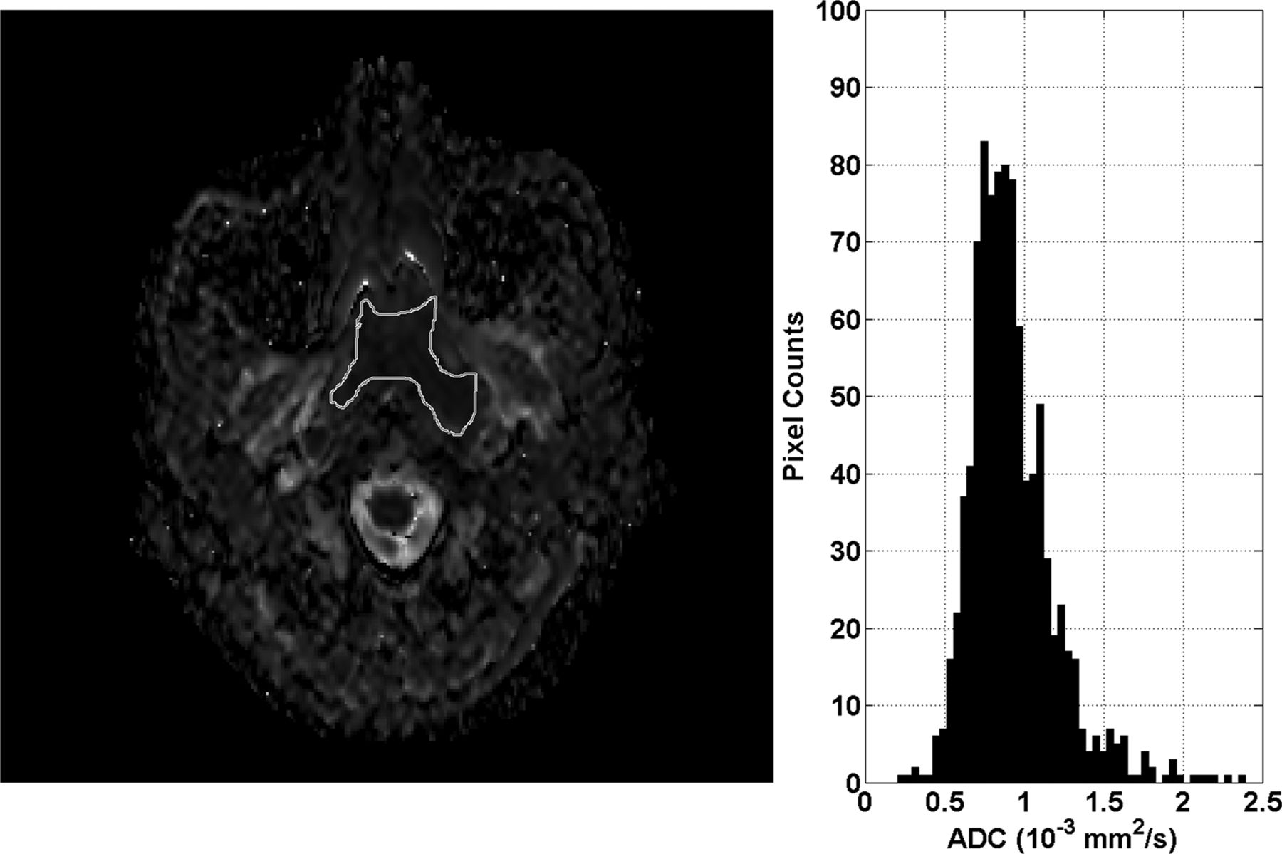
ADC map and histogram of primary NPC before treatment in a 50-year-old woman with local failure. The histogram shows that the greatest frequency of ADC values is shifted toward the central ADC range (ADC skewness = 0.11) and the peak is broadened (ADC kurtosis = 3.89).

Fig 2.
  • Download figure
  • Open in new tab
  • Download powerpoint
Fig 2.

ADC map and histogram of primary NPC before treatment in a 43-year-old man with local control. The histogram shows that the greatest frequency of ADC values is shifted toward the lower ADC range (ADC skewness = 1.42) and the peak is more acute (ADC kurtosis = 8.77).

Fig 3.
  • Download figure
  • Open in new tab
  • Download powerpoint
Fig 3.

Side-by-side boxplots of ADC skewness comparing local control and local failure.

Fig 4.
  • Download figure
  • Open in new tab
  • Download powerpoint
Fig 4.

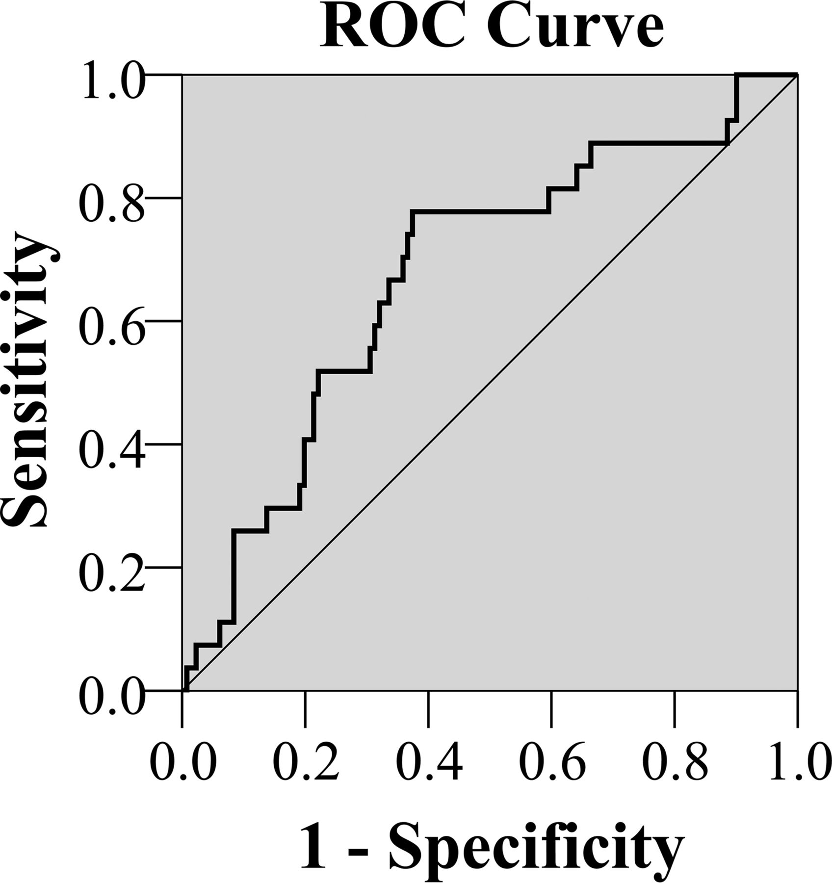
ROC analysis curve for ADC skewness.

View this table:
  • View inline
  • View popup
Table 2:

ADC skewness threshold obtained from ROC curve analysis to predict local failure

Discussion

On the basis of long-term clinical follow-up of patients treated for NPC, pretreatment DWI showed that the ADC distribution curves of primary tumors with LF had significantly lower ADC skewness than primary tumors with LC (ie, the ADC distribution curve of tumors with LF showed that the greatest frequencies of ADC values were shifted away from the lower ADC range). Therefore, visually, compared with tumors with LC, those with LF showed a shift of the curve peak away from the left side of the curve (greater frequency of lower ADC values) toward the center (symmetric frequency distribution of low and high ADC values) or right side of the curve (greater frequency of higher ADC values). ADC skewness remained significant on multivariate analysis, and a threshold of ≤0.55 produced a relatively high negative predictive value (93%) for LF, though the positive predictive value was low (30%), which could limit the clinical value of using DWI to predict NPC response.

Analysis of the shape of the ADC histogram curve pretreatment also showed that primary tumors with LF had significantly lower ADC kurtosis values than tumors with LC (ie, their ADC histogram peak was broader and less acute compared with the ADC histogram peak of the tumors with LC). A broader peak suggests that the tumor is more heterogeneous; this finding supports the view that those tumors with greater heterogeneity are more likely to fail treatment.16 In this study, there was also a trend toward tumors with higher volume having LF, but neither volume difference nor T-stage was significant. These results highlight the potential value of ADC skewness and ADC kurtosis because they stand out as the only parameters that may be able to identify resistant tumors over a range of tumor volumes and T-stages.

Pretreatment tumor ADC values have been shown to be predictors of treatment response in head and neck cancers. Previous head and neck squamous cell carcinoma studies have found significantly higher tumor ADC values in patients with LF,5⇓–7,10 nodal failure,4 or poor treatment response,8–9 while other studies have shown a similar trend in the ADC values that did not reach statistical significance.17⇓–19 It has been postulated that poor outcomes of some squamous cell carcinomas are due to tumor factors that are known to increase ADC values, such as micronecrosis, lower cellularity, and, more recently, negative human papillomavirus status and high stromal content.3,20–21 Of note, the ratio of stroma to tumor cells is recognized as an important determinant of outcome in head and neck squamous cell carcinoma. In regard to NPC, research now also shows that stroma-rich NPCs are associated with poor prognosis and an increased risk of relapse.22 Therefore, it could be postulated that NPCs with high ADCs are more likely to have a poor outcome compared with those with low ADCs. Currently, there are only a few NPC DWI studies that have correlated diffusion parameters with tumor characteristics at diagnosis, such as T-stage,23,24 early intratreatment response,25 and posttreatment response.12⇓⇓–15,26 Regarding using pretreatment tumor ADC to predict posttreatment response,12⇓⇓–15 3 of these studies were based on short-term outcome at the end or 3 months after the end of treatment.12⇓–14 These 3 studies showed mixed results with both low12–13 and high14 pretreatment ADCs reported in tumors with poor outcomes, with the results being significant in only 1 of the studies.12 However, a recent NPC study by Zhang et al15 with long-term follow-up and a large sample size of 541 patients showed a significant association between a high mean ADC in primary tumors pretreatment and poor survival at 3 years. That study measured the pretreatment mean ADC at the level of the largest primary tumor diameter and used ROC curve analysis to identify an optimal cutoff ADC for LF of ≥0.747 × 10⁻³ mm2/s (area under the ROC curve = 0.68, P = .004), which was shown to correlate with both local relapse-free survival and disease-free survival.

Our study broadly supports the findings of Zhang et al,15 with high primary tumor ADC values being associated with poor local response. However, we were unable to show this correlation by using the ADC mean and could only show such a correlation by using the ADC skewness. This discrepancy between our results and those of Zhang et al15 may be related to the longer follow-up period in our study, with a subsequently greater incidence of failure at the primary site, 17% (27/158) compared with 4.3% (23/541), of which 44% (13/27) in our study occurred beyond 3 years.

We postulate that more sophisticated ADC measurements such as ADC skewness may be needed to identify primary tumors that will relapse at a longer time after the end of treatment. The assessment of ADC skewness in this study used histogram analysis of the distribution of the ADC values from the entire tumor volume and had an advantage over the ADC mean in that it took tumor heterogeneity into account. Most tumors are heterogeneous, and the proportion of the cancer cell population with high ADC values may influence the final treatment outcome. From the results, we postulate that tumors that are likely to have resistant tumor cells are those in which the proportion of high ADC cells is similar or greater than the proportion of low ADC cells. However, other reasons for the discrepancy in the significance of the ADC mean between the 2 studies could include the smaller sample size in our study and the difference in the outcome measures used to denote response. Specifically, we did not use survival data such as local relapse-free survival to assess primary site response; instead, we took a simple approach and directly compared the ADC values of the primary tumors with LC against the ADC values of primary tumors with LF.

Previous NPC studies have shown that larger tumor volumes are associated with more unfavorable outcomes at the primary site.27⇓⇓⇓⇓⇓⇓⇓–35 This finding was also a trend in this study but did not reach significance, possibly because the mean volume of our primary tumors (24.6 mL) was in the lower range of previously reported cutoff thresholds, which have ranged from 13 to 60 mL.27⇓⇓⇓⇓⇓⇓⇓–35 In addition, the T-stage was not significant. This finding is possibly explained by the better treatment outcomes as a result of intensity-modulated radiation therapy36 and also the wide use of MR imaging for staging, which can lead to upstaging to T3 disease as a result of greater sensitivity to bone invasion compared with CT.37 Histologic NPC subtype also has a major influence on treatment outcome, whereby the undifferentiated form of NPC has a better prognosis than the other subtypes.38 In our fairly large study of 158 patients, we think that the histologic subtype did not influence the results because most (98%) were of the same undifferentiated carcinoma subtype, with only 2% (3 tumors) being poorly differentiated, none of which showed LF.

Use of ADC measures of tumor heterogeneity such as ADC skewness and kurtosis is fairly new to the DWI research, but a few cancer studies in head and neck squamous cell carcinoma and tumors of the ovary/peritoneum and brain have shown that pretreatment39,40 or intratreatment19,39,40 ADC skewness and kurtosis may predict treatment outcome.

NPC may relapse many years after treatment; therefore, one of the main strengths of this study was the long-term clinical follow-up of the primary site (mean, ∼7.5 years and maximum, ∼10 years) with a minimum of 5 years for patients with LC. However, because of the long clinical follow-up required for this study, one of the main limitations was that the DWI protocol was set up some time ago when, to reduce susceptibility artifacts at the skull base, the fitted 6 b-values used were up to a maximum of 500 s/mm2. It has been shown subsequently that more advanced non-Gaussian models for ADC analysis influence the ADC in NPC.41 In head and neck squamous cell carcinoma, the choice of b-values may also influence the accuracy of ADC for the prediction of treatment response, and some authors have proposed using ADCs calculated from the mid/high b-range (300/500-1000 s/mm2)9,10 to predict locoregional response.

Conclusions

This study correlated the ADC values of the pretreatment primary NPC with treatment outcome at the primary site on the basis of long-term clinical follow-up. Compared with primary tumors with LC, those with LF had lower ADC skewness and kurtosis. The ADC skewness remained significant on multivariate analysis. The simple ADC measurement using the mean value was not a predictor of outcome in this study, suggesting that more sophisticated measurements, such as skewness, may be needed to reflect the predictive value of high ADC cancer cell populations in heterogeneous tumors. The primary tumor volume and T-stage of NPC were not significant parameters in this study for predicting treatment response at the primary site, suggesting that ADC skewness and kurtosis may have the potential to predict tumor response, even in smaller volume or earlier stage tumors.

Acknowledgments

We acknowledge the assistance of H.L.E. Chan, S.T. Chan, C.H. Chen, Z. He, F.Y. Lam, and L.L. Leung.

References

  1. 1.↵
    1. Lee AW,
    2. Sze WM,
    3. Au JS, et al
    . Treatment results for nasopharyngeal carcinoma in the modern era: the Hong Kong experience. Int J Radiat Oncol Biol Phys 2005;61:1107–16 doi:10.1016/j.ijrobp.2004.07.702 pmid:15752890
    CrossRefPubMed
  2. 2.↵
    1. Yu KH,
    2. Leung SF,
    3. Tung SY, et al
    ; Hong Kong Nasopharyngeal Carcinoma Study Group. Survival outcome of patients with nasopharyngeal carcinoma with first local failure: a study by the Hong Kong Nasopharyngeal Carcinoma Study Group. Head Neck 2005;27:397–405 doi:10.1002/hed.20161 pmid:15726589
    CrossRefPubMed
  3. 3.↵
    1. Driessen JP,
    2. Caldas-Magalhaes J,
    3. Janssen LM, et al
    . Diffusion-weighted MR imaging in laryngeal and hypopharyngeal carcinoma: association between apparent diffusion coefficient and histologic findings. Radiology 2014;272:456–63 doi:10.1148/radiol.14131173 pmid:24749712
    CrossRefPubMed
  4. 4.↵
    1. Kim S,
    2. Loevner L,
    3. Quon H, et al
    . Diffusion-weighted magnetic resonance imaging for predicting and detecting early response to chemoradiation therapy of squamous cell carcinomas of the head and neck. Clin Cancer Res 2009;15:986–94 doi:10.1158/1078-0432.CCR-08-1287 pmid:19188170
    Abstract/FREE Full Text
  5. 5.↵
    1. Hatakenaka M,
    2. Nakamura K,
    3. Yabuuchi H, et al
    . Pretreatment apparent diffusion coefficient of the primary lesion correlates with local failure in head-and-neck cancer treated with chemoradiotherapy or radiotherapy. Int J Radiat Oncol Biol Phys 2011;81:339–45 doi:10.1016/j.ijrobp.2010.05.051 pmid:20832179
    CrossRefPubMed
  6. 6.↵
    1. Hatakenaka M,
    2. Shioyama Y,
    3. Nakamura K, et al
    . Apparent diffusion coefficient calculated with relatively high b-values correlates with local failure of head and neck squamous cell carcinoma treated with radiotherapy. AJNR Am J Neuroradiol 2011;32:1904–10 doi:10.3174/ajnr.A2610 pmid:21778248
    Abstract/FREE Full Text
  7. 7.↵
    1. Ohnishi K,
    2. Shioyama Y,
    3. Hatakenaka M, et al
    . Prediction of local failures with a combination of pretreatment tumor volume and apparent diffusion coefficient in patients treated with definitive radiotherapy for hypopharyngeal or oropharyngeal squamous cell carcinoma. J Radiat Res 2011;52:522–30 doi:10.1269/jrr.10178 pmid:21905311
    Abstract/FREE Full Text
  8. 8.↵
    1. Srinivasan A,
    2. Chenevert TL,
    3. Dwamena BA, et al
    . Utility of pretreatment mean apparent diffusion coefficient and apparent diffusion coefficient histograms in prediction of outcome to chemoradiation in head and neck squamous cell carcinoma. J Comput Assist Tomogr 2012;36:131–37 doi:10.1097/RCT.0b013e3182405435 pmid:22261783
    CrossRefPubMed
  9. 9.↵
    1. Lambrecht M,
    2. Van Calster B,
    3. Vandecaveye V, et al
    . Integrating pretreatment diffusion weighted MRI into a multivariable prognostic model for head and neck squamous cell carcinoma. Radiother Oncol 2014;110:429–34 doi:10.1016/j.radonc.2014.01.004 pmid:24630535
    CrossRefPubMed
  10. 10.↵
    1. Hatakenaka M,
    2. Nakamura K,
    3. Yabuuchi H, et al
    . Apparent diffusion coefficient is a prognostic factor of head and neck squamous cell carcinoma treated with radiotherapy. Jpn J Radiol 2014;32:80–89 doi:10.1007/s11604-013-0272-y pmid:24408077
    CrossRefPubMed
  11. 11.↵
    1. Lee AW,
    2. Foo W,
    3. Law SC, et al
    . Recurrent nasopharyngeal carcinoma: the puzzles of long latency. Int J Radiat Oncol Biol Phys 1999;44:149–56 doi:10.1016/S0360-3016(98)00524-0 pmid:10219808
    CrossRefPubMed
  12. 12.↵
    1. Zheng D,
    2. Chen Y,
    3. Chen Y, et al
    . Early assessment of induction chemotherapy response of nasopharyngeal carcinoma by pretreatment diffusion-weighted magnetic resonance imaging. J Comput Assist Tomogr 2013;37:673–80 doi:10.1097/RCT.0b013e31829a2599 pmid:24045239
    CrossRefPubMed
  13. 13.↵
    1. Chen Y,
    2. Liu X,
    3. Zheng D, et al
    . Diffusion-weighted magnetic resonance imaging for early response assessment of chemoradiotherapy in patients with nasopharyngeal carcinoma. Magn Reson Imaging 2014;32:630–37 doi:10.1016/j.mri.2014.02.009 pmid:24703576
    CrossRefPubMed
  14. 14.↵
    1. Hong J,
    2. Yao Y,
    3. Zhang Y, et al
    . Value of magnetic resonance diffusion-weighted imaging for the prediction of radiosensitivity in nasopharyngeal carcinoma. Otolaryngol Head Neck Surg 2013;149:707–13 doi:10.1177/0194599813496537 pmid:23884282
    Abstract/FREE Full Text
  15. 15.↵
    1. Zhang Y,
    2. Liu X,
    3. Zhang Y, et al
    . Prognostic value of the primary lesion apparent diffusion coefficient (ADC) in nasopharyngeal carcinoma: a retrospective study of 541 cases. Sci Rep 2015;5:12242 doi:10.1038/srep12242 pmid:26184509
    CrossRefPubMed
  16. 16.↵
    1. Mroz EA,
    2. Tward AD,
    3. Pickering CR, et al
    . High intratumor genetic heterogeneity is related to worse outcome in patients with head and neck squamous cell carcinoma. Cancer 2013;119:3034–42 doi:10.1002/cncr.28150 pmid:23696076
    CrossRefPubMed
  17. 17.↵
    1. Matoba M,
    2. Tuji H,
    3. Shimode Y, et al
    . Fractional change in apparent diffusion coefficient as an imaging biomarker for predicting treatment response in head and neck cancer treated with chemoradiotherapy. AJNR Am J Neuroradiol 2014;35:379–85 doi:10.3174/ajnr.A3706 pmid:24029391
    Abstract/FREE Full Text
  18. 18.↵
    1. Chawla S,
    2. Kim S,
    3. Dougherty L, et al
    . Pretreatment diffusion-weighted and dynamic contrast-enhanced MRI for prediction of local treatment response in squamous cell carcinomas of the head and neck. AJR Am J Roentgenol 2013;200:35–43 doi:10.2214/AJR.12.9432 pmid:23255739
    CrossRefPubMed
  19. 19.↵
    1. King AD,
    2. Chow KK,
    3. Yu KH, et al
    . Head and neck squamous cell carcinoma: diagnostic performance of diffusion-weighted MR imaging for the prediction of treatment response. Radiology 2013;266:531–38 doi:10.1148/radiol.12120167 pmid:23151830
    CrossRefPubMed
  20. 20.↵
    1. Driessen JP,
    2. van Bemmel AJ,
    3. van Kempen PM, et al
    . Correlation of human papillomavirus status with apparent diffusion coefficient of diffusion-weighted MRI in head and neck squamous cell carcinomas. Head Neck 2015 Mar 17. [Epub ahead of print] doi:10.1002/hed.24051 pmid:25783872
    CrossRefPubMed
  21. 21.↵
    1. Nakahira M,
    2. Saito N,
    3. Yamaguchi H, et al
    . Use of quantitative diffusion-weighted magnetic resonance imaging to predict human papilloma virus status in patients with oropharyngeal squamous cell carcinoma. Eur Arch Otorhinolaryngol 2014;271:1219–25 doi:10.1007/s00405-013-2641-7 pmid:23880924
    CrossRefPubMed
  22. 22.↵
    1. Zhang XL,
    2. Jiang C,
    3. Zhang ZX, et al
    . The tumor-stroma ratio is an independent predictor for survival in nasopharyngeal cancer. Oncol Res Treat 2014;37:480–84 doi:10.1159/000365165 pmid:25231688
    CrossRefPubMed
  23. 23.↵
    1. Abdel Razek AA,
    2. Kamal E
    . Nasopharyngeal carcinoma: correlation of apparent diffusion coefficient value with prognostic parameters. Radiol Med 2013;118:534–39 doi:10.1007/s11547-012-0890-x pmid:23090251
    CrossRefPubMed
  24. 24.↵
    1. Lai V,
    2. Li X,
    3. Lee VH, et al
    . Nasopharyngeal carcinoma: comparison of diffusion and perfusion characteristics between different tumour stages using intravoxel incoherent motion MR imaging. Eur Radiol 2014;24:176–83 doi:10.1007/s00330-013-2995-7 pmid:23990005
    CrossRefPubMed
  25. 25.↵
    1. Chen Y,
    2. Ren W,
    3. Zheng D, et al
    . Diffusion kurtosis imaging predicts neoadjuvant chemotherapy responses within 4 days in advanced nasopharyngeal carcinoma patients. J Magn Reson Imaging 2015;42:1354–61 doi:10.1002/jmri.24910 pmid:25873208
    CrossRefPubMed
  26. 26.↵
    1. Xu JF,
    2. Wu XW,
    3. Wang WQ, et al
    . Value of diffusion-weighted magnetic resonance imaging on the follow-up of nasopharyngeal carcinoma after radiotherapy. J Xray Sci Technol 2014;22:605–12 doi:10.3233/XST-140448 pmid:25265921
    CrossRefPubMed
  27. 27.↵
    1. Wu Z,
    2. Su Y,
    3. Zeng RF, et al
    . Prognostic value of tumor volume for patients with nasopharyngeal carcinoma treated with concurrent chemotherapy and intensity-modulated radiotherapy. J Cancer Res Clin Oncol 2014;140:69–76 doi:10.1007/s00432-013-1542-x pmid:24173695
    CrossRefPubMed
  28. 28.↵
    1. Feng M,
    2. Wang W,
    3. Fan Z, et al
    . Tumor volume is an independent prognostic indicator of local control in nasopharyngeal carcinoma patients treated with intensity-modulated radiotherapy. Radiat Oncol 2013;8:208 doi:10.1186/1748-717X-8-208 pmid:24007375
    CrossRefPubMed
  29. 29.↵
    1. Guo R,
    2. Sun Y,
    3. Yu XL, et al
    . Is primary tumor volume still a prognostic factor in intensity modulated radiation therapy for nasopharyngeal carcinoma? Radiother Oncol 2012;104:294–99 doi:10.1016/j.radonc.2012.09.001 pmid:22998947
    CrossRefPubMed
  30. 30.↵
    1. Lee CC,
    2. Huang TT,
    3. Lee MS, et al
    . Clinical application of tumor volume in advanced nasopharyngeal carcinoma to predict outcome. Radiat Oncol 2010;5:20 doi:10.1186/1748-717X-5-20 pmid:20222940
    CrossRefPubMed
  31. 31.↵
    1. Shen C,
    2. Lu JJ,
    3. Gu Y, et al
    . Prognostic impact of primary tumor volume in patients with nasopharyngeal carcinoma treated by definitive radiation therapy. Laryngoscope 2008;118:1206–10 doi:10.1097/MLG.0b013e31816ed587 pmid:18418278
    CrossRefPubMed
  32. 32.↵
    1. Kim JH,
    2. Lee JK
    . Prognostic value of tumor volume in nasopharyngeal carcinoma. Yonsei Med J 2005;46:221–27 doi:10.3349/ymj.2005.46.2.221 pmid:15861494
    CrossRefPubMed
  33. 33.↵
    1. Sze WM,
    2. Lee AW,
    3. Yau TK, et al
    . Primary tumor volume of nasopharyngeal carcinoma: prognostic significance for local control. Int J Radiat Oncol Biol Phys 2004;59:21–27 doi:10.1016/j.ijrobp.2003.10.027 pmid:15093895
    CrossRefPubMed
  34. 34.↵
    1. Chua DT,
    2. Sham JS,
    3. Leung LH, et al
    . Tumor volume is not an independent prognostic factor in early-stage nasopharyngeal carcinoma treated by radiotherapy alone. Int J Radiat Oncol Biol Phys 2004;58:1437–44 doi:10.1016/j.ijrobp.2003.09.075 pmid:15050321
    CrossRefPubMed
  35. 35.↵
    1. Chua DT,
    2. Sham JS,
    3. Kwong DL, et al
    . Volumetric analysis of tumor extent in nasopharyngeal carcinoma and correlation with treatment outcome. Int J Radiat Oncol Biol Phys 1997;39:711–19 doi:10.1016/S0360-3016(97)00374-X pmid:9336154
    CrossRefPubMed
  36. 36.↵
    1. Lai SZ,
    2. Li WF,
    3. Chen L, et al
    . How does intensity-modulated radiotherapy versus conventional two-dimensional radiotherapy influence the treatment results in nasopharyngeal carcinoma patients? Int J Radiat Oncol Biol Phys 2011;80:661–68 doi:10.1016/j.ijrobp.2010.03.024 pmid:20643517
    CrossRefPubMed
  37. 37.↵
    1. Liao XB,
    2. Mao YP,
    3. Liu LZ, et al
    . How does magnetic resonance imaging influence staging according to AJCC staging system for nasopharyngeal carcinoma compared with computed tomography? Int J Radiat Oncol Biol Phys 2008;72:1368–77 doi:10.1016/j.ijrobp.2008.03.017 pmid:18455329
    CrossRefPubMed
  38. 38.↵
    1. Cheng SH,
    2. Tsai SY,
    3. Horng CF, et al
    . A prognostic scoring system for locoregional control in nasopharyngeal carcinoma following conformal radiotherapy. Int J Radiat Oncol Biol Phys 2006;66:992–1003 doi:10.1016/j.ijrobp.2006.06.006 pmid:16979832
    CrossRefPubMed
  39. 39.↵
    1. Kyriazi S,
    2. Collins DJ,
    3. Messiou C, et al
    . Metastatic ovarian and primary peritoneal cancer: assessing chemotherapy response with diffusion-weighted MR imaging–value of histogram analysis of apparent diffusion coefficients. Radiology 2011;261:182–92 doi:10.1148/radiol.11110577 pmid:21828186
    CrossRefPubMed
  40. 40.↵
    1. Nowosielski M,
    2. Recheis W,
    3. Goebel G, et al
    . ADC histograms predict response to anti-angiogenic therapy in patients with recurrent high-grade glioma. Neuroradiology 2011;53:291–302 doi:10.1007/s00234-010-0808-0 pmid:21125399
    CrossRefPubMed
  41. 41.↵
    1. Yuan J,
    2. Yeung DK,
    3. Mok GS, et al
    . Non-Gaussian analysis of diffusion weighted imaging in head and neck at 3T: a pilot study in patients with nasopharyngeal carcinoma. PLoS One 2014;9:e87024 doi:10.1371/journal.pone.0087024 pmid:24466318
    CrossRefPubMed
  • Received November 30, 2015.
  • Accepted after revision February 27, 2016.
  • © 2016 by American Journal of Neuroradiology
View Abstract
PreviousNext
Back to top

In this issue

American Journal of Neuroradiology: 37 (9)
American Journal of Neuroradiology
Vol. 37, Issue 9
1 Sep 2016
  • Table of Contents
  • Index by author
  • Complete Issue (PDF)
Advertisement
Print
Download PDF
Email Article

Thank you for your interest in spreading the word on American Journal of Neuroradiology.

NOTE: We only request your email address so that the person you are recommending the page to knows that you wanted them to see it, and that it is not junk mail. We do not capture any email address.

Enter multiple addresses on separate lines or separate them with commas.
Diffusion-Weighted Imaging of Nasopharyngeal Carcinoma: Can Pretreatment DWI Predict Local Failure Based on Long-Term Outcome?
(Your Name) has sent you a message from American Journal of Neuroradiology
(Your Name) thought you would like to see the American Journal of Neuroradiology web site.
CAPTCHA
This question is for testing whether or not you are a human visitor and to prevent automated spam submissions.
Cite this article
B.K.H. Law, A.D. King, K.S. Bhatia, A.T. Ahuja, M.K.M. Kam, B.B. Ma, Q.Y. Ai, F.K.F. Mo, J. Yuan, D.K.W. Yeung
Diffusion-Weighted Imaging of Nasopharyngeal Carcinoma: Can Pretreatment DWI Predict Local Failure Based on Long-Term Outcome?
American Journal of Neuroradiology Sep 2016, 37 (9) 1706-1712; DOI: 10.3174/ajnr.A4792

Citation Manager Formats

  • BibTeX
  • Bookends
  • EasyBib
  • EndNote (tagged)
  • EndNote 8 (xml)
  • Medlars
  • Mendeley
  • Papers
  • RefWorks Tagged
  • Ref Manager
  • RIS
  • Zotero
0 Responses
Respond to this article
Share
Bookmark this article
Diffusion-Weighted Imaging of Nasopharyngeal Carcinoma: Can Pretreatment DWI Predict Local Failure Based on Long-Term Outcome?
B.K.H. Law, A.D. King, K.S. Bhatia, A.T. Ahuja, M.K.M. Kam, B.B. Ma, Q.Y. Ai, F.K.F. Mo, J. Yuan, D.K.W. Yeung
American Journal of Neuroradiology Sep 2016, 37 (9) 1706-1712; DOI: 10.3174/ajnr.A4792
del.icio.us logo Twitter logo Facebook logo Mendeley logo
  • Tweet Widget
  • Facebook Like
  • Google Plus One
Purchase

Jump to section

  • Article
    • Abstract
    • ABBREVIATIONS:
    • Materials and Methods
    • Results
    • Discussion
    • Conclusions
    • Acknowledgments
    • References
  • Figures & Data
  • Info & Metrics
  • Responses
  • References
  • PDF

Related Articles

  • No related articles found.
  • PubMed
  • Google Scholar

Cited By...

  • Reply:
  • Treatment Response Prediction of Nasopharyngeal Carcinoma Based on Histogram Analysis of Diffusional Kurtosis Imaging
  • Crossref (34)
  • Google Scholar

This article has been cited by the following articles in journals that are participating in Crossref Cited-by Linking.

  • A Prognostic Predictive System Based on Deep Learning for Locoregionally Advanced Nasopharyngeal Carcinoma
    Mengyun Qiang, Chaofeng Li, Yuyao Sun, Ying Sun, Liangru Ke, Chuanmiao Xie, Tao Zhang, Yujian Zou, Wenze Qiu, Mingyong Gao, Yingxue Li, Xiang Li, Zejiang Zhan, Kuiyuan Liu, Xi Chen, Chixiong Liang, Qiuyan Chen, Haiqiang Mai, Guotong Xie, Xiang Guo, Xing Lv
    JNCI: Journal of the National Cancer Institute 2021 113 5
  • Radiomics Analysis of PET and CT Components of PET/CT Imaging Integrated with Clinical Parameters: Application to Prognosis for Nasopharyngeal Carcinoma
    Wenbing Lv, Qingyu Yuan, Quanshi Wang, Jianhua Ma, Qianjin Feng, Wufan Chen, Arman Rahmim, Lijun Lu
    Molecular Imaging and Biology 2019 21 5
  • State of the art MRI in head and neck cancer
    Y.L. Dai, A.D. King
    Clinical Radiology 2018 73 1
  • ADC-histogram analysis in head and neck squamous cell carcinoma. Associations with different histopathological features including expression of EGFR, VEGF, HIF-1α, Her 2 and p53. A preliminary study
    Hans Jonas Meyer, Leonard Leifels, Gordian Hamerla, Anne Kathrin Höhn, Alexey Surov
    Magnetic Resonance Imaging 2018 54
  • Pre-treatment intravoxel incoherent motion diffusion-weighted imaging predicts treatment outcome in nasopharyngeal carcinoma
    Sahrish Qamar, Ann D. King, Qi-Yong H. Ai, Tiffany Y. So, Frankie Kwok Fai Mo, Weitian Chen, Darren M.C. Poon, Macy Tong, Brigette B. Ma, Edwin P. Hui, David Ka-Wai Yeung, Yi-Xiang Wang, Jing Yuan
    European Journal of Radiology 2020 129
  • Diffusion magnetic resonance imaging: A molecular imaging tool caught between hope, hype and the real world of “personalized oncology”
    Abhishek Mahajan, Sneha S Deshpande, Meenakshi H Thakur
    World Journal of Radiology 2017 9 6
  • Investigation of the feasibility of synthetic MRI in the differential diagnosis of non-keratinising nasopharyngeal carcinoma and benign hyperplasia using different contoured methods for delineation of the region of interest
    T. Meng, H. He, H. Liu, X. Lv, C. Huang, L. Zhong, K. Liu, L. Qian, L. Ke, C. Xie
    Clinical Radiology 2021 76 3
  • Volumetric histogram analysis of apparent diffusion coefficient for predicting pathological complete response and survival in esophageal cancer patients treated with chemoradiotherapy
    Atsushi Hirata, Koichi Hayano, Gaku Ohira, Shunsuke Imanishi, Toshiharu Hanaoka, Kentaro Murakami, Tomoyoshi Aoyagi, Kiyohiko Shuto, Hisahiro Matsubara
    The American Journal of Surgery 2020 219 6
  • Diffusion-weighted imaging of nasopharyngeal carcinoma to predict distant metastases
    Qi-Yong Ai, Ann D. King, Benjamin King Hong Law, David Ka-Wai Yeung, Kunwar S. Bhatia, Jing Yuan, Anil T. Ahuja, Lok Yiu Sheila Wong, Brigette B. Ma, Frankie Kwok Fai Mo, Michael K. M. Kam
    European Archives of Oto-Rhino-Laryngology 2017 274 2
  • Acquisition repeatability of MRI radiomics features in the head and neck: a dual-3D-sequence multi-scan study
    Cindy Xue, Jing Yuan, Yihang Zhou, Oi Lei Wong, Kin Yin Cheung, Siu Ki Yu
    Visual Computing for Industry, Biomedicine, and Art 2022 5 1

More in this TOC Section

  • Chondrosarcoma vs Synovial Chondromatosis: Imaging
  • WHO Classification Update: Nasal&Skull Base Tumors
  • Peritumoral Signal in Vestibular Schwannomas
Show more HEAD & NECK

Similar Articles

Advertisement

Indexed Content

  • Current Issue
  • Accepted Manuscripts
  • Article Preview
  • Past Issues
  • Editorials
  • Editor's Choice
  • Fellows' Journal Club
  • Letters to the Editor
  • Video Articles

Cases

  • Case Collection
  • Archive - Case of the Week
  • Archive - Case of the Month
  • Archive - Classic Case

Special Collections

  • AJNR Awards
  • ASNR Foundation Special Collection
  • Most Impactful AJNR Articles
  • Photon-Counting CT
  • Spinal CSF Leak Articles (Jan 2020-June 2024)

More from AJNR

  • Trainee Corner
  • Imaging Protocols
  • MRI Safety Corner

Multimedia

  • AJNR Podcasts
  • AJNR Scantastics

Resources

  • Turnaround Time
  • Submit a Manuscript
  • Submit a Video Article
  • Submit an eLetter to the Editor/Response
  • Manuscript Submission Guidelines
  • Statistical Tips
  • Fast Publishing of Accepted Manuscripts
  • Graphical Abstract Preparation
  • Imaging Protocol Submission
  • Evidence-Based Medicine Level Guide
  • Publishing Checklists
  • Author Policies
  • Become a Reviewer/Academy of Reviewers
  • News and Updates

About Us

  • About AJNR
  • Editorial Board
  • Editorial Board Alumni
  • Alerts
  • Permissions
  • Not an AJNR Subscriber? Join Now
  • Advertise with Us
  • Librarian Resources
  • Feedback
  • Terms and Conditions
  • AJNR Editorial Board Alumni

American Society of Neuroradiology

  • Not an ASNR Member? Join Now

© 2025 by the American Society of Neuroradiology All rights, including for text and data mining, AI training, and similar technologies, are reserved.
Print ISSN: 0195-6108 Online ISSN: 1936-959X

Powered by HighWire