Advertisement

Main menu

  • Home
  • Content
    • Current Issue
    • Accepted Manuscripts
    • Article Preview
    • Past Issue Archive
    • Video Articles
    • AJNR Case Collection
    • Case of the Week Archive
    • Case of the Month Archive
    • Classic Case Archive
  • Special Collections
    • AJNR Awards
    • ASNR Foundation Special Collection
    • Most Impactful AJNR Articles
    • Photon-Counting CT
    • Spinal CSF Leak Articles (Jan 2020-June 2024)
  • Multimedia
    • AJNR Podcasts
    • AJNR SCANtastic
    • Trainee Corner
    • MRI Safety Corner
    • Imaging Protocols
  • For Authors
    • Submit a Manuscript
    • Submit a Video Article
    • Submit an eLetter to the Editor/Response
    • Manuscript Submission Guidelines
    • Statistical Tips
    • Fast Publishing of Accepted Manuscripts
    • Graphical Abstract Preparation
    • Imaging Protocol Submission
    • Author Policies
  • About Us
    • About AJNR
    • Editorial Board
    • Editorial Board Alumni
  • More
    • Become a Reviewer/Academy of Reviewers
    • Subscribers
    • Permissions
    • Alerts
    • Feedback
    • Advertisers
    • ASNR Home

User menu

  • Alerts
  • Log in

Search

  • Advanced search
American Journal of Neuroradiology

American Journal of Neuroradiology

ASHNR American Society of Functional Neuroradiology ASHNR American Society of Pediatric Neuroradiology ASSR
  • Alerts
  • Log in

Advanced Search

  • Home
  • Content
    • Current Issue
    • Accepted Manuscripts
    • Article Preview
    • Past Issue Archive
    • Video Articles
    • AJNR Case Collection
    • Case of the Week Archive
    • Case of the Month Archive
    • Classic Case Archive
  • Special Collections
    • AJNR Awards
    • ASNR Foundation Special Collection
    • Most Impactful AJNR Articles
    • Photon-Counting CT
    • Spinal CSF Leak Articles (Jan 2020-June 2024)
  • Multimedia
    • AJNR Podcasts
    • AJNR SCANtastic
    • Trainee Corner
    • MRI Safety Corner
    • Imaging Protocols
  • For Authors
    • Submit a Manuscript
    • Submit a Video Article
    • Submit an eLetter to the Editor/Response
    • Manuscript Submission Guidelines
    • Statistical Tips
    • Fast Publishing of Accepted Manuscripts
    • Graphical Abstract Preparation
    • Imaging Protocol Submission
    • Author Policies
  • About Us
    • About AJNR
    • Editorial Board
    • Editorial Board Alumni
  • More
    • Become a Reviewer/Academy of Reviewers
    • Subscribers
    • Permissions
    • Alerts
    • Feedback
    • Advertisers
    • ASNR Home
  • Follow AJNR on Twitter
  • Visit AJNR on Facebook
  • Follow AJNR on Instagram
  • Join AJNR on LinkedIn
  • RSS Feeds

AJNR is seeking candidates for the AJNR Podcast Editor. Read the position description.

Research ArticleINTERVENTIONAL

Quantifying the Cerebral Hemodynamics of Dural Arteriovenous Fistula in Transverse Sigmoid Sinus Complicated by Sinus Stenosis: A Retrospective Cohort Study

W.-Y. Guo, C.-C.J. Lee, C.-J. Lin, H.-C. Yang, H.-M. Wu, C.-C. Wu, W.-Y. Chung and K.-D. Liu
American Journal of Neuroradiology January 2017, 38 (1) 132-138; DOI: https://doi.org/10.3174/ajnr.A4960
W.-Y. Guo
aFrom the Department of Radiology (W.-Y.G., C.-J.L., H.-M.W., C.-C.W.)
cSchool of Medicine (W.-Y.G., C.-C.J.L., C.-J.L., H.-C.Y., H.-M.W., C.-C.W., W.-Y.C., K.-D.L.), National Yang-Ming University, Taipei, Taiwan.
  • Find this author on Google Scholar
  • Find this author on PubMed
  • Search for this author on this site
  • ORCID record for W.-Y. Guo
C.-C.J. Lee
bDepartment of Neurosurgery (C.-C.J.L., H.-C.Y., W.-Y.C., K.-D.L.), Neurological Institute, Taipei Veterans General Hospital, Taipei, Taiwan
cSchool of Medicine (W.-Y.G., C.-C.J.L., C.-J.L., H.-C.Y., H.-M.W., C.-C.W., W.-Y.C., K.-D.L.), National Yang-Ming University, Taipei, Taiwan.
  • Find this author on Google Scholar
  • Find this author on PubMed
  • Search for this author on this site
  • ORCID record for C.-C.J. Lee
C.-J. Lin
aFrom the Department of Radiology (W.-Y.G., C.-J.L., H.-M.W., C.-C.W.)
cSchool of Medicine (W.-Y.G., C.-C.J.L., C.-J.L., H.-C.Y., H.-M.W., C.-C.W., W.-Y.C., K.-D.L.), National Yang-Ming University, Taipei, Taiwan.
  • Find this author on Google Scholar
  • Find this author on PubMed
  • Search for this author on this site
  • ORCID record for C.-J. Lin
H.-C. Yang
bDepartment of Neurosurgery (C.-C.J.L., H.-C.Y., W.-Y.C., K.-D.L.), Neurological Institute, Taipei Veterans General Hospital, Taipei, Taiwan
cSchool of Medicine (W.-Y.G., C.-C.J.L., C.-J.L., H.-C.Y., H.-M.W., C.-C.W., W.-Y.C., K.-D.L.), National Yang-Ming University, Taipei, Taiwan.
  • Find this author on Google Scholar
  • Find this author on PubMed
  • Search for this author on this site
  • ORCID record for H.-C. Yang
H.-M. Wu
aFrom the Department of Radiology (W.-Y.G., C.-J.L., H.-M.W., C.-C.W.)
cSchool of Medicine (W.-Y.G., C.-C.J.L., C.-J.L., H.-C.Y., H.-M.W., C.-C.W., W.-Y.C., K.-D.L.), National Yang-Ming University, Taipei, Taiwan.
  • Find this author on Google Scholar
  • Find this author on PubMed
  • Search for this author on this site
  • ORCID record for H.-M. Wu
C.-C. Wu
aFrom the Department of Radiology (W.-Y.G., C.-J.L., H.-M.W., C.-C.W.)
cSchool of Medicine (W.-Y.G., C.-C.J.L., C.-J.L., H.-C.Y., H.-M.W., C.-C.W., W.-Y.C., K.-D.L.), National Yang-Ming University, Taipei, Taiwan.
  • Find this author on Google Scholar
  • Find this author on PubMed
  • Search for this author on this site
  • ORCID record for C.-C. Wu
W.-Y. Chung
bDepartment of Neurosurgery (C.-C.J.L., H.-C.Y., W.-Y.C., K.-D.L.), Neurological Institute, Taipei Veterans General Hospital, Taipei, Taiwan
cSchool of Medicine (W.-Y.G., C.-C.J.L., C.-J.L., H.-C.Y., H.-M.W., C.-C.W., W.-Y.C., K.-D.L.), National Yang-Ming University, Taipei, Taiwan.
  • Find this author on Google Scholar
  • Find this author on PubMed
  • Search for this author on this site
  • ORCID record for W.-Y. Chung
K.-D. Liu
bDepartment of Neurosurgery (C.-C.J.L., H.-C.Y., W.-Y.C., K.-D.L.), Neurological Institute, Taipei Veterans General Hospital, Taipei, Taiwan
cSchool of Medicine (W.-Y.G., C.-C.J.L., C.-J.L., H.-C.Y., H.-M.W., C.-C.W., W.-Y.C., K.-D.L.), National Yang-Ming University, Taipei, Taiwan.
  • Find this author on Google Scholar
  • Find this author on PubMed
  • Search for this author on this site
  • ORCID record for K.-D. Liu
  • Article
  • Figures & Data
  • Info & Metrics
  • Responses
  • References
  • PDF
Loading

Abstract

BACKGROUND AND PURPOSE: Sinus stenosis occasionally occurs in dural arteriovenous fistulas. Sinus stenosis impedes venous outflow and aggravates intracranial hypertension by reversing cortical venous drainage. This study aimed to analyze the likelihood of sinus stenosis and its impact on cerebral hemodynamics of various types of dural arteriovenous fistulas.

MATERIALS AND METHODS: Forty-three cases of dural arteriovenous fistula in the transverse-sigmoid sinus were reviewed and divided into 3 groups: Cognard type I, type IIa, and types with cortical venous drainage. Sinus stenosis and the double peak sign (occurrence of 2 peaks in the time-density curve of the ipsilateral drainage of the internal jugular vein) in dural arteriovenous fistula were evaluated. “TTP” was defined as the time at which a selected angiographic point reached maximum concentration. TTP of the vein of Labbé, TTP of the ipsilateral normal transverse sinus, trans-fistula time, and trans-stenotic time were compared across the 3 groups.

RESULTS: Thirty-six percent of type I, 100% of type IIa, and 84% of types with cortical venous drainage had sinus stenosis. All sinus stenosis cases demonstrated loss of the double peak sign that occurs in dural arteriovenous fistula. Trans-fistula time (2.09 seconds) and trans-stenotic time (0.67 seconds) in types with cortical venous drainage were the most prolonged, followed by those in type IIa and type I. TTP of the vein of Labbé was significantly shorter in types with cortical venous drainage. Six patients with types with cortical venous drainage underwent venoplasty and stent placement, and 4 were downgraded to type IIa.

CONCLUSIONS: Sinus stenosis indicated dysfunction of venous drainage and is more often encountered in dural arteriovenous fistula with more aggressive types. Venoplasty ameliorates cortical venous drainage in dural arteriovenous fistulas and serves as a bridge treatment to stereotactic radiosurgery in most cases.

ABBREVIATIONS:

CVD
cortical venous drainage
DAVF
dural arteriovenous fistula
SRS
stereotactic radiosurgery
SS
sinus stenosis
TFT
trans-fistula time
TST
trans-stenotic time
TTPPV
TTP for the parietal vein
TTPVL
TTP of the vein of Labbé

Dural arteriovenous fistulas (DAVFs) account for 10%–15% of intracranial vascular malformations.1,2 The most common location of an intracranial DAVF is the cavernous sinus, followed by the transverse-sigmoid sinus.1⇓–3 Major DAVF classification systems, such as the Cognard and Borden systems, grade DAVFs on the basis of venous drainage patterns, in which the presence of retrograde cortical venous drainage (CVD) indicates a higher risk of hemorrhage.4⇓⇓–7 Cases of venous outlet obstruction playing a role in transforming benign (without CVD) into malignant DAVFs (with CVD) have been reported in the literature.8 Sinus stenosis (SS) is frequently associated with idiopathic intracranial hypertension.9,10 Nevertheless, the incidence of SS and its association with DAVFs have not been thoroughly explored. SS can be found in DAVFs with retrograde or antegrade sinus flow, but its impact on cerebral hemodynamics has rarely been discussed. Theoretically, stenotic and thrombosed sinuses impede the venous outflow, and a DAVF itself increases overall blood volume in the affected sinus; the combination of the 2 hemodynamic disorders adversely affects venous flow and subsequently increases intracranial pressure and the risk of intracranial hemorrhage.

Current treatment strategies for DAVFs in the transverse sinus include microsurgery, endovascular treatment, stereotactic radiosurgery (SRS), or their combinations.11⇓–13 Endovascular treatment has been the treatment of choice for DAVFs with CVD because it provides immediate curative results and minimizes the risk of hemorrhage.14⇓–16 Nevertheless, the complication rate of endovascular treatment is higher than that of SRS.14,17,18 By contrast, SRS has hardly any periprocedural risks and achieves DAVF cure rates of between 58% and 73%. Although SRS can reduce the bleeding rate from 20% to 2% after shunting has been totally closed,19 the latent period for SRS ranges from 1 to 3 years and carries a 4.1% hemorrhagic rate in DAVFs with CVD.3,20 Therefore, SRS is usually preferred for cases without CVD, and endovascular treatment is more suitable for immediately minimizing the risk of hemorrhage.

Several studies have proposed a reconstructive method by using venoplasty and stent placement in combination with transarterial embolization to ameliorate or even cure DAVFs with venous outlet obstruction.21⇓–23 We wondered whether this approach could downgrade DAVFs with CVD—that is, to restore their normal cortical venous drainage and make them eligible for SRS, thereby minimizing the risk of hemorrhage during the latent period. Therefore, the purpose of the current study was to clarify the following: 1) the incidence of SS in different grades of DAVF in the transverse sigmoid sinus, 2) the impact of SS on DAVF hemodynamics by using quantitative DSA, and 3) the initial treatment results of venoplasty and/or stent placement followed by SRS.

Materials and Methods

Patient Population

The institutional review board of Taipei Veterans General Hospital approved this study. From January 2011 to December 2015, we consecutively recruited cases of angiography-proved DAVFs from the angiosuite logbook. In terms of our inclusion and exclusion criteria, DAVFs involving either side of the transverse-sigmoid sinus were included, while cases already treated before visiting our hospital, DAVFs located elsewhere, and those that did not receive SRS as treatment at all were excluded.

Clinical Presentation, Treatment, and Follow-Up

Data for symptoms at initial presentation, such as tinnitus, bruit, headaches, previous hemorrhage, and neurologic deficits (ie, visual disturbance, seizure, ataxia, and memory decline), and treatment results during follow-up were based on chart review. There were no missing data for the initial presentation. All patients received SRS and the same follow-up protocol: outpatient department visit and MR imaging at 6-month intervals. Adjunctive endovascular treatment was also recorded. The “primary end point” was complete regression, defined as disappearance of abnormal vasculature on follow-up MR imaging with or without additional angiography.20 “Partial regression” was defined as decreased abnormal vascularity compared with the baseline imaging. “Hemorrhage” was defined as any new hemorrhage in the follow-up MR imaging. The “composite end point” (ie, favorable outcome) was defined as complete regression without hemorrhage radiologically. We used the Kaplan-Meier method to handle missing data, treating it as censored. “Restenosis” was defined as a 50% narrowing of the immediate postvenoplasty sinus diameter during MR imaging follow-up.

Imaging Protocol and Data Analysis

DSA acquisition with a standard, clinically routine protocol was performed in all 43 cases. A power injector (Liebel-Flarsheim Angiomat; Illumena, San Diego, California) created a contrast bolus by placing a 4F angiocatheter in the common carotid artery at the C4 vertebral body level. A bolus of 12–14 mL of 60% diluted contrast medium (340 mg I/mL) was administered within 1.5 seconds. Neither extra contrast medium nor extra radiation was used. The acquisition parameters were 7.5 frames/s for the first 5 seconds, followed by 4 frames/s for 3 seconds, 3 frames/s for 2 seconds, and finally 2 frames/s for 2 seconds. The entire DSA acquisition process thus normally lasted for 12 seconds, although it was manually prolonged in cases of slow intracranial circulation to allow visualization of internal jugular vein opacification.24 All DSAs were performed with the same biplane angiosuite scanner (Axiom Artis; Siemens, Erlangen, Germany). “SS” was defined as the diameter at the stenotic site <50% of that of the proximal normal sinus in the lateral view.25 All DSA analyses were performed on a workstation equipped with the software syngo iFlow (Siemens). On the basis of the time-density curve, syngo iFlow extracts the time-to-peak of user-selected vascular ROIs on DSA. With the internal carotid artery as a reference, “TTP” was defined as the time point at which the ROI reached the maximum concentration. The “difference in TTP between 2 ROIs” was defined as the time for blood flow to travel between the 2 ROIs; this measure has been validated as a successful surrogate for the pathologic hemodynamics of cerebrovascular disease.24,26⇓–28

Definition of Different Time Parameters

ROIs were placed on the internal carotid artery, ipsilateral transverse sinus, internal jugular vein in the anteroposterior view and the parietal vein, vein of Labbé, and prestenotic and the poststenotic segments of the sinus on lateral views for circulation-time analysis (Fig 1). “Trans-fistula time” (TFT) was defined as the time difference of TTPs between the ICA and internal jugular vein (ie, TTPJV). “Trans-stenotic time” (TST) was defined as the time difference between the TTPs of pre- and poststenotic ROIs. The TTP for the parietal vein (TTPPV) indicated the normal circulation time of normal brain parenchyma.29 The TTP for the vein of Labbé (TTPVL) indicated the drainage function of the transverse-sigmoid sinus. The ROI placement was standardized to avoid overlapping anatomic structures and inhomogeneous areas. The caliber of the target vessel was used as the diameter for the ROIs.24 The determination of ROIs was performed by a neuroradiologist with 10 years' experience (reader A) and an angiographic technician with 30 years' experience (reader B) who were unaware of the condition of sinus stenosis and clinical features.

Fig 1.
  • Download figure
  • Open in new tab
  • Download powerpoint
Fig 1.

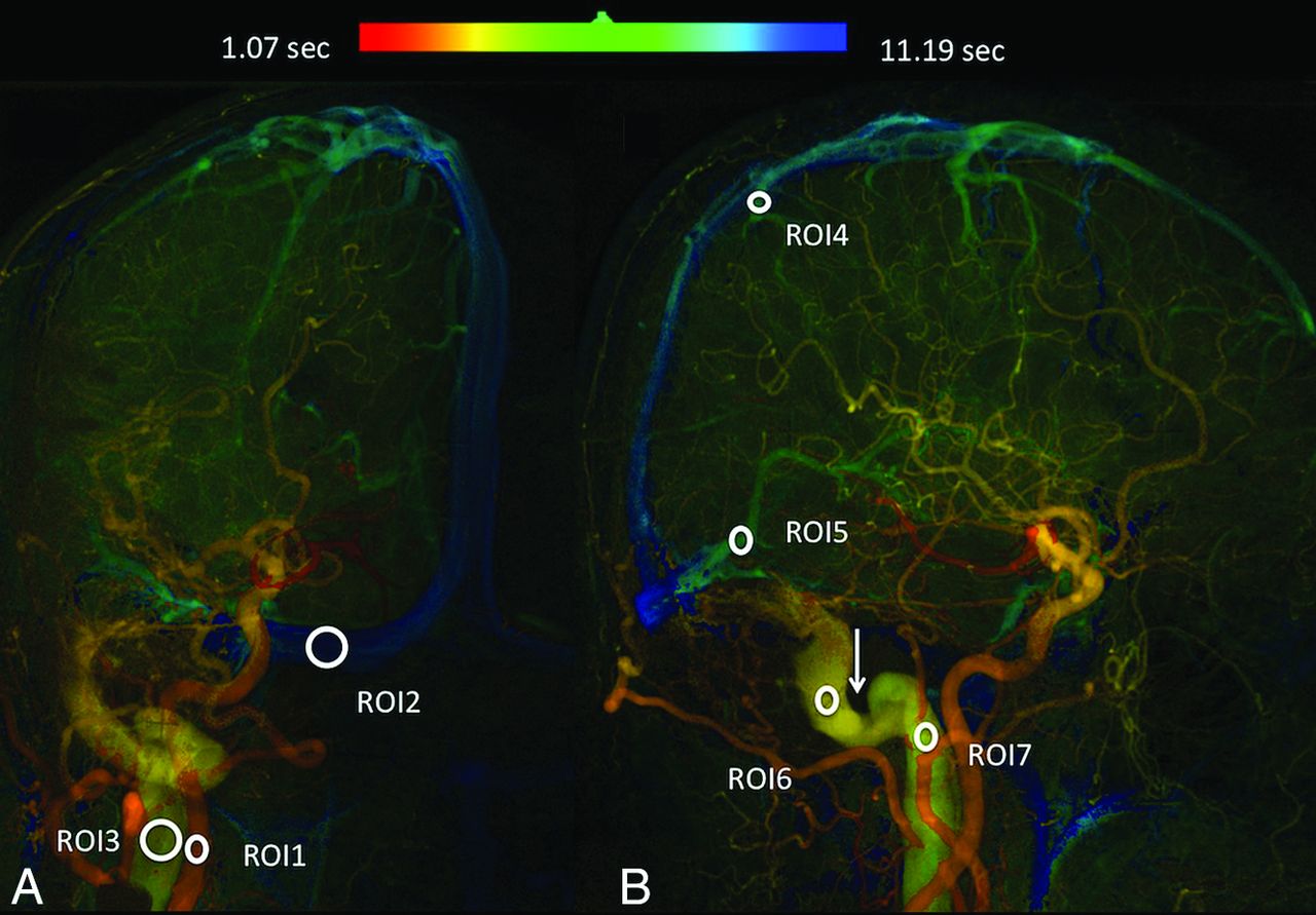
Quantitative color-coded digital subtraction angiography of the anteroposterior (A) and lateral (B) views of a Cognard type I DAVF. ROI1: internal carotid artery; ROI2: ipsilateral normal transverse sinus; ROI3: internal jugular vein; ROI4: parietal vein; ROI5: vein of Labbé; ROI6: prestenotic segment; ROI7: poststenotic segment. The Arrow indicates the stenotic sinus segment.

Definition of Angiographic Signs

We modified the method of Riggeal et al9 and defined “venous stenosis” as a diameter <50% of the diameter of the normal sigmoid sinus at the stenotic segment. The “double peak sign” refers to the occurrence of 2 peaks in the time-density curve of the ipsilateral drainage internal jugular vein of the DAVF (Fig 2). The first peak indicates shunting arterial blood from the arteriovenous fistula, and the second peak indicates the returning venous blood flows from brain parenchyma. Loss of a double peak with a solitary early peak in cases of DAVF suggests a dysfunction of venous drainage stagnation and may cause venous congestion or venous hypertension. Determinations of sinus stenosis and the double peak sign were made by reader A 1 month after ROI measurement.

Fig 2.
  • Download figure
  • Open in new tab
  • Download powerpoint
Fig 2.

A, Quantitative digital subtraction angiography of a healthy subject. B, One single peak appears at 9.87 seconds (venous phase) in the internal jugular vein. C, Quantitative digital subtraction angiography of a Cognard type I DAVF in the left transverse sinus in a 50-year-old woman. D, Time-density curve of the internal jugular vein demonstrates 2 peaks. The first peak comes from arterial flow from the DAVF shunt; the second peak comes from blood flow from normal brain parenchyma. E, Quantitative digital subtraction of a case of Cognard type IIa+b in the left transverse sigmoid sinus in a 73-year-old man. F, Only a single peak can be depicted in a time-density curve of the ipsilateral jugular vein, indicating that it lacks the drainage function of the normal brain.

Statistical Analysis

All statistical analyses were performed by using SPSS 20 (2010; IBM, Armonk, New York). The differences in various clinical symptoms, incidences of venous stenosis, and loss of the double peak sign among different DAVF types were compared by using a χ2 test; differences in age and various time parameters among different DAVF types were compared by using an ANOVA test. Inter- and intraobserver variations were evaluated by intraclass classification. The complete regression rate and favorable outcomes were estimated via the Kaplan-Meier method with a log-rank test. Bonferroni adjustment was applied for post hoc intergroup difference analysis. Significance was set at P < .018 for all statistical tests except intraclass classification (P < .05).

Results

One hundred twenty-six intracranial DAVFs were initially identified from the angiosuite logbook. After excluding 5 patients treated in other hospitals before visiting our hospital, 70 cases of DAVFs in locations other than the transverse-sigmoid sinus, and 8 patients who had undergone endovascular treatment as the sole treatment, there were 43 DAVFs available for analysis (Fig 3). The cohort consisted of 23 men and 18 women (mean, 56.7 years of age); there were 22 Cognard type I, 8 Cognard type IIa, and 13 Cognard types IIa+b or higher DAVFs. Two patients presented with simultaneous cases of bilateral DAVF IIa+b with SS. CVD occurred in all 8 patients who had previous hemorrhage or neurologic deficits other than tinnitus and bruit. No previous hemorrhage or other neurologic deficits occurred in any of the patients with type I and type IIa. Headaches were observed significantly more often in type IIa and types with CVD (Table 1). Previous hemorrhage was significantly more frequent in patients with SS. However, neither headaches nor neurologic deficits differed significantly between patients with and without SS.

Fig 3.
  • Download figure
  • Open in new tab
  • Download powerpoint
Fig 3.

The process of case selection from the angiosuite logbook for our study cohort.

View this table:
  • View inline
  • View popup
Table 1:

Comparison of patient characteristics in 3 different groups: type I, type IIa, and types with CVDa

SS occurred least in Cognard type I (n = 8, 36%), followed by types with CVD (n = 11, 85%) and type IIa (n = 8, 100%). Seven of 8 patients (88%) with type I and SS demonstrated a loss of double peaks in their time-density curves. All patients with type IIa and SS had a loss of double peaks. Nine of 13 patients with CVD had a loss of double peaks (Table 1). The other 2 patients with CVD and SS showing double peaks were classified as having types III and IV, respectively, indicating that their venous outlets were still functioning.

The intraclass classification of reader A in different time parameters ranged from 0.95 to 0.98; the intraclass classification of reader B ranged from 0.92 to 0.97. The interobserver variation ranged from 0.91 to 0.94 (Table 2). TFT was significantly prolonged in cases with CVD. Trans-stenotic time differed significantly across the 3 groups: longest in types with CVD (2.09 seconds), followed by type IIa (0.42 seconds), with type I (1.04 seconds) having the shortest times. TTPVL was significantly reduced in types with CVD (1.4 seconds) compared with type IIa (4.40 seconds) and type I (4.20 seconds). There was no significant difference in TTPPV among the 3 groups. TTP of the ipsilateral transverse sinus was significantly shortened in type IIa and types with CVD (Table 3).

View this table:
  • View inline
  • View popup
Table 2:

Intra- and interobserver variability in different time parametersa

View this table:
  • View inline
  • View popup
Table 3:

Comparison of different time parameters among the 3 groupsa

Six of 13 patients with CVD underwent venoplasty with (n = 4) or without (n = 2) stent placement before SRS. Peri-stent placement medication consisted of aspirin, 300 mg, and clopidogrel, 100 mg daily for 3 days before the procedure and life-long after stent implantation. For those who underwent angioplasty only, the medication was the same as with stent implantation except that the duration of after-procedure medication was shortened to 3 months. Two received post-SRS transarterial embolization due to the presence of new hemorrhage. Only 1 patient received adjunct transarterial embolization before SRS due to existing hemorrhage. Otherwise, transarterial embolization was not performed before SRS in the remaining 12 patients not showing aggressive clinical symptoms and signs.4 One type I patient and 2 patients with CVD did not return for follow-up; these 3 patients were not included in subsequent analyses. The average follow-up time of the SRS was 34 ± 11.5 months. The complete regression rate was 54.5% (12/22) in type I, 38% (3/8) in type IIa, and 23% (3/13) in types with CVD. There was no significant difference in complete regression (P = .176) and favorable outcomes (P = .079) among type I, type IIa, type IIa+b, or higher (Fig 4).

Fig 4.
  • Download figure
  • Open in new tab
  • Download powerpoint
Fig 4.

Kaplan-Meier analysis of complete regression (A) and favorable outcomes (B) among types I, IIa, and IIa+b or higher.

All patients experienced improvement of existing pulsatile tinnitus and headaches. Four of the 6 were downgraded to type IIa after combined venoplasty and/or stent placement before SRS treatment. Only 1 patient experienced asymptomatic hemorrhage after the combined treatment, resulting in an annual hemorrhagic rate of 4.0% after treatment. Follow-up DSA of this patient showed reocclusion of the draining sinus. MR imaging detected 2 cases of restenosis in 4 patients undergoing venoplasty and SRS (Table 4). We kept the 2 patients with restenosis under observation due to their asymptomatic clinical course.

View this table:
  • View inline
  • View popup
Table 4:

Clinical characteristics, treatment strategy, and response in 13 patients with DAVF types with CVD

Discussion

Sinus stenosis is a common associated finding in DAVF in the transverse sigmoid sinus, especially in Cognard types IIa and IIa+b. The venous-return from normal parenchyma is predominantly drained via the contralateral normal transverse-sigmoid sinus in all patients with type I with SS. Those patients failed to demonstrate passage of normal brain parenchymal returning blood flow in their ipsilateral jugular veins, which makes them distinct from patients with type I without SS, in which the normal brain parenchyma was still drained via the ipsilateral “healthy” sinus. This finding appears to favor the hypothesis that the stenotic venous outlet plays a role in the progressive development of CVD, pathophysiologically. The hypothesis is supported by Satomi et al,8 who reported 2 DAVFs that were longitudinally deteriorated by the development of CVD and venous thrombosis.

In general, the faster the intravascular flow, the shorter the TFT will be. The prolonged TFT in types with CVD in the current study was due to stenotic-induced stagnant flow. In type I, the venous outlet received arterialized antegrade flow and showed no time difference in the peristenotic segment. When the flow reversed in Cognard type II, the TST was prolonged. As SS progressed and CVD developed, the TST was further prolonged (Fig 5). The significantly shorter TTPVL in patients with CVD compared with patients with types I and IIa DAVFs without CVD quantitatively reflects the severity of refluxed arterialized venous flow. It could serve as a real-time quantitative regional hemodynamic surrogate marker for treatment strategies used inside the angiosuite. In other words, normalization of TTPVL after successful venoplasty and/or stent placement in SS indicates that CVD was caused by sinus outlet obstruction before treatment and was relieved after the interventional procedures.

Fig 5.
  • Download figure
  • Open in new tab
  • Download powerpoint
Fig 5.

Quantitative DSA of cases of Cognard type I (A), Cognard type IIa (B), and Cognard type IIa+b (C). Severe sinus stenosis is more commonly encountered in more aggressive DAVF types. The TFT (TTP of the internal jugular vein) was largest in type IIa+b (green), followed by types IIa (yellow-green) and I (yellow). Progressive shortening of the TTP in the superior sagittal sinuses in type IIa and type IIa+b is also depicted.

Several previous reports also described symptomatic relief after venoplasty or stent placement to recanalize the stenotic venous outlets in patients with sinus thrombosis and/or tumor compression.23,30⇓–32 The exact etiology of DAVF remains an enigma and might be multifactorial, though most hypotheses hold that it is an acquired disease.33 The first hypothesis suggests that DAVF develops from reopening of the existing potential arteriovenous communication due to an increment of sinus pressure.34 The second hypothesis asserts that de novo shunts (ie, angiogenesis) develop in response to the stimulation of vascular growth factors in the presence of hypoxia, trauma, or otitis.35,36 Venous hypertension induced by an obstruction of the venous outflow may reduce cerebral perfusion and lead to hypoxia with de novo formation of a DAVF. On the basis of these theories, a correction of the venous hypertension in the sinus should reduce cerebral venous edema and reverse the vicious cycle of creating DAVFs.

None of our patients had a cure of DAVFs by venoplasty and/or stent placement alone. We hypothesize that the application of overlapping stents or stents with a finer network may result in completely blocking the arterial-venous shunts that occur on the sinus wall.21,22 Embolization or resection of the sinus is conventionally an option for DAVF types with CVD. The obliteration rate of transarterial embolization in DAVF is 80%.37 If the arterial route to embolize the DAVF also supplies cranial nerves or is technically inaccessible, transvenous embolization might be an option for curing DAVFs.38,39 However, sacrifice of a sinus is reserved for cases of an isolated (nonfunctioning) sinus, because sacrificing a functioning sinus might cause deterioration of the CVD and hemorrhage.36 We preferred venoplasty and/or stent placement to transarterial embolization followed by SRS because embolizing agents such as Onyx would likely make optimal targeting challenging in both MR imaging and DSA and because tissue ischemia may render the vascular bed less sensitive to radiation and stimulate angiogenesis leading to lesion growth.40,41

Nevertheless this multidisciplinary treatment approach will potentially increase the number of unfavorable outcomes in patients before SRS takes effect if a hemorrhage were to occur in the unprotected timeframe. The relative risks of curative embolization attempts alone versus venoplasty and stent placement adjunctive to SRS should be very thoroughly weighed because the latent hemorrhage risk for this combined approach was 4.0% according to our study. There are several options when facing restenosis: If patients are asymptomatic, they can be kept under observation. If the symptoms persist or are aggravated, then re-stent placement in the sinus, a transarterial approach, or microsurgery can be tried.42 In our case, we managed to achieve the benefits of both SRS and endovascular treatment without increasing the periprocedural risks of treating DAVFs with CVD.

There were several limitations to the current study. First, because ours is a tertiary referral medical center for neurologic vascular disorders, the incidences of sinus stenosis might be unusually high due to referring bias. Second, the overall efficacy of combined treatments of venoplasty and/or stent placement followed by SRS warrant a larger scale study with a longer follow-up. Moreover, the aggressiveness of DAVF may also serve as an indicator of response to SRS and warrants individualization of dose selection in SRS. Currently, 2D quantitative DSA merely provides time-based parameters such as time-to-peak to reflect intravascular flow changes. Genuine velocity estimation in DSA relies on 3D acquisitions with an iterative reconstruction algorithm and therefore is not ready for a clinical scenario.43 3D quantitative assessment of angiographic morphology and DAVF hemodynamics might further improve the accuracy, whatever treatment strategy is taken.43

Conclusions

Sinus stenosis is present in nearly one-third of cases of Cognard type I DAVF in the transverse-sigmoid sinus and is more frequently encountered in patients with more aggressive types. Loss of the double peak time-density curve of ipsilateral sinus flow in a DAVF suggests dysfunction of venous drainage and warrants urgent treatment. Venoplasty and/or stent placement of the dysfunctional sinus may downgrade the DAVF and make it amenable to SRS with less risk of hemorrhage in the latent period in most cases.

Acknowledgments

We thank Chung-Hsien Lin for his assistance with the statistical analysis.

Footnotes

  • Disclosures: Wan-Yuo Guo—RELATED: Grant: Siemens, Comments: This work is supported in part by a collaboration contract between Taipei Veterans General Hospital and Siemens.* Chung-Jung Lin—RELATED: Grant: Taipei Veterans General Hospital (No. V104-C-012).* *Money paid to the institution.

  • Dr Chung-Jung Lin is supported by the Taipei Veterans General Hospital (No. V104-C-012), and Dr Wan-Yuo Guo receives a collaboration grant from Siemens and Taipei Veterans General Hospital (No. T1100200).

References

  1. 1.↵
    1. Piippo A,
    2. Niemelä M,
    3. van Popta J, et al
    . Characteristics and long-term outcome of 251 patients with dural arteriovenous fistulas in a defined population. J Neurosurg 2013;118:923–34 doi:10.3171/2012.11.JNS111604 pmid:23259821
    CrossRefPubMed
  2. 2.↵
    1. Kim MS,
    2. Han DH,
    3. Kwon O-K, et al
    . Clinical characteristics of dural arteriovenous fistula. J Clin Neurosci 2002;9:147–55 doi:10.1054/jocn.2001.1029 pmid:11922702
    CrossRefPubMed
  3. 3.↵
    1. Chen CJ,
    2. Lee CC,
    3. Ding D, et al
    . Stereotactic radiosurgery for intracranial dural arteriovenous fistulas: a systematic review. J Neurosurg 2015;122:353–62 doi:10.3171/2014.10.JNS14871 pmid:25479123
    CrossRefPubMed
  4. 4.↵
    1. Söderman M,
    2. Pavic L,
    3. Edner G, et al
    . Natural history of dural arteriovenous shunts. Stroke 2008;39:1735–39 doi:10.1161/STROKEAHA.107.506485 pmid:18388337
    Abstract/FREE Full Text
  5. 5.↵
    1. Davies MA,
    2. Ter Brugge K,
    3. Willinsky R, et al
    . The natural history and management of intracranial dural arteriovenous fistulae: part 2, aggressive lesions. Interv Neuroradiol 1997;3:303–11 pmid:20678361
    Abstract/FREE Full Text
  6. 6.↵
    1. van Rooij WJ,
    2. Sluzewski M,
    3. Beute GN
    . Dural arteriovenous fistulas with cortical venous drainage: incidence, clinical presentation, and treatment. AJNR Am J Neuroradiol 2007;28:651–55 pmid:17416815
    Abstract/FREE Full Text
  7. 7.↵
    1. van Dijk JM,
    2. terBrugge KG,
    3. Willinsky RA, et al
    . Clinical course of cranial dural arteriovenous fistulas with long-term persistent cortical venous reflux. Stroke 2002;33:1233–36 doi:10.1161/01.STR.0000014772.02908.44 pmid:11988596
    Abstract/FREE Full Text
  8. 8.↵
    1. Satomi J,
    2. van Dijk JM,
    3. Terbrugge KG, et al
    . Benign cranial dural arteriovenous fistulas: outcome of conservative management based on the natural history of the lesion. J Neurosurg 2002;97:767–70 doi:10.3171/jns.2002.97.4.0767 pmid:12405361
    CrossRefPubMed
  9. 9.↵
    1. Riggeal BD,
    2. Bruce BB,
    3. Saindane AM, et al
    . Clinical course of idiopathic intracranial hypertension with transverse sinus stenosis. Neurology 2013;80:289–95 doi:10.1212/WNL.0b013e31827debd6 pmid:23269597
    Abstract/FREE Full Text
  10. 10.↵
    1. Degnan AJ,
    2. Levy LM
    . Pseudotumor cerebri: brief review of clinical syndrome and imaging findings. AJNR Am J Neuroradiol 2011;32:1986–93 doi:10.3174/ajnr.A2404 pmid:21680652
    Abstract/FREE Full Text
  11. 11.↵
    1. Vanlandingham M,
    2. Fox B,
    3. Hoit D, et al
    . Endovascular treatment of intracranial dural arteriovenous fistulas. Neurosurgery 2014;74(suppl 1):S42–49 doi:10.1227/NEU.0000000000000180 pmid:24402491
    CrossRefPubMed
  12. 12.↵
    1. Söderman M,
    2. Dodoo E,
    3. Karlsson B
    . Dural arteriovenous fistulas and the role of gamma knife stereotactic radiosurgery: the Stockholm experience. Prog Neurol Surg 2013;27:205–17 doi:10.1159/000341798 pmid:23258524
    CrossRefPubMed
  13. 13.↵
    1. Yang H,
    2. Kano H,
    3. Kondziolka D, et al
    . Stereotactic radiosurgery with or without embolization for intracranial dural arteriovenous fistulas. Prog Neurol Surg 2013;27:195–204 doi:10.1159/000341796 pmid:23258523
    CrossRefPubMed
  14. 14.↵
    1. van Rooij WJ,
    2. Sluzewski M
    . Curative embolization with Onyx of dural arteriovenous fistulas with cortical venous drainage. AJNR Am J Neuroradiol 2010;31:1516–20 doi:10.3174/ajnr.A2101 pmid:20395390
    Abstract/FREE Full Text
  15. 15.↵
    1. Stiefel MF,
    2. Albuquerque FC,
    3. Park MS, et al
    . Endovascular treatment of intracranial dural arteriovenous fistulae using Onyx: a case series. Neurosurgery 2009;65:132–39; discussion 139–40 pmid:19934987
    CrossRefPubMed
  16. 16.↵
    1. Macdonald JH,
    2. Millar JS,
    3. Barker CS
    . Endovascular treatment of cranial dural arteriovenous fistulae: a single-centre, 14-year experience and the impact of Onyx on local practise. Neuroradiology 2010;52:387–95 doi:10.1007/s00234-009-0620-x pmid:19894041
    CrossRefPubMed
  17. 17.↵
    1. Yoshida K,
    2. Melake M,
    3. Oishi H, et al
    . Transvenous embolization of dural carotid cavernous fistulas: a series of 44 consecutive patients. AJNR Am J Neuroradiol 2010;31:651–55 doi:10.3174/ajnr.A1882 pmid:19959773
    Abstract/FREE Full Text
  18. 18.↵
    1. Zenteno M,
    2. Santos-Franco J,
    3. Rodríguez-Parra V, et al
    . Management of direct carotid-cavernous sinus fistulas with the use of ethylene-vinyl alcohol (Onyx) only: preliminary results. J Neurosurg 2010;112:595–602 doi:10.3171/2009.6.JNS09440 pmid:19663551
    CrossRefPubMed
  19. 19.↵
    1. Plasencia AR,
    2. Santillan A
    . Embolization and radiosurgery for arteriovenous malformations. Surg Neurol Int 2012;3(suppl 2):S90–S104 doi:10.4103/2152-7806.95420 pmid:22826821
    CrossRefPubMed
  20. 20.↵
    1. Pan DH,
    2. Chung WY,
    3. Guo WY, et al
    . Stereotactic radiosurgery for the treatment of dural arteriovenous fistulas involving the transverse-sigmoid sinus. J Neurosurg 2002;96:823–29 doi:10.3171/jns.2002.96.5.0823 pmid:12005389
    CrossRefPubMed
  21. 21.↵
    1. Liebig T,
    2. Henkes H,
    3. Brew S, et al
    . Reconstructive treatment of dural arteriovenous fistulas of the transverse and sigmoid sinus: transvenous angioplasty and stent deployment. Neuroradiology 2005;47:543–51 doi:10.1007/s00234-005-1377-5 pmid:15906020
    CrossRefPubMed
  22. 22.↵
    1. Levrier O,
    2. Métellus P,
    3. Fuentes S, et al
    . Use of a self-expanding stent with balloon angioplasty in the treatment of dural arteriovenous fistulas involving the transverse and/or sigmoid sinus: functional and neuroimaging-based outcome in 10 patients. J Neurosurg 2006;104:254–63 doi:10.3171/jns.2006.104.2.254 pmid:16509499
    CrossRefPubMed
  23. 23.↵
    1. Xu K,
    2. Yu T,
    3. Yuan Y, et al
    . Current status of the application of intracranial venous sinus stenting. Int J Med Sci 2015;12:780–89 doi:10.7150/ijms.12604 pmid:26516306
    CrossRefPubMed
  24. 24.↵
    1. Lin CJ,
    2. Hung SC,
    3. Guo WY, et al
    . Monitoring peri-therapeutic cerebral circulation time: a feasibility study using color-coded quantitative DSA in patients with steno-occlusive arterial disease. AJNR Am J Neuroradiol 2012;33:1685–90 doi:10.3174/ajnr.A3049 pmid:22499839
    Abstract/FREE Full Text
  25. 25.↵
    1. Lin CJ,
    2. Chang FC,
    3. Tsai FY, et al
    . Stenotic transverse sinus predisposes to poststenting hyperperfusion syndrome as evidenced by quantitative analysis of peritherapeutic cerebral circulation time. AJNR Am J Neuroradiol 2014;35:1132–36 doi:10.3174/ajnr.A3838 pmid:24436346
    Abstract/FREE Full Text
  26. 26.↵
    1. Levitt MR,
    2. Morton RP,
    3. Haynor DR, et al
    . Angiographic perfusion imaging: real-time assessment of endovascular treatment for cerebral vasospasm. J Neuroimaging 2014;24:387–92 doi:10.1111/jon.12046 pmid:24015728
    CrossRefPubMed
  27. 27.↵
    1. Gölitz P,
    2. Struffert T,
    3. Lücking H, et al
    . Parametric color coding of digital subtraction angiography in the evaluation of carotid cavernous fistulas. Clin Neuroradiol 2013;23:113–20 doi:10.1007/s00062-012-0184-8 pmid:23241650
    CrossRefPubMed
  28. 28.↵
    1. Strother CM,
    2. Bender F,
    3. Deuerling-Zheng Y, et al
    . Parametric color coding of digital subtraction angiography. AJNR Am J Neuroradiol 2010;31:919–24 doi:10.3174/ajnr.A2020 pmid:20167651
    Abstract/FREE Full Text
  29. 29.↵
    1. Greitz T
    . A radiologic study of the brain circulation by rapid serial angiography of the carotid artery. Acta Radiol Suppl 1956;140:1–123 pmid:13372389
    PubMed
  30. 30.↵
    1. Tsumoto T,
    2. Miyamoto T,
    3. Shimizu M, et al
    . Restenosis of the sigmoid sinus after stenting for treatment of intracranial venous hypertension: case report. Neuroradiology 2003;45:911–15 doi:10.1007/s00234-003-1112-z pmid:14605788
    CrossRefPubMed
  31. 31.↵
    1. Ganesan D,
    2. Higgins JN,
    3. Harrower T, et al
    . Stent placement for management of a small parasagittal meningioma: technical note. J Neurosurg 2008;108:377–81 doi:10.3171/JNS/2008/108/2/0377 pmid:18240939
    CrossRefPubMed
  32. 32.↵
    1. Hirata E,
    2. Higashi T,
    3. Iwamuro Y, et al
    . Angioplasty and stent deployment in acute sinus thrombosis following endovascular treatment of dural arteriovenous fistulae. J Clin Neurosci 2009;16:725–27 doi:10.1016/j.jocn.2008.07.071 pmid:19264487
    CrossRefPubMed
  33. 33.↵
    1. Gupta A,
    2. Periakaruppan A
    . Intracranial dural arteriovenous fistulas: a review. Indian J Radiol Imaging 2009;19:43–48 doi:10.4103/0971-3026.45344 pmid:19774139
    CrossRefPubMed
  34. 34.↵
    1. Houser OW,
    2. Campbell JK,
    3. Campbell RJ, et al
    . Arteriovenous malformation affecting the transverse dural venous sinus: an acquired lesion. Mayo Clin Proc 1979;54:651–61 pmid:480990
    PubMed
  35. 35.↵
    1. Tirakotai W,
    2. Bertalanffy H,
    3. Liu-Guan B, et al
    . Immunohistochemical study in dural arteriovenous fistulas and possible role of local hypoxia for the de novo formation of dural arteriovenous fistulas. Clin Neurol Neurosurg 2005;107:455–60 doi:10.1016/j.clineuro.2004.11.014 pmid:16202817
    CrossRefPubMed
  36. 36.↵
    1. Lasjaunias P,
    2. Brenstein A,
    3. ter Brugge KG
    . Surgical Neuroangiography. Berlin: Springer; 2004:565–607
  37. 37.↵
    1. Cognard C,
    2. Januel AC,
    3. Silva NA Jr., et al
    . Endovascular treatment of intracranial dural arteriovenous fistulas with cortical venous drainage: new management using Onyx. AJNR Am J Neuroradiol 2008;29:235–41 doi:10.3174/ajnr.A0817 pmid:17989374
    Abstract/FREE Full Text
  38. 38.↵
    1. Natarajan SK,
    2. Ghodke B,
    3. Kim LJ, et al
    . Multimodality treatment of intracranial dural arteriovenous fistulas in the Onyx era: a single center experience. World Neurosurg 2010;73:365–79 doi:10.1016/j.wneu.2010.01.009 pmid:20849795
    CrossRefPubMed
  39. 39.↵
    1. Lekkhong E,
    2. Pongpech S,
    3. Ter Brugge K, et al
    . Transvenous embolization of intracranial dural arteriovenous shunts through occluded venous segments: experience in 51 patients. AJNR Am J Neuroradiol 2011;32:1738–44 doi:10.3174/ajnr.A2566 pmid:21799036
    Abstract/FREE Full Text
  40. 40.↵
    1. Akakin A,
    2. Ozkan A,
    3. Akgun E, et al
    . Endovascular treatment increases but gamma knife radiosurgery decreases angiogenic activity of arteriovenous malformations: an in vivo experimental study using a rat cornea model. Neurosurgery 2010;66:121–29; discussion 129–30 doi:10.1227/01.NEU.0000363154.88768.34 pmid:20023542
    CrossRefPubMed
  41. 41.↵
    1. Mullan S,
    2. Mojtahedi S,
    3. Johnson DL, et al
    . Embryological basis of some aspects of cerebral vascular fistulas and malformations. J Neurosurg 1996;85:1–8 doi:10.3171/jns.1996.85.1.0001 pmid:8683257
    CrossRefPubMed
  42. 42.↵
    1. Choi BJ,
    2. Lee TH,
    3. Kim CW, et al
    . Reconstructive treatment using a stent graft for a dural arteriovenous fistula of the transverse sinus in the case of hypoplasia of the contralateral venous sinuses: technical case report. Neurosurgery 2009;65:E994–96; discussion E996 doi:10.1227/01.NEU.0000351772.45417.92 pmid:19834391
    CrossRefPubMed
  43. 43.↵
    1. Chen GH,
    2. Li Y
    . Synchronized multiartifact reduction with tomographic reconstruction (SMART-RECON): a statistical model based iterative image reconstruction method to eliminate limited-view artifacts and to mitigate the temporal-average artifacts in time-resolved CT. Med Phys 2015;42:4698–707 doi:10.1118/1.4926430 pmid:26233197
    CrossRefPubMed
  • Received April 1, 2016.
  • Accepted after revision August 18, 2016.
  • © 2017 by American Journal of Neuroradiology
View Abstract
PreviousNext
Back to top

In this issue

American Journal of Neuroradiology: 38 (1)
American Journal of Neuroradiology
Vol. 38, Issue 1
1 Jan 2017
  • Table of Contents
  • Index by author
  • Complete Issue (PDF)
Advertisement
Print
Download PDF
Email Article

Thank you for your interest in spreading the word on American Journal of Neuroradiology.

NOTE: We only request your email address so that the person you are recommending the page to knows that you wanted them to see it, and that it is not junk mail. We do not capture any email address.

Enter multiple addresses on separate lines or separate them with commas.
Quantifying the Cerebral Hemodynamics of Dural Arteriovenous Fistula in Transverse Sigmoid Sinus Complicated by Sinus Stenosis: A Retrospective Cohort Study
(Your Name) has sent you a message from American Journal of Neuroradiology
(Your Name) thought you would like to see the American Journal of Neuroradiology web site.
CAPTCHA
This question is for testing whether or not you are a human visitor and to prevent automated spam submissions.
Cite this article
W.-Y. Guo, C.-C.J. Lee, C.-J. Lin, H.-C. Yang, H.-M. Wu, C.-C. Wu, W.-Y. Chung, K.-D. Liu
Quantifying the Cerebral Hemodynamics of Dural Arteriovenous Fistula in Transverse Sigmoid Sinus Complicated by Sinus Stenosis: A Retrospective Cohort Study
American Journal of Neuroradiology Jan 2017, 38 (1) 132-138; DOI: 10.3174/ajnr.A4960

Citation Manager Formats

  • BibTeX
  • Bookends
  • EasyBib
  • EndNote (tagged)
  • EndNote 8 (xml)
  • Medlars
  • Mendeley
  • Papers
  • RefWorks Tagged
  • Ref Manager
  • RIS
  • Zotero
0 Responses
Respond to this article
Share
Bookmark this article
Quantifying the Cerebral Hemodynamics of Dural Arteriovenous Fistula in Transverse Sigmoid Sinus Complicated by Sinus Stenosis: A Retrospective Cohort Study
W.-Y. Guo, C.-C.J. Lee, C.-J. Lin, H.-C. Yang, H.-M. Wu, C.-C. Wu, W.-Y. Chung, K.-D. Liu
American Journal of Neuroradiology Jan 2017, 38 (1) 132-138; DOI: 10.3174/ajnr.A4960
del.icio.us logo Twitter logo Facebook logo Mendeley logo
  • Tweet Widget
  • Facebook Like
  • Google Plus One
Purchase

Jump to section

  • Article
    • Abstract
    • ABBREVIATIONS:
    • Materials and Methods
    • Results
    • Discussion
    • Conclusions
    • Acknowledgments
    • Footnotes
    • References
  • Figures & Data
  • Info & Metrics
  • Responses
  • References
  • PDF

Related Articles

  • No related articles found.
  • PubMed
  • Google Scholar

Cited By...

  • No citing articles found.
  • Crossref (22)
  • Google Scholar

This article has been cited by the following articles in journals that are participating in Crossref Cited-by Linking.

  • Stasis index from hemodynamic analysis using quantitative DSA correlates with hemorrhage of supratentorial arteriovenous malformation: a cross-sectional study
    Te Ming Lin, Huai Che Yang, Cheng Chia Lee, Hsiu Mei Wu, Yong Sin Hu, Chao Bao Luo, Wan Yuo Guo, Yi Hsuan Kao, Wen Yuh Chung, Chung Jung Lin
    Journal of Neurosurgery 2020 132 5
  • Stagnant Venous Outflow Predicts Brain Arteriovenous Malformation Obliteration After Gamma Knife Radiosurgery Without Prior Intervention
    Yong-Sin Hu, Cheng-Chia Lee, Hsiu-Mei Wu, Huai-Che Yang, Te-Ming Lin, Chao-Bao Luo, Wan-Yuo Guo, Wen-Yuh Chung, Chung-Jung Lin
    Neurosurgery 2020 87 2
  • Hybrid Operating Room Settings for Treatment of Complex Dural Arteriovenous Fistulas
    Basil E. Grüter, Fabio Strange, Felice Burn, Luca Remonda, Michael Diepers, Javier Fandino, Serge Marbacher
    World Neurosurgery 2018 120
  • Automatic flow analysis of digital subtraction angiography using independent component analysis in patients with carotid stenosis
    Han-Jui Lee, Jia-Sheng Hong, Chung-Jung Lin, Yi-Hsuan Kao, Feng-Chi Chang, Chao-Bao Luo, Wei-Fa Chu, Stephan Meckel
    PLOS ONE 2017 12 9
  • Machine Learning Application With Quantitative Digital Subtraction Angiography for Detection of Hemorrhagic Brain Arteriovenous Malformations
    Jia-Sheng Hong, Chung-Jung Lin, Yue-Hsin Lin, Cheng-Chia Lee, Huai-Che Yang, Ling-Hsuan Meng, Te-Ming Lin, Yong-Sin Hu, Wan-Yuo Guo, Wei-Fa Chu, Yu-Te Wu
    IEEE Access 2020 8
  • Clinical outcomes of adjunct sinus stenting in dural arteriovenous fistulas
    Yen-Heng Lin, Chung-Wei Lee, Hon-Man Liu
    Clinical Neuroradiology 2022 32 2
  • Stagnant venous outflow in ruptured arteriovenous malformations revealed by delayed quantitative digital subtraction angiography
    Yong-Sin Hu, Te-Ming Lin, Hsiu-Mei Wu, Cheng-Chia Lee, Huai-Che Yang, Chao-Bao Luo, Wan-Yuo Guo, Wen-Yuh Chung, Chung-Jung Lin
    European Journal of Radiology 2021 134
  • Gradual dilatation of an occluded transverse sinus associated with dural arteriovenous fistula after balloon angioplasty with sinus packing: A case report
    Takuya Osuki, Hiroyuki Ikeda, Tomoko Hayashi, Silsu Park, Minami Uezato, Masanori Kinosada, Yoshitaka Kurosaki, Akira Handa, Masaki Chin
    The Neuroradiology Journal 2022 35 3
  • Peri-therapeutic multi-modal hemodynamic assessment and detection of predictors for symptomatic in-stent restenosis after percutaneous transluminal angioplasty and stenting
    Xiaowen Song, Hancheng Qiu, Shuo Yang, Yuqi Liu, Yong Cao, Shuo Wang, Jizong Zhao
    Frontiers in Neurology 2023 14
  • Complex torcular dural arteriovenous fistula leading to cortical venous reflux-induced severe varix and subsequent bilateral cerebral hemispheric hemorrhage: a case report
    Ziqi Li, Pengwei Hou, Qizuan Chen, Shuwen Mu, Jun Li, Yi Fang, Wenting Jiang, Xinhua Tian, Shousen Wang
    Frontiers in Neurology 2023 14

More in this TOC Section

  • SAVE vs. Solumbra Techniques for Thrombectomy
  • Contrast-Induced Encephalopathy after NeuroIR
  • CT Perfusion&Reperfusion in Acute Ischemic Stroke
Show more Interventional

Similar Articles

Advertisement

Indexed Content

  • Current Issue
  • Accepted Manuscripts
  • Article Preview
  • Past Issues
  • Editorials
  • Editor's Choice
  • Fellows' Journal Club
  • Letters to the Editor
  • Video Articles

Cases

  • Case Collection
  • Archive - Case of the Week
  • Archive - Case of the Month
  • Archive - Classic Case

Special Collections

  • AJNR Awards
  • ASNR Foundation Special Collection
  • Most Impactful AJNR Articles
  • Photon-Counting CT
  • Spinal CSF Leak Articles (Jan 2020-June 2024)

More from AJNR

  • Trainee Corner
  • Imaging Protocols
  • MRI Safety Corner

Multimedia

  • AJNR Podcasts
  • AJNR Scantastics

Resources

  • Turnaround Time
  • Submit a Manuscript
  • Submit a Video Article
  • Submit an eLetter to the Editor/Response
  • Manuscript Submission Guidelines
  • Statistical Tips
  • Fast Publishing of Accepted Manuscripts
  • Graphical Abstract Preparation
  • Imaging Protocol Submission
  • Evidence-Based Medicine Level Guide
  • Publishing Checklists
  • Author Policies
  • Become a Reviewer/Academy of Reviewers
  • News and Updates

About Us

  • About AJNR
  • Editorial Board
  • Editorial Board Alumni
  • Alerts
  • Permissions
  • Not an AJNR Subscriber? Join Now
  • Advertise with Us
  • Librarian Resources
  • Feedback
  • Terms and Conditions
  • AJNR Editorial Board Alumni

American Society of Neuroradiology

  • Not an ASNR Member? Join Now

© 2025 by the American Society of Neuroradiology All rights, including for text and data mining, AI training, and similar technologies, are reserved.
Print ISSN: 0195-6108 Online ISSN: 1936-959X

Powered by HighWire