
Article Figures & Data
Figures
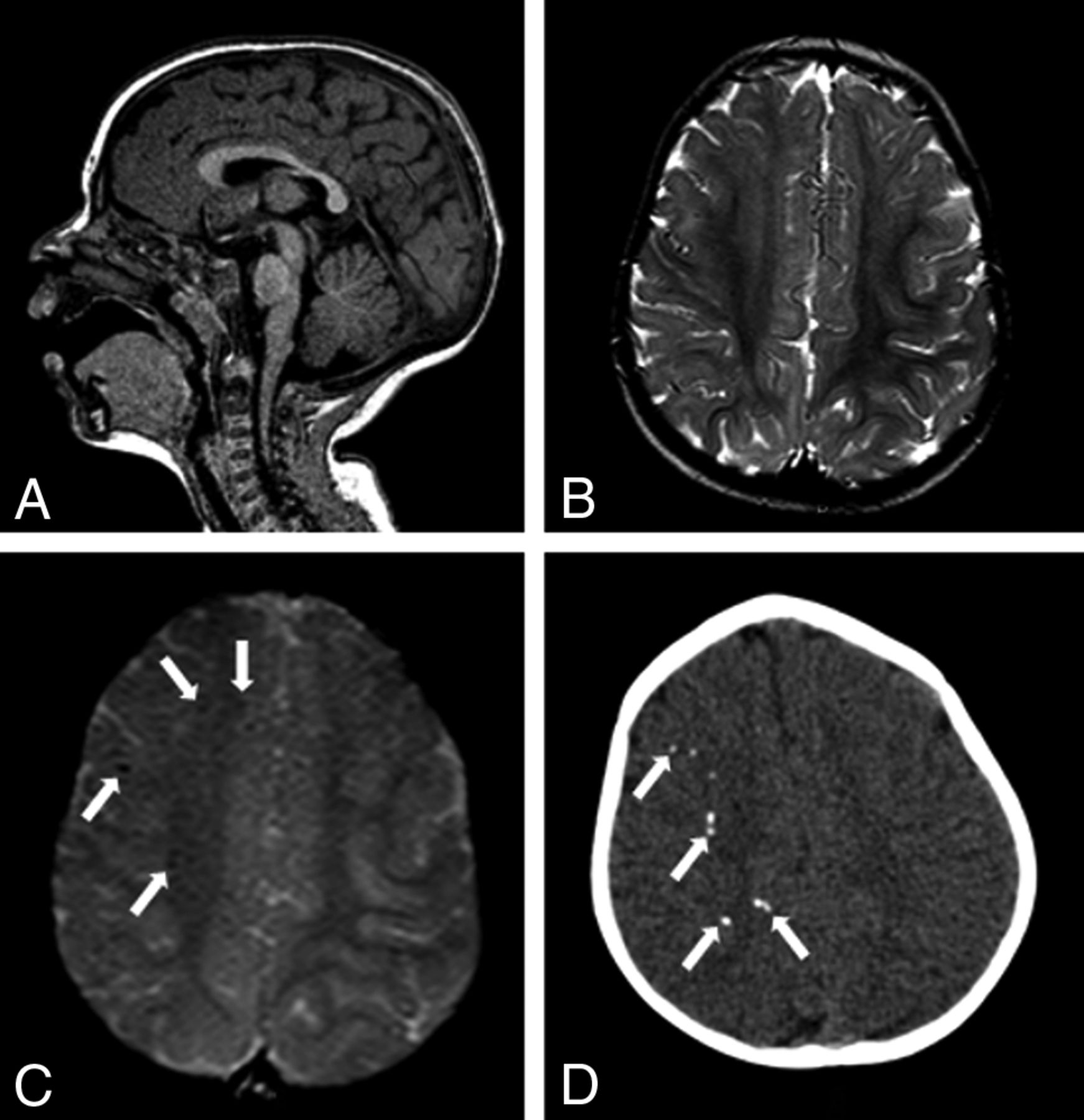
- Fig 1.
MR imaging and CT of an 8-month-old girl without microcephaly with probable congenital Zika syndrome. Sagittal T1-weighted image shows a normal corpus callosum and cisterna magna (A) and a small hyperintense focus of dystrophic calcification in the junction between the cortical and subcortical white matter (long white arrow) and left frontal polymicrogyria (short white arrow) (B). Axial T2-weighted image (C) shows right polymicrogyria (white arrows) and mildly decreased right hemisphere volume. CT scans show asymmetric hemispheres, with an enlarged right lateral ventricle (white arrow) (D) and small punctate foci, representing calcifications, at the cortico-subcortical white matter junction bilaterally in the frontal lobes (white arrows) (E and F).
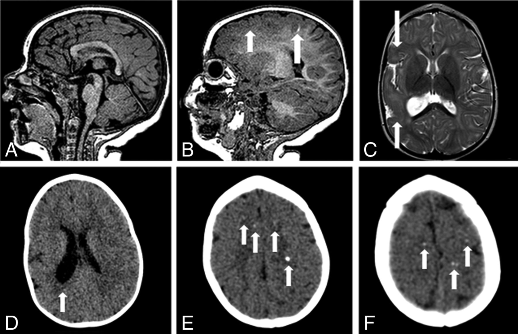
- Fig 2.
MR imaging and CT of an 11-month-old girl without microcephaly with probable congenital Zika syndrome. Sagittal T1-weighted image shows a normal corpus callosum and cisterna magna (A) and left polymicrogyria (white arrows) (B). Axial T2-weighted image (C) shows left polymicrogyria (white arrow) and mild left ventriculomegaly. A gradient-echo image (D) shows very few small and subtle punctate foci, representing calcifications, at the cortico-subcortical white matter junction (white arrow). Axial CT scans (E and F) show right frontal and left parietal punctate foci (white arrows).
- Fig 3.
MR imaging and CT of an 11-month-old girl without microcephaly with probable congenital Zika syndrome. Sagittal T1-weighted image shows a normal corpus callosum and cisterna magna (A). Axial T2-weighted image (B) shows a normal cortex. A gradient-echo image (C) shows small and subtle punctate foci, representing calcifications, at the cortico-subcortical white matter junction (white arrows). Axial CT scan (D) shows punctate foci in the right hemisphere (white arrows).
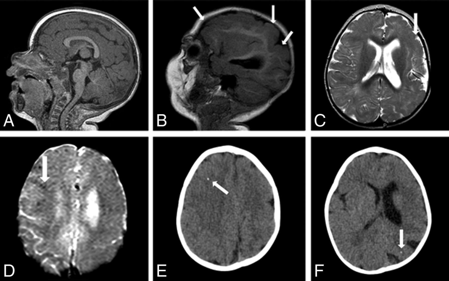
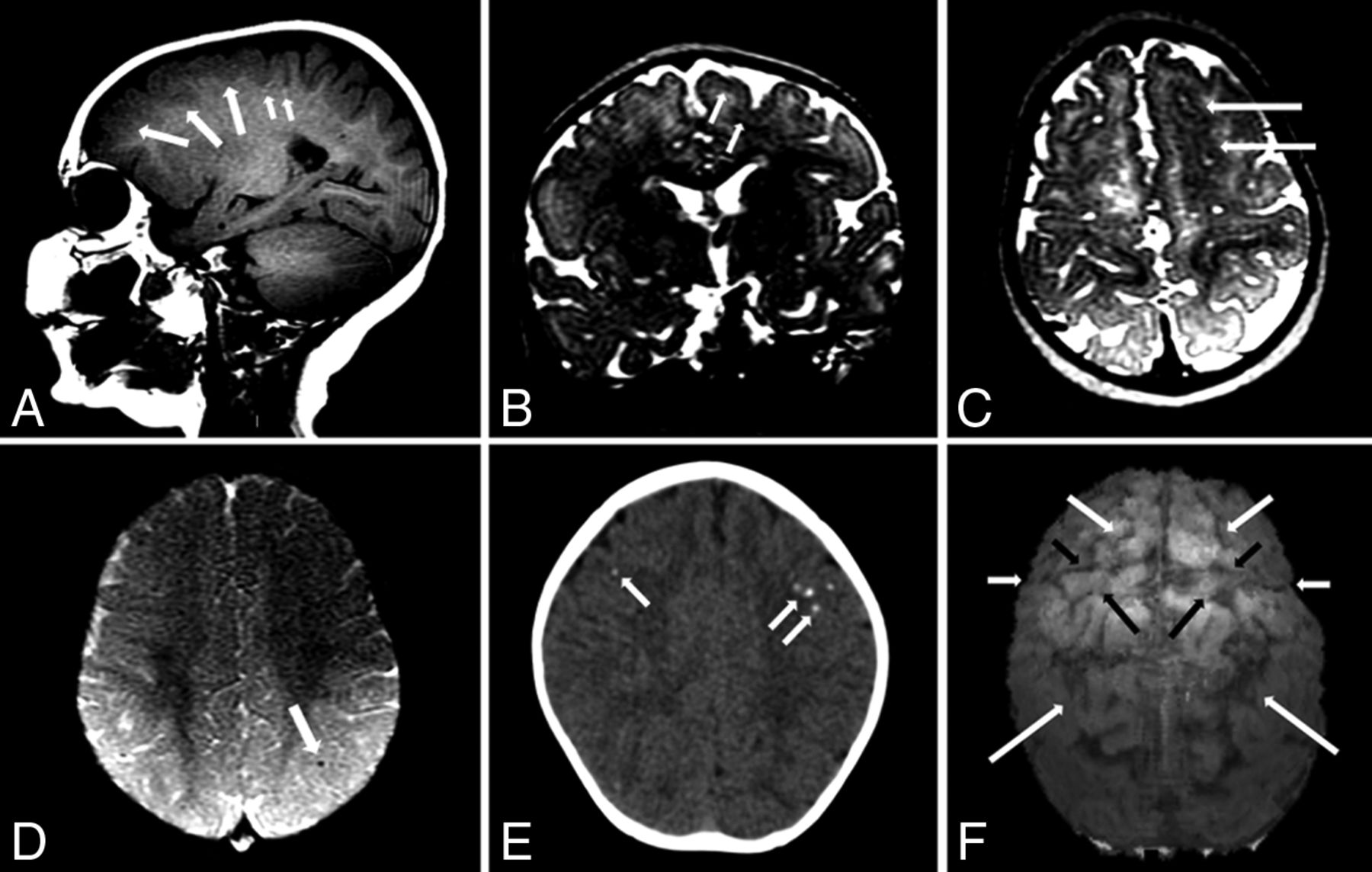
- Fig 4.
MR imaging and CT of a 10-month-old girl with microcephaly developed postnatally with possible congenital Zika syndrome. Sagittal T1-weighted image shows frontal polymicrogyria (medium white arrows) and very subtle hyperintense punctate foci, representing calcifications at the cortico-subcortical white matter junction (small white arrows). Coronal and axial T2-weighted images (B and C, respectively) show the thick and irregular cortex at the superior frontal sulcus (white arrows). Axial SWI (D) shows a small punctate focus, representing calcification at the cortico-subcortical white matter junction (white arrow). Axial CT scan (E) shows punctate foci in both frontal lobes. An echo-spoiled gradient-echo volumetric (3D reconstruction) image (F) shows malformation and prominence of brain high frontoparietal convexity gyri and sulci, predominantly at the left hemisphere: The superior frontal sulcus (medium white arrows) is well-identified bilaterally, as well as precentral (short black arrows), central (short white arrows), and intraparietal (long white arrows) sulci. The left hemisphere is more reduced than the right hemisphere; the left precentral gyrus (large black arrows) seems to be the most reduced in volume. These findings are located probably where the polymicrogyria is most severe, according to the T2-weighted images (B and C). These findings are better seen in a 3D reconstruction (F) than in sectional images (A–C), despite the presence of movement artifacts in the former.
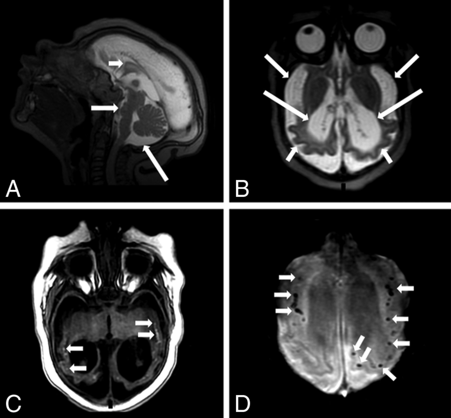
- Fig 5.
MR imaging of a 4-month-old boy with microcephaly with confirmed congenital Zika syndrome. Sagittal T2-weighted image (A) shows a hypogenetic corpus callosum (short white arrow), pons hypoplasia (medium white arrow), and an enlarged cisterna magna (long white arrow). An axial T2-weighted image (B) shows a diffuse simplified gyral pattern (note the thin cortex) (short white arrows), an enlarged extra-axial CSF space (medium white arrows), and severe ventriculomegaly (long white arrows). An axial T1-weighted image (C) shows hyperintense punctate foci, representing calcifications, at the cortico-subcortical white matter junction (small white arrows). Axial SWI (D) shows several punctate foci at the cortico-subcortical white matter junction (short white arrows).


Tables
Comparison of brain MRI findings between microcephalic and normocephalic infants with congenital Zika syndrome
MRI Findings Microcephaly Without Microcephaly (n = 3) P1 P2 P3 P4 At Birth (n = 9) Postnatally (n = 7) Reduced brain volume 8 (88.9%) 5 (71.4%) 0 (0.0%) .018a .550 .141 .021a Brain stem hypoplasia 6 (66.7%) 1 (14.3%) 0 (0.0%) .182 .060 .020a .263 Cerebellum hypoplasia 3 (33.3%) 0 (0.0%) 0 (0.0%) .509 .213 .087 1.000 Malformations of cortical developmentb Simplified gyral pattern 6 (66.7%) 0 (0.0%) 0 (0.0%) .182 .028a .009a .515 Pachygyria 6 (66.7%) 4 (66.7%) 0 (0.0%) .182 1.000 .637 .069 Polymicrogyria 0 (0.0%) 4 (66.7%) 2 (66.7%) .045a .011a .009a .245 Normal cortex 1 (11.1%) 0 (0.0%) 1 (33.3%) .455 1.000 1.000 .314 Brain calcifications Cortico-subcortical white matter junction 8 (88.9%) 7 (100.0%) 3 (100.0%) 1.000 1.000 .474 1.000 Calcifications at other sites 7 (77.8%) 0 (0.0%) 0 (0.0%) .046a .003a .001a .263 Basal ganglia 4 (44.4%) 0 (0.0%) 0 (0.0%) .491 .103 .082 1.000 Thalamus 3 (33.3%) 0 (0.0%) 0 (0.0%) .509 .229 .206 1.000 Periventricular 3 (33.3%) 0 (0.0%) 0 (0.0%) .509 .229 .206 1.000 Brain stem 4 (44.4%) 0 (0.0%) 0 (0.0%) .491 .103 .082 1.000 Cerebellum 2 (22.2%) 0 (0.0%) 0 (0.0%) 1.000 .475 .471 1.000 Corpus callosum abnormalities 9 (100.0%) 2 (28.6%) 0 (0.0%) .005a .005a .001a .058 Moderate-to-severe ventriculomegaly 8 (88.9%) 2 (28.6%) 0 (0.0%) .018a .035a .005a .087 Enlarged extra-axial CSF space 9 (100.0%) 3 (42.9%) 0 (0.0%) .005a .019a .003a .036a Enlarged cisterna magna 9 (100.0%) 4 (57.1%) 0 (0.0%) .005a .063 .008a .021a Delayed myelinationb 7 (77.8%) 2 (33.3%) 2 (66.7%) 1.000 .136 .335 1.000 Symmetry of abnormalities 8 (88.9%) 3 (42.9%) 1 (33.3%) .127 .106 .057 .523 Note:—P indicates Fisher exact test; P1, microcephaly at birth vs without microcephaly; P2, microcephaly at birth vs microcephaly postnatally; P3, microcephaly at birth vs without microcephaly at birth (microcephaly postnatally + without microcephaly); P4, microcephaly (microcephaly at birth + microcephaly postnatally) vs without microcephaly. Microcephaly postnatally vs without microcephaly yielded no statistically significant results.
↵a Statistically significant results.
↵b Not assessed in Infant 14 (without MRI). For myelination assessment, infant 15 was considered born at term.


Jump to section
Related Articles
Cited By...
- Saxitoxin potentiates Zika virus-induced cell death in human neurons but not in neural progenitors and astrocytes
- A Zika virus protein expression screen in Drosophila to investigate targeted host pathways during development
- Neurodevelopment in normocephalic children with and without prenatal Zika virus exposure
- Zika Virus Congenital Microcephaly Severity Classification and the Association of Severity with Neuropsychomotor Development
- Neuroanatomical abnormalities in a nonhuman primate model of congenital Zika virus infection
- Zika Virus Infection: Review Of Neuroimage Studies And The Relationship Between Findings And Time Of Infection
- Life-long persistence of infectious Zika virus: inflammation and behavioral sequela
- Long-term alterations in brain and behavior after postnatal Zika virus infections in infant macaques
- Mouse Strain and Sex-Dependent Differences in Long-term Behavioral Abnormalities and Neuropathologies after Developmental Zika Infection
- CImP: Cellular Imprinting Proteomics applied to ocular disorders elicited by Congenital Zika virus Syndrome
- Zika Virus Infection at Different Pregnancy Stages: Anatomopathological Findings, Target Cells and Viral Persistence in Placental Tissues
- Postnatal Zika virus infection is associated with persistent abnormalities in brain structure, function, and behavior in infant macaques
- Zika Virus Iceberg: Very Large