
Article Figures & Data
Figures
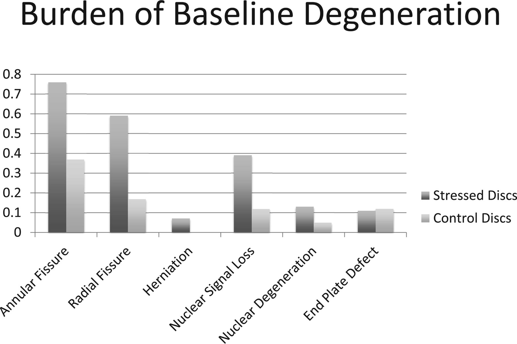
- Fig 1.
A bar diagram demonstrating the baseline burden of degenerative parameters in the individual components of the lumbar intervertebral discs of patients <25 years old and presenting with low back pain caused by stress reaction in the posterior elements of the lumbar vertebrae. Stressed discs refer to intervertebral discs attached to the vertebra with increased bony stresses. Control discs refer to discs not in contact with stressed vertebra, but with otherwise equivalent axial loading.
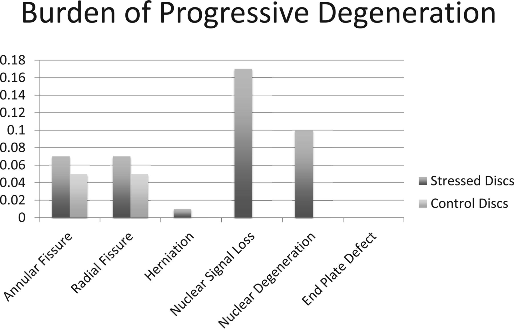
- Fig 2.
A bar diagram demonstrating the burden of new degenerative changes in stressed and control discs as observed on follow-up MR imaging acquired >6 months after the baseline scan.
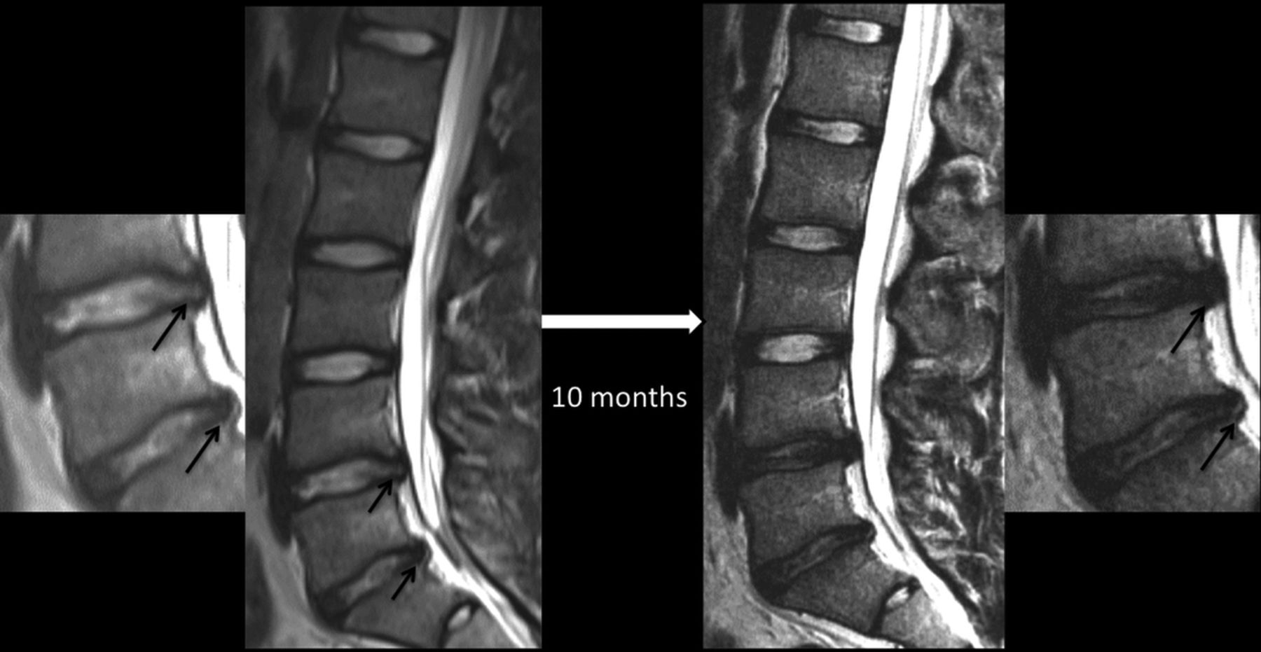
- Fig 3.
Sequential MR images of the lumbar spine showing the evolution of lumbar disc degeneration in a 16-year-old female patient presenting with low back pain and evidence for stress reaction in both L5 pedicles (not shown) at the time of presentation. Both intervertebral discs (L4–5 and L5–S1; also shown in magnified images) around the stressed segment demonstrated the presence of annular fissures (arrows), disc herniation, and nuclear degeneration at baseline. The control disc in the lower half of the lumbar spine (L3–4) had an intact annulus and preserved nuclear signal intensity at baseline. Follow-up MR imaging 10 months later reveals stability in the appearance of the control disc, but interval loss of signal intensity, signifying progressive nuclear degeneration of both stressed discs. The annular fissure at the L4–5 level has become less conspicuous, but one at the L5–S1 level is more easily recognizable on a follow-up scan.
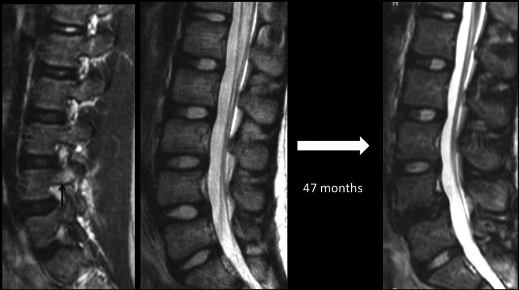
- Fig 4.
Sequential lumbar MR imaging over a 47-month period in a 12-year-old female patient presenting with low back pain caused by a partial L4 stress fracture as indicated by edema in the right pedicle (arrow) on a parasagittal STIR image. Initial and follow-up midsagittal T2-weighted images highlight marked progressive nuclear degeneration in the inferior stressed disc at the L4–5 level, but preserved appearance of a more caudally located control disc at the L5–S1 level.
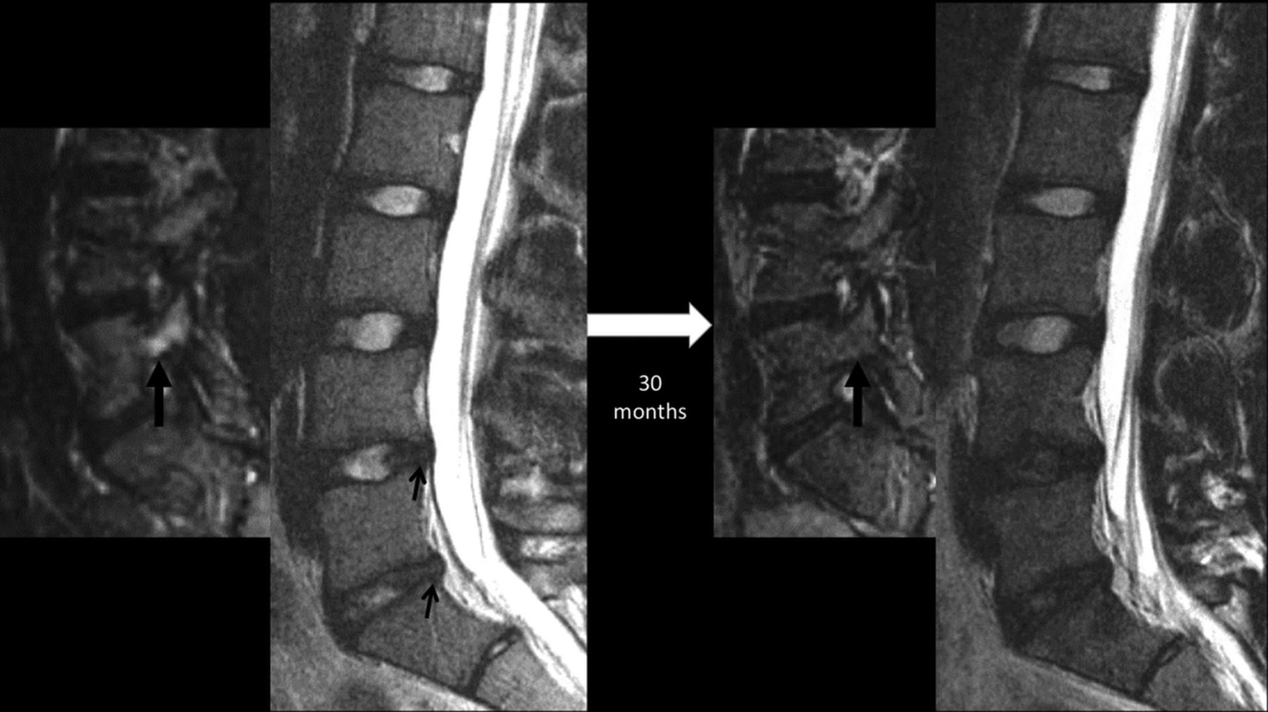
- Fig 5.
Sequential lumbar MR images obtained 30 months apart in a 15-year-old male patient with back pain, highlighting progressive disc degeneration despite the resolution of bony stress reaction. Initial MR imaging revealed edema in the right L5 pedicle (thick arrow) as shown here on a parasagittal STIR image, consistent with a stress reaction. Sagittal T2WI at this time revealed subtle annular fissures at the L4–5 and L5–S1 discs (small arrows) and signs of nuclear degeneration at the L5–S1 level. Follow-up MR imaging obtained 30 months later reveals the resolution of pedicular edema (thick arrow), but progressive loss of nuclear signal intensity of both stressed discs at the L4–5 and L5–S1 levels. Note that the control disc at the L3–4 level remains normal.

Tables
- Table 1:
Definitions of stressed discs and control discs for each vertebral segment showing bony stress
Location of Bony Stress Changes Stressed Discs Control Discs T12 T12–LI L1–2, L2–3 L1 T12–L1, L1–2 L2–3 L2 L1–2, L2–3 T12–L1 L3 L2–3, L3–4 T12–L1, L1–2, L4–5, L5–S1 L4 L3–4, L4–5 L5–S1 L5 L4–5, L5–S1 L3–4 S1 L5–S1 L3–4, L4–5 Baseline Degeneration Progressive Degeneration Stressed Discs Control Discs Stressed Discs Control Discs Annular fissurea,b 0.76 ± 0.35 0.37 ± 0.48 0.07 ± 0.18 0.05 ± 0.22 37 (88.1) 16 (38.1) 6 (14.3) 2 (4.9) Radial fissurea,b 0.59 ± 0.43 0.17 ± 0.38 0.07 ± 0.18 0.05 ± 0.22 30 (71.4) 8 (19.1) 6 (14.3) 2 (4.9) Herniationa,b 0.07 ± 0.20 0.00 ± 0.00 0.01 ± 0.08 0.00 ± 0.00 5 (11.9) 0 (0.0) 1 (2.4) 0 (0.0) Nuclear degenerationa,b,c 0.13 ± 0.30 0.05 ± 0.22 0.10 ± 0.28 0.00 ± 0.00 8 (19.1) 2 (4.8) 5 (11.9) 0 (0.0) Nuclear SI lossa,b,d 0.39 ± 0.42 0.12 ± 0.33 0.17 ± 0.48 0.00 ± 0.00 22 (52.4) 5 (11.9) 6 (14.3) 0 (0.0) EPDa,b 0.11 ± 0.24 0.12 ± 0.33 0.00 ± 0.00 0.00 ± 0.00 8 (19.1) 5 (11.9) 0 (0.0) 0 (0.0) Note:—EPD indicates endplate burden; SI, signal intensity.
↵a Burden (mean ± SD) of degenerative parameter calculated on per disc basis.
↵b Number (%) of patients with specified degenerative parameters in stressed or control segments of lumbar spine; “Baseline Degeneration” refers to changes on MR scan obtained at the time of presentation and “Progressive Degeneration” refers to appearance of new changes noted on a follow-up MR scan obtained >6 months later.
↵c Defined as discs graded higher than 2 on Pfirrmann grading system.
↵d Discs graded higher than 1 on a 6-point scale based on SI on T2-weighted images indicating any loss of normal hyperintense signal.


Jump to section
Related Articles
Cited By...
- No citing articles found.