
Article Figures & Data
Figures
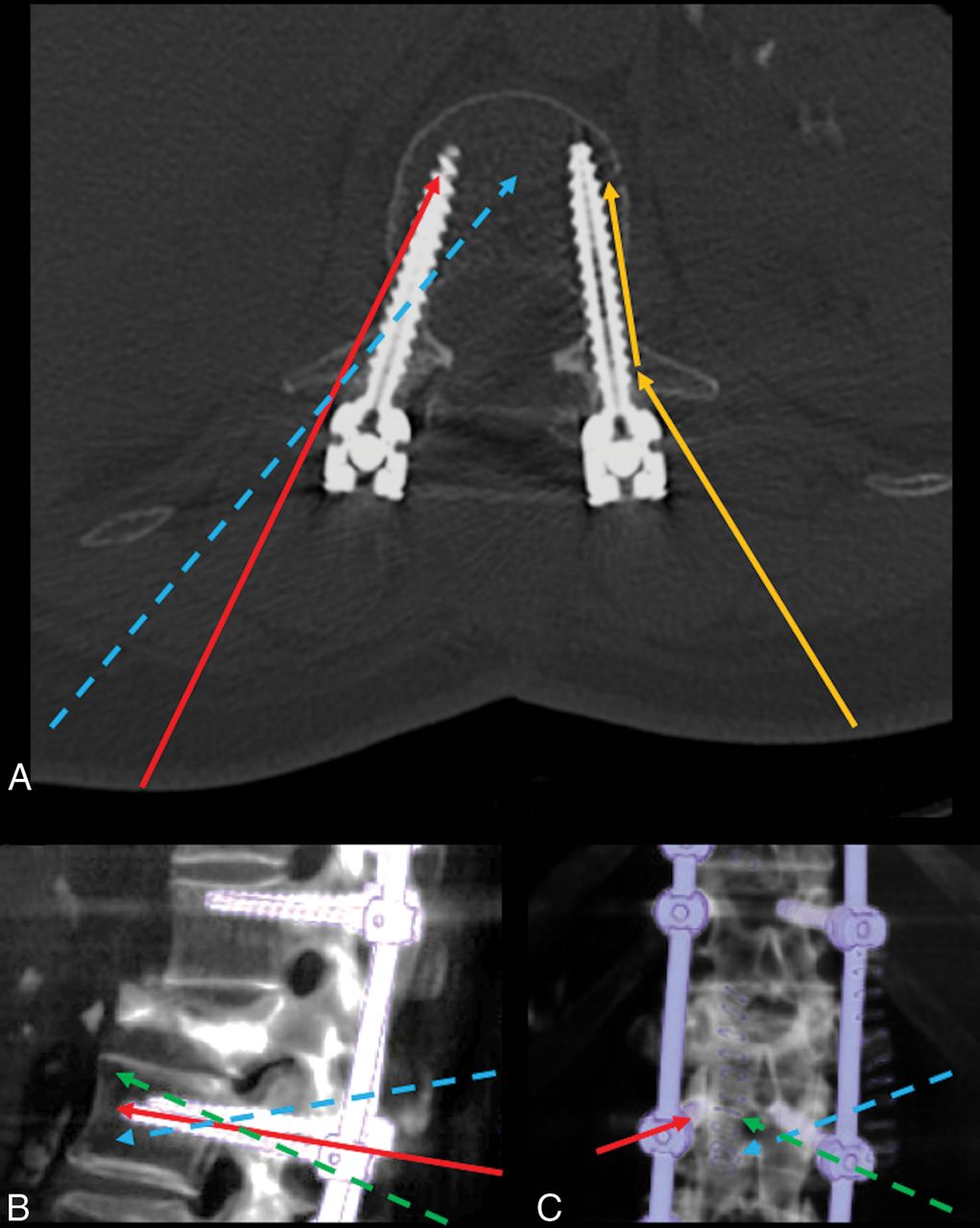
- Fig 1.
Schematic representation of different access approaches to the instrumented vertebra. A, Axial CT image of a vertebra instrumented with bilateral pedicular screws. Yellow arrows represent transpedicular access, with a thin, flexible, beveled needle contacting the proximal screw shaft, then bending and sliding along the screw shaft. The red arrow represents transpedicular access targeting the tip of the screw, with a slightly more oblique course than the screw path. The dashed blue arrow represents extrapedicular access targeting the anterior third of the vertebral body along the midline, crossing the course of the screw at the level of posterior wall with an obliquity from lateral to medial. B and C, Volume-rendered CT lateral and posteroanterior views of an instrumented spine segment. Red arrows in B and C show transpedicular access parallel to the screw used to augment loose screws, while dashed arrows represent extrapedicular accesses to the vertebral body used to augment vertebral body fractures, coursing lateral to medial to the screw, traversing the screw course from cranial to caudal (blue dashed arrows) or from caudal to cranial (green dashed arrow), respectively, passing cranial or caudal to the transverse process.
- Fig 3.
Cage subsidence/fracture and multiple targets. A and B, CT images of an L1 fracture treated with corpectomy, cage grafting, and T11–L3 posterior stabilization in a patient with osteoporosis. Due to bone compaction/fracture cranial and caudal to the cage, there is cage subsidence and focal kyphosis (arrows in B). Another fracture is noted at T11 (arrowhead in B), and there is bone resorption and screw loosening at L3 (not shown), with initial screw pullout. C and D, Anteroposterior and lateral fluoroscopy views after placement of multiple needles to perform cement augmentation at the cranial and caudal bone-metal cage interface in T12 and L2 (arrowheads), in the T11 fracture (arrow), in L3 (arrow) to augment the screw osseous purchase, and in T10 to perform prophylactic augmentation (arrow). E, Postprocedural sagittal MIP CT image demonstrates satisfactory cement filling of the target levels. F, Standing plain film at 12-month follow-up, with stable results.
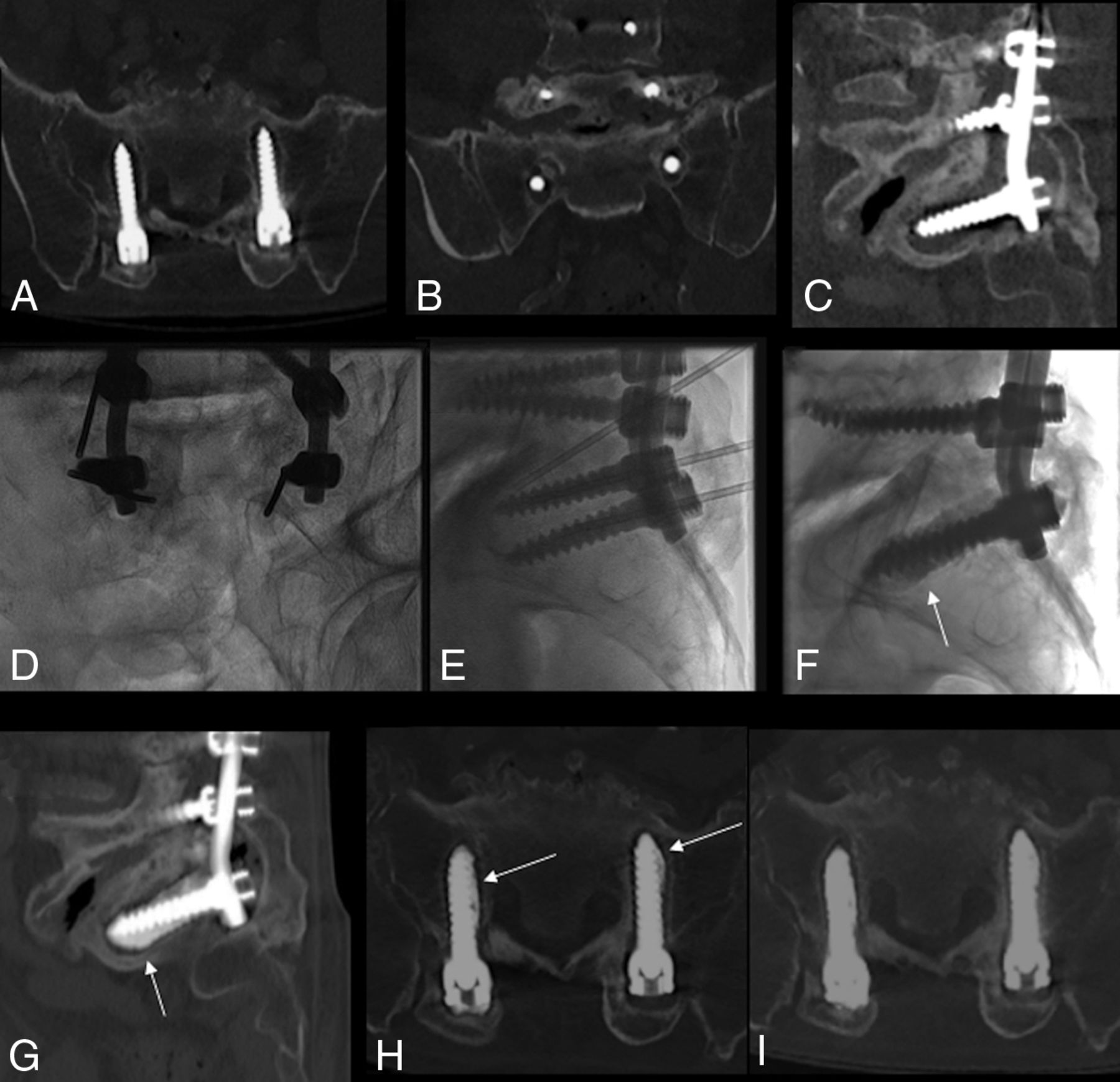
- Fig 2.
Cement augmentation of bilateral S1 screw loosening. A–C, Multiplanar preprocedural CT shows circumferential osteolysis around the screws in S1. Frontal (D) and lateral oblique views (E) of S1 screws, with bilateral placement of needles along the screws and the needle tip in bone osteolysis around the screw. F–H, Fluoroscopic and CT MIP images post-cement augmentation, demonstrating optimal filling of the osteolytic area (arrows), acting as screw oversizing, and potentially reducing hypermobility. I, Follow-up CT 3 months postaugmentation shows stable results in this patient reporting clinical amelioration.


