Advertisement

Main menu

  • Home
  • Content
    • Current Issue
    • Accepted Manuscripts
    • Article Preview
    • Past Issue Archive
    • Video Articles
    • AJNR Case Collection
    • Case of the Week Archive
    • Case of the Month Archive
    • Classic Case Archive
  • Special Collections
    • AJNR Awards
    • ASNR Foundation Special Collection
    • Most Impactful AJNR Articles
    • Photon-Counting CT
    • Spinal CSF Leak Articles (Jan 2020-June 2024)
  • Multimedia
    • AJNR Podcasts
    • AJNR SCANtastic
    • Trainee Corner
    • MRI Safety Corner
    • Imaging Protocols
  • For Authors
    • Submit a Manuscript
    • Submit a Video Article
    • Submit an eLetter to the Editor/Response
    • Manuscript Submission Guidelines
    • Statistical Tips
    • Fast Publishing of Accepted Manuscripts
    • Graphical Abstract Preparation
    • Imaging Protocol Submission
    • Author Policies
  • About Us
    • About AJNR
    • Editorial Board
    • Editorial Board Alumni
  • More
    • Become a Reviewer/Academy of Reviewers
    • Subscribers
    • Permissions
    • Alerts
    • Feedback
    • Advertisers
    • ASNR Home

User menu

  • Alerts
  • Log in

Search

  • Advanced search
American Journal of Neuroradiology

American Journal of Neuroradiology

ASHNR American Society of Functional Neuroradiology ASHNR American Society of Pediatric Neuroradiology ASSR
  • Alerts
  • Log in

Advanced Search

  • Home
  • Content
    • Current Issue
    • Accepted Manuscripts
    • Article Preview
    • Past Issue Archive
    • Video Articles
    • AJNR Case Collection
    • Case of the Week Archive
    • Case of the Month Archive
    • Classic Case Archive
  • Special Collections
    • AJNR Awards
    • ASNR Foundation Special Collection
    • Most Impactful AJNR Articles
    • Photon-Counting CT
    • Spinal CSF Leak Articles (Jan 2020-June 2024)
  • Multimedia
    • AJNR Podcasts
    • AJNR SCANtastic
    • Trainee Corner
    • MRI Safety Corner
    • Imaging Protocols
  • For Authors
    • Submit a Manuscript
    • Submit a Video Article
    • Submit an eLetter to the Editor/Response
    • Manuscript Submission Guidelines
    • Statistical Tips
    • Fast Publishing of Accepted Manuscripts
    • Graphical Abstract Preparation
    • Imaging Protocol Submission
    • Author Policies
  • About Us
    • About AJNR
    • Editorial Board
    • Editorial Board Alumni
  • More
    • Become a Reviewer/Academy of Reviewers
    • Subscribers
    • Permissions
    • Alerts
    • Feedback
    • Advertisers
    • ASNR Home
  • Follow AJNR on Twitter
  • Visit AJNR on Facebook
  • Follow AJNR on Instagram
  • Join AJNR on LinkedIn
  • RSS Feeds

AJNR is seeking candidates for the AJNR Podcast Editor. Read the position description.

Research ArticleHead & Neck

Comparison of MR Imaging and Dual-Energy CT for the Evaluation of Cartilage Invasion by Laryngeal and Hypopharyngeal Squamous Cell Carcinoma

H. Kuno, K. Sakamaki, S. Fujii, K. Sekiya, K. Otani, R. Hayashi, T. Yamanaka, O. Sakai and M. Kusumoto
American Journal of Neuroradiology March 2018, 39 (3) 524-531; DOI: https://doi.org/10.3174/ajnr.A5530
H. Kuno
aFrom the Departments of Diagnostic Radiology (H.K., K.S., M.K.)
cDepartments of Radiology (H.K., O.S.)
  • Find this author on Google Scholar
  • Find this author on PubMed
  • Search for this author on this site
  • ORCID record for H. Kuno
K. Sakamaki
aFrom the Departments of Diagnostic Radiology (H.K., K.S., M.K.)
fDepartment of Biostatistics (K.S., T.Y.), Yokohama City University, Yokohama, Kanagawa, Japan
  • Find this author on Google Scholar
  • Find this author on PubMed
  • Search for this author on this site
  • ORCID record for K. Sakamaki
S. Fujii
gDivision of Pathology (S.F.), Exploratory Oncology Research and Clinical Trial Center, National Cancer Center, Kashiwa, Chiba, Japan
  • Find this author on Google Scholar
  • Find this author on PubMed
  • Search for this author on this site
  • ORCID record for S. Fujii
K. Sekiya
aFrom the Departments of Diagnostic Radiology (H.K., K.S., M.K.)
  • Find this author on Google Scholar
  • Find this author on PubMed
  • Search for this author on this site
  • ORCID record for K. Sekiya
K. Otani
hAdvanced Therapies Innovation Department (K.O.), Siemens Healthcare K.K., Shinagawa-ku, Tokyo, Japan.
  • Find this author on Google Scholar
  • Find this author on PubMed
  • Search for this author on this site
  • ORCID record for K. Otani
R. Hayashi
bHead and Neck Surgery (R.H.), National Cancer Center Hospital East, Kashiwa, Chiba, Japan
  • Find this author on Google Scholar
  • Find this author on PubMed
  • Search for this author on this site
  • ORCID record for R. Hayashi
T. Yamanaka
fDepartment of Biostatistics (K.S., T.Y.), Yokohama City University, Yokohama, Kanagawa, Japan
  • Find this author on Google Scholar
  • Find this author on PubMed
  • Search for this author on this site
  • ORCID record for T. Yamanaka
O. Sakai
cDepartments of Radiology (H.K., O.S.)
dOtolaryngology–Head and Neck Surgery (O.S.)
eRadiation Oncology (O.S.), Boston Medical Center, Boston University School of Medicine, Boston, Massachusetts
  • Find this author on Google Scholar
  • Find this author on PubMed
  • Search for this author on this site
  • ORCID record for O. Sakai
M. Kusumoto
aFrom the Departments of Diagnostic Radiology (H.K., K.S., M.K.)
  • Find this author on Google Scholar
  • Find this author on PubMed
  • Search for this author on this site
  • ORCID record for M. Kusumoto
  • Article
  • Figures & Data
  • Supplemental
  • Info & Metrics
  • Responses
  • References
  • PDF
Loading

Article Figures & Data

Figures

  • Tables
  • Fig 1.
    • Download figure
    • Open in new tab
    • Download powerpoint
    Fig 1.

    False-positive findings for thyroid cartilage invasion on MR imaging in a 59-year-old man with hypopharyngeal cancer. A, T2-weighted MR image obtained at the glottis level shows a tumor (T) arising from the right piriform sinus with intermediate signal intensity. The adjacent posterior right thyroid lamina also shows an area of intermediate signal intensity (arrow). B, T1-weighted image shows that the tumor (T) has low signal intensity, whereas adjacent thyroid cartilage has similar signal intensity (arrow). C, Contrast-enhanced fat-suppressed T1-weighted MR image shows similar contrast enhancement of the tumor (T) and adjacent thyroid cartilage (arrow). A weighted-average image does not show erosion or lysis at the same level (D, soft-tissue window; E, bone window). F, Iodine overlay image shows enhancement of tumor (T) more clearly and is not used for the diagnosis of cartilage according to the findings of the WA image. G, A micrograph of the corresponding axial slice of the surgical specimen at the same level shows that the squamous cell carcinoma cells do not permeate into the right thyroid cartilage lamina (hematoxylin-eosin stain; original magnification, ×5). H, Magnified photograph (square in G) of the posterior part of the right thyroid cartilage lamina with enhancement on MR imaging shows moderate infiltration of lymphocytes into the medullary space, accompanied by fibrosis and aggregation of macrophages, without tumor (H&E stain; original magnification, ×200).

  • Fig 2.
    • Download figure
    • Open in new tab
    • Download powerpoint
    Fig 2.

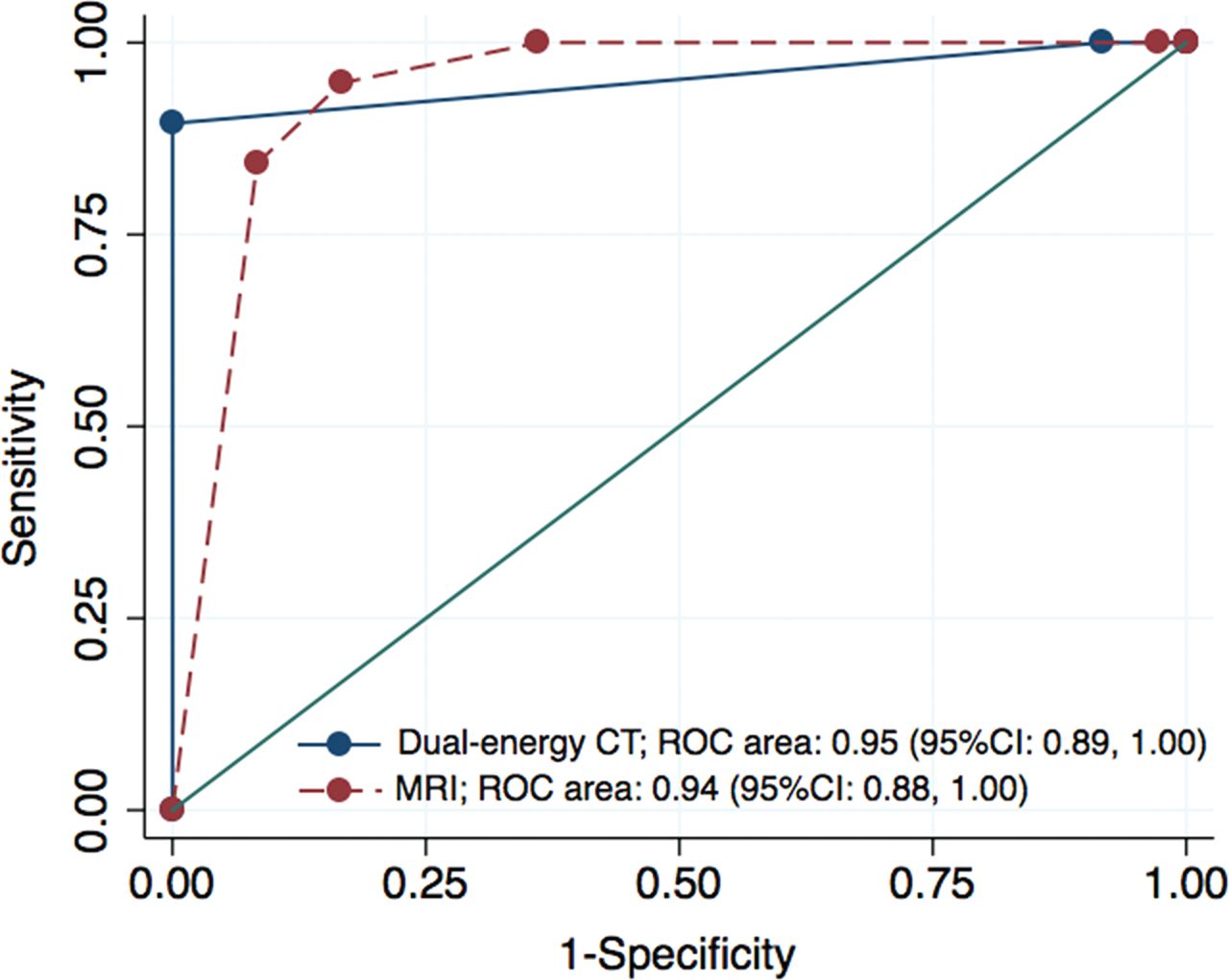
    Graph shows 2 crossing ROC curves and corresponding AUCs in the prediction of thyroid cartilage invasion. There was no evidence of differences in the average areas under the ROC curve between MR imaging and dual-energy CT (0.938 versus 0.952, respectively; P = .70).

Tables

  • Figures
    • View popup
    Table 1:

    Summary of motion artifact grading scores for MRI and dual-energy CTa

    12345Mean Score (SE)P Value
    All patients (n = 55)<.001b
        MRI0348403.02 (0.05)
        Dual-energy CT0004694.16 (0.05)
    T1 or T2 stage (n = 6).050
        MRI003303.50 (0.22)
        Dual-energy CT000424.33 (0.21)
    T3 stage (n = 13)<.001b
        MRI0111103.00 (0.11)
        Dual-energy CT0001214.08 (0.08)
    T4a stage (n = 36)<.0001b
        MRI0234002.94 (0.04)
        Dual-energy CT0003064.17 (0.06)
    • Note:—SE indicates standard error.

    • ↵a Data are number of patients. The rating grade is as follows: grade 1, nondiagnostic with major artifacts; grade 2, major artifacts with most organs depicted with diagnostic image quality; grade 3, moderate artifacts with image quality low but diagnostic; grade 4, minor artifacts with good image quality; grade 5, no artifacts with excellent image quality.

    • ↵b Indicates a significant difference using the Wilcoxon signed rank test (P < .05).

    • View popup
    Table 2:

    Relationship between dual-energy CT/MR imaging and histopathologic findings for the detection of cartilage invasiona

    TPTNFNFPSensitivity (%)Specificity (%)PPV (%)NPV (%)
    Thyroid cartilage (n = 55)
        MRI1923013100 (82–100)64 (46–79)59 (41–76)100 (85–100)
        Dual-energy CT17362089 (67–99)100 (90–100)100 (80–100)95 (82–99)
        P valueb.5<.001
    Cricoid cartilage (n = 55)
        MRI84106100 (63–100)87 (74–95)57 (29–82)100 (91–100)
        Dual-energy CT6462175 (35–97)98 (89–100)86 (42–100)96 (86–99)
        P valueb.5.06
    Arytenoid cartilage (n = 110)
        MRI89111089 (52–100)91 (83–95)44 (22–69)99 (94–100)
        Dual-energy CT6983367 (30–93)97 (92–100)67 (30–93)97 (92–99)
        P valuec.43.09
    All cartilage (n = 220)
        MRI3515512997 (85–100)84 (78–89)55 (42–67)99 (96–100)
        Dual-energy CT291807481 (64–92)98 (95–99)88 (72–97)96 (92–98)
        P valuec.16<.004
    • Note:—TP indicates true positive; TN, true negative; FP, false positive; FN, false negative; PPV, positive predictive value; NPV, negative predictive value.

    • ↵a Numbers in parentheses are 95% confidence intervals. Negative findings for cartilage invasion are scores 1 and 2; positive findings are scores 3–5.

    • ↵b As determined with the McNemar test.

    • ↵c According to the generalized linear mixed model that accounted for the multiple observations within patients.

PreviousNext
Back to top

In this issue

American Journal of Neuroradiology: 39 (3)
American Journal of Neuroradiology
Vol. 39, Issue 3
1 Mar 2018
  • Table of Contents
  • Index by author
  • Complete Issue (PDF)
Advertisement
Print
Download PDF
Email Article

Thank you for your interest in spreading the word on American Journal of Neuroradiology.

NOTE: We only request your email address so that the person you are recommending the page to knows that you wanted them to see it, and that it is not junk mail. We do not capture any email address.

Enter multiple addresses on separate lines or separate them with commas.
Comparison of MR Imaging and Dual-Energy CT for the Evaluation of Cartilage Invasion by Laryngeal and Hypopharyngeal Squamous Cell Carcinoma
(Your Name) has sent you a message from American Journal of Neuroradiology
(Your Name) thought you would like to see the American Journal of Neuroradiology web site.
CAPTCHA
This question is for testing whether or not you are a human visitor and to prevent automated spam submissions.
Cite this article
H. Kuno, K. Sakamaki, S. Fujii, K. Sekiya, K. Otani, R. Hayashi, T. Yamanaka, O. Sakai, M. Kusumoto
Comparison of MR Imaging and Dual-Energy CT for the Evaluation of Cartilage Invasion by Laryngeal and Hypopharyngeal Squamous Cell Carcinoma
American Journal of Neuroradiology Mar 2018, 39 (3) 524-531; DOI: 10.3174/ajnr.A5530

Citation Manager Formats

  • BibTeX
  • Bookends
  • EasyBib
  • EndNote (tagged)
  • EndNote 8 (xml)
  • Medlars
  • Mendeley
  • Papers
  • RefWorks Tagged
  • Ref Manager
  • RIS
  • Zotero
0 Responses
Respond to this article
Share
Bookmark this article
Comparison of MR Imaging and Dual-Energy CT for the Evaluation of Cartilage Invasion by Laryngeal and Hypopharyngeal Squamous Cell Carcinoma
H. Kuno, K. Sakamaki, S. Fujii, K. Sekiya, K. Otani, R. Hayashi, T. Yamanaka, O. Sakai, M. Kusumoto
American Journal of Neuroradiology Mar 2018, 39 (3) 524-531; DOI: 10.3174/ajnr.A5530
del.icio.us logo Twitter logo Facebook logo Mendeley logo
  • Tweet Widget
  • Facebook Like
  • Google Plus One
Purchase

Jump to section

  • Article
    • Abstract
    • ABBREVIATIONS:
    • Materials and Methods
    • Results
    • Discussion
    • Conclusions
    • Footnotes
    • References
  • Figures & Data
  • Supplemental
  • Info & Metrics
  • Responses
  • References
  • PDF

Related Articles

  • PubMed
  • Google Scholar

Cited By...

  • CT of the Larynx: Is an Additional High-Resolution Acquisition Necessary for Diagnostic Accuracy?
  • Dual-Energy Parathyroid 4D-CT: Improved Discrimination of Parathyroid Lesions from Thyroid Tissue Using Noncontrast 40-keV Virtual Monoenergetic Images
  • Preoperative MRI Evaluation of Thyroid Cartilage Invasion in Patients with Laryngohypopharyngeal Cancer: Comparison of Contrast-Enhanced 2D Spin-Echo and 3D T1-Weighted Radial Gradient Recalled-Echo Techniques
  • Reply:
  • Laryngeal Cartilage Invasion
  • Crossref (53)
  • Google Scholar

This article has been cited by the following articles in journals that are participating in Crossref Cited-by Linking.

  • An update on advanced dual-energy CT for head and neck cancer imaging
    Reza Forghani
    Expert Review of Anticancer Therapy 2019 19 7
  • CT-based radiomics features in the prediction of thyroid cartilage invasion from laryngeal and hypopharyngeal squamous cell carcinoma
    Ran Guo, Jian Guo, Lichen Zhang, Xiaoxia Qu, Shuangfeng Dai, Ruchen Peng, Vincent F. H. Chong, Junfang Xian
    Cancer Imaging 2020 20 1
  • In vivo efficacy of 3D‐printed elastin–gelatin–hyaluronic acid scaffolds for regeneration of nasal septal cartilage defects
    Abbas Shokri, Kousar Ramezani, Mohammad Reza Jamalpour, Chiman Mohammadi, Farshid Vahdatinia, Amin Doosti Irani, Esmaeel Sharifi, Rasool Haddadi, Shokoofeh Jamshidi, Leila Mohammadi Amirabad, Sanaz Tajik, Amir Yadegari, Lobat Tayebi
    Journal of Biomedical Materials Research Part B: Applied Biomaterials 2022 110 3
  • Comparison of diagnostic performance between CT and MRI for detection of cartilage invasion for primary tumor staging in patients with laryngo-hypopharyngeal cancer: a systematic review and meta-analysis
    Se Jin Cho, Jeong Hyun Lee, Chong Hyun Suh, Jung Youn Kim, Donghyun Kim, Jung Bin Lee, Min Kyoung Lee, Sae Rom Chung, Young Jun Choi, Jung Hwan Baek
    European Radiology 2020 30 7
  • Prediction of Posterior Paraglottic Space and Cricoarytenoid Unit Involvement in Endoscopically T3 Glottic Cancer with Arytenoid Fixation by Magnetic Resonance with Surface Coils
    Marco Ravanelli, Alberto Paderno, Francesca Del Bon, Nausica Montalto, Carlotta Pessina, Simonetta Battocchio, Davide Farina, Piero Nicolai, Roberto Maroldi, Cesare Piazza
    Cancers 2019 11 1
  • Detecting abnormal thyroid cartilages on CT using deep learning
    M. Santin, C. Brama, H. Théro, E. Ketheeswaran, I. El-Karoui, F. Bidault, R. Gillet, P. Gondim Teixeira, A. Blum
    Diagnostic and Interventional Imaging 2019 100 4
  • Dual Energy Computed Tomography in Head and Neck Imaging
    Thiparom Sananmuang, Mohit Agarwal, Farhad Maleki, Nikesh Muthukrishnan, Juan Camilo Marquez, Jeffrey Chankowsky, Reza Forghani
    Neuroimaging Clinics of North America 2020 30 3
  • Automated segmentation of the larynx on computed tomography images: a review
    Divya Rao, Prakashini K, Rohit Singh, Vijayananda J
    Biomedical Engineering Letters 2022 12 2
  • Dual-Energy Parathyroid 4D-CT: Improved Discrimination of Parathyroid Lesions from Thyroid Tissue Using Noncontrast 40-keV Virtual Monoenergetic Images
    P.M. Bunch, A.A. Pavlina, M.E. Lipford, J.R. Sachs
    American Journal of Neuroradiology 2021 42 11
  • Qualitative and Quantitative Diagnosis in Head and Neck Cancer
    Fernando López, Antti Mäkitie, Remco de Bree, Alessandro Franchi, Pim de Graaf, Juan C. Hernández-Prera, Primoz Strojan, Nina Zidar, Margareta Strojan Fležar, Juan P. Rodrigo, Alessandra Rinaldo, Barbara A. Centeno, Alfio Ferlito
    Diagnostics 2021 11 9

More in this TOC Section

  • WHO Classification Update: Nasal&Skull Base Tumors
  • Peritumoral Signal in Vestibular Schwannomas
  • NI-RADS for HEAD&NECK Cancer Recurrence
Show more HEAD & NECK

Similar Articles

Advertisement

Indexed Content

  • Current Issue
  • Accepted Manuscripts
  • Article Preview
  • Past Issues
  • Editorials
  • Editor's Choice
  • Fellows' Journal Club
  • Letters to the Editor
  • Video Articles

Cases

  • Case Collection
  • Archive - Case of the Week
  • Archive - Case of the Month
  • Archive - Classic Case

Special Collections

  • AJNR Awards
  • ASNR Foundation Special Collection
  • Most Impactful AJNR Articles
  • Photon-Counting CT
  • Spinal CSF Leak Articles (Jan 2020-June 2024)

More from AJNR

  • Trainee Corner
  • Imaging Protocols
  • MRI Safety Corner

Multimedia

  • AJNR Podcasts
  • AJNR Scantastics

Resources

  • Turnaround Time
  • Submit a Manuscript
  • Submit a Video Article
  • Submit an eLetter to the Editor/Response
  • Manuscript Submission Guidelines
  • Statistical Tips
  • Fast Publishing of Accepted Manuscripts
  • Graphical Abstract Preparation
  • Imaging Protocol Submission
  • Evidence-Based Medicine Level Guide
  • Publishing Checklists
  • Author Policies
  • Become a Reviewer/Academy of Reviewers
  • News and Updates

About Us

  • About AJNR
  • Editorial Board
  • Editorial Board Alumni
  • Alerts
  • Permissions
  • Not an AJNR Subscriber? Join Now
  • Advertise with Us
  • Librarian Resources
  • Feedback
  • Terms and Conditions
  • AJNR Editorial Board Alumni

American Society of Neuroradiology

  • Not an ASNR Member? Join Now

© 2025 by the American Society of Neuroradiology All rights, including for text and data mining, AI training, and similar technologies, are reserved.
Print ISSN: 0195-6108 Online ISSN: 1936-959X

Powered by HighWire