
Article Figures & Data
Figures
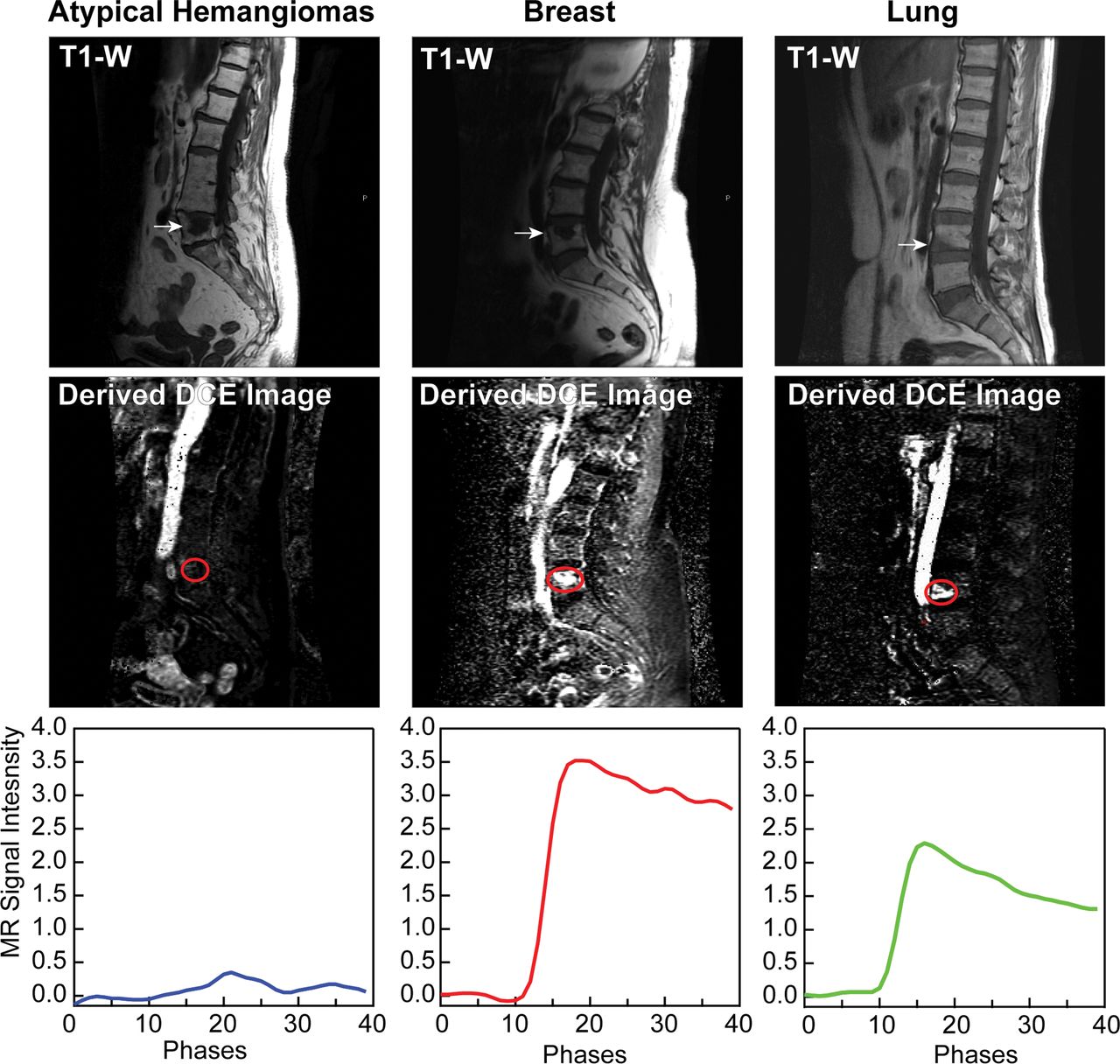
- Fig 1.
Representative sagittal T1-weighted and dynamic images derived from DCE MR imaging for atypical hemangiomas and spinal lesions originating from primary breast and lung carcinomas. The lower panel corresponds to the MR imaging signal enhancement curve as a function of phases (time) obtained for each representative lesion shown.
- Fig 2.
A, Enhancement curve for all 34 atypical hemangiomas. Curves highlighted in cyan show the 4 cases with elevated enhancement. B, Average enhancement curves for all atypical hemangiomas (cyan) excluding atypical hemangiomas with elevated enhancement (blue). C, Average enhancement curves for all atypical hemangiomas excluding enhancement curve outliers (blue) compared with breast (red) and lung (green) metastases.
- Fig 3.
Representative sagittal T1-weighted imaging and the corresponding perfusion maps for Vp and Ktrans parameters for atypical hemangiomas and metastasis from lung carcinoma. Arrows indicate the level of the lesion in T1WI, and the cyan circle highlights the region on Vp and Ktrans maps where the lesion is located.
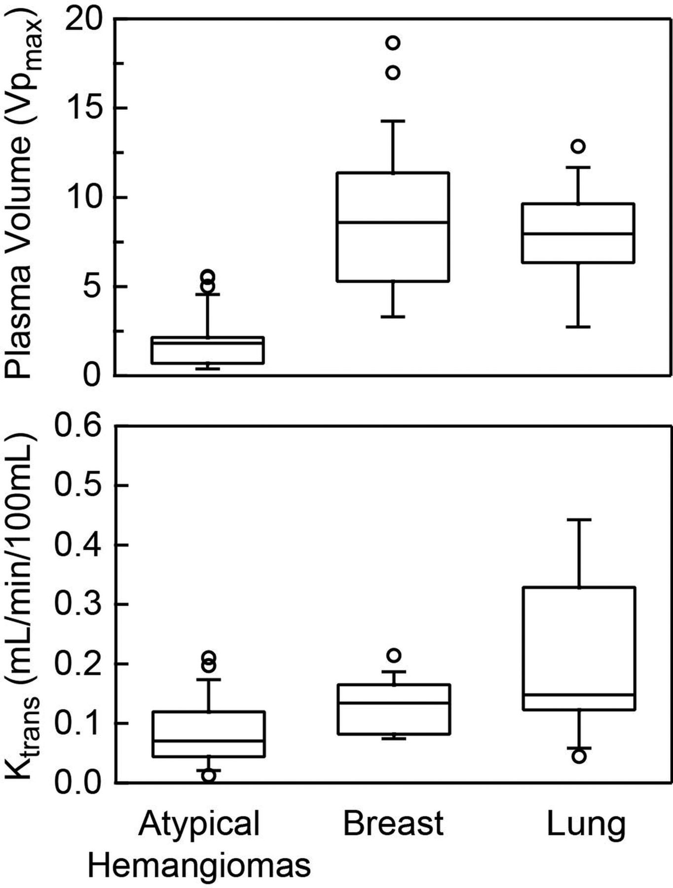
- Fig 4.
Vp and Ktrans parameters obtained for atypical hemangiomas and metastatic lesions from breast and lung carcinoma. Metastases show significantly higher values (P < .05) for both Vp and Ktrans compared with atypical vertebral hemangiomas. Note that the 4 outlier values representing aggressive hemangiomas account for the higher Vp values in the atypical hemangioma group that demonstrate minimal overlap with the metastatic group and remain low compared with other metastases.


Jump to section
Related Articles
Cited By...
- Comprehensive Review of the Utility of Dynamic Contrast-Enhanced MRI for the Diagnosis and Treatment Assessment of Spinal Benign and Malignant Osseous Disease
- T1-Weighted, Dynamic Contrast-Enhanced MR Perfusion Imaging Can Differentiate between Treatment Success and Failure in Spine Metastases Undergoing Radiation Therapy
- Differentiation of Skull Base Chondrosarcomas, Chordomas, and Metastases: Utility of DWI and Dynamic Contrast-Enhanced Perfusion MR Imaging
- Reply:
- Quality-Control Assessment to Improve the Accuracy of Dynamic Contrast-Enhanced MR Imaging Perfusion