Advertisement

Main menu

  • Home
  • Content
    • Current Issue
    • Accepted Manuscripts
    • Article Preview
    • Past Issue Archive
    • Video Articles
    • AJNR Case Collection
    • Case of the Week Archive
    • Case of the Month Archive
    • Classic Case Archive
  • Special Collections
    • AJNR Awards
    • ASNR Foundation Special Collection
    • Most Impactful AJNR Articles
    • Photon-Counting CT
    • Spinal CSF Leak Articles (Jan 2020-June 2024)
  • Multimedia
    • AJNR Podcasts
    • AJNR SCANtastic
    • Trainee Corner
    • MRI Safety Corner
    • Imaging Protocols
  • For Authors
    • Submit a Manuscript
    • Submit a Video Article
    • Submit an eLetter to the Editor/Response
    • Manuscript Submission Guidelines
    • Statistical Tips
    • Fast Publishing of Accepted Manuscripts
    • Graphical Abstract Preparation
    • Imaging Protocol Submission
    • Author Policies
  • About Us
    • About AJNR
    • Editorial Board
    • Editorial Board Alumni
  • More
    • Become a Reviewer/Academy of Reviewers
    • Subscribers
    • Permissions
    • Alerts
    • Feedback
    • Advertisers
    • ASNR Home

User menu

  • Alerts
  • Log in

Search

  • Advanced search
American Journal of Neuroradiology

American Journal of Neuroradiology

ASHNR American Society of Functional Neuroradiology ASHNR American Society of Pediatric Neuroradiology ASSR
  • Alerts
  • Log in

Advanced Search

  • Home
  • Content
    • Current Issue
    • Accepted Manuscripts
    • Article Preview
    • Past Issue Archive
    • Video Articles
    • AJNR Case Collection
    • Case of the Week Archive
    • Case of the Month Archive
    • Classic Case Archive
  • Special Collections
    • AJNR Awards
    • ASNR Foundation Special Collection
    • Most Impactful AJNR Articles
    • Photon-Counting CT
    • Spinal CSF Leak Articles (Jan 2020-June 2024)
  • Multimedia
    • AJNR Podcasts
    • AJNR SCANtastic
    • Trainee Corner
    • MRI Safety Corner
    • Imaging Protocols
  • For Authors
    • Submit a Manuscript
    • Submit a Video Article
    • Submit an eLetter to the Editor/Response
    • Manuscript Submission Guidelines
    • Statistical Tips
    • Fast Publishing of Accepted Manuscripts
    • Graphical Abstract Preparation
    • Imaging Protocol Submission
    • Author Policies
  • About Us
    • About AJNR
    • Editorial Board
    • Editorial Board Alumni
  • More
    • Become a Reviewer/Academy of Reviewers
    • Subscribers
    • Permissions
    • Alerts
    • Feedback
    • Advertisers
    • ASNR Home
  • Follow AJNR on Twitter
  • Visit AJNR on Facebook
  • Follow AJNR on Instagram
  • Join AJNR on LinkedIn
  • RSS Feeds

AJNR is seeking candidates for the AJNR Podcast Editor. Read the position description.

Research ArticleInterventional

Adjunctive Efficacy of Intra-Arterial Conebeam CT Angiography Relative to DSA in the Diagnosis and Surgical Planning of Micro-Arteriovenous Malformations

A.S. Al-Smadi, A. Elmokadem, A. Shaibani, M.C. Hurley, M.B. Potts, B.S. Jahromi and S.A. Ansari
American Journal of Neuroradiology September 2018, 39 (9) 1689-1695; DOI: https://doi.org/10.3174/ajnr.A5745
A.S. Al-Smadi
aFrom the Departments of Radiology (A.S.A.-S., A.E., A.S., M.C.H., M.B.P., B.S.J., S.A.A.)
  • Find this author on Google Scholar
  • Find this author on PubMed
  • Search for this author on this site
  • ORCID record for A.S. Al-Smadi
A. Elmokadem
aFrom the Departments of Radiology (A.S.A.-S., A.E., A.S., M.C.H., M.B.P., B.S.J., S.A.A.)
  • Find this author on Google Scholar
  • Find this author on PubMed
  • Search for this author on this site
  • ORCID record for A. Elmokadem
A. Shaibani
aFrom the Departments of Radiology (A.S.A.-S., A.E., A.S., M.C.H., M.B.P., B.S.J., S.A.A.)
cNeurological Surgery (A.S., M.C.H., M.B.P., B.S.J., S.A.A.), Northwestern University, Feinberg School of Medicine, Chicago, Illinois.
  • Find this author on Google Scholar
  • Find this author on PubMed
  • Search for this author on this site
  • ORCID record for A. Shaibani
M.C. Hurley
aFrom the Departments of Radiology (A.S.A.-S., A.E., A.S., M.C.H., M.B.P., B.S.J., S.A.A.)
cNeurological Surgery (A.S., M.C.H., M.B.P., B.S.J., S.A.A.), Northwestern University, Feinberg School of Medicine, Chicago, Illinois.
  • Find this author on Google Scholar
  • Find this author on PubMed
  • Search for this author on this site
  • ORCID record for M.C. Hurley
M.B. Potts
aFrom the Departments of Radiology (A.S.A.-S., A.E., A.S., M.C.H., M.B.P., B.S.J., S.A.A.)
cNeurological Surgery (A.S., M.C.H., M.B.P., B.S.J., S.A.A.), Northwestern University, Feinberg School of Medicine, Chicago, Illinois.
  • Find this author on Google Scholar
  • Find this author on PubMed
  • Search for this author on this site
  • ORCID record for M.B. Potts
B.S. Jahromi
aFrom the Departments of Radiology (A.S.A.-S., A.E., A.S., M.C.H., M.B.P., B.S.J., S.A.A.)
cNeurological Surgery (A.S., M.C.H., M.B.P., B.S.J., S.A.A.), Northwestern University, Feinberg School of Medicine, Chicago, Illinois.
  • Find this author on Google Scholar
  • Find this author on PubMed
  • Search for this author on this site
  • ORCID record for B.S. Jahromi
S.A. Ansari
aFrom the Departments of Radiology (A.S.A.-S., A.E., A.S., M.C.H., M.B.P., B.S.J., S.A.A.)
bNeurology (S.A.A.)
cNeurological Surgery (A.S., M.C.H., M.B.P., B.S.J., S.A.A.), Northwestern University, Feinberg School of Medicine, Chicago, Illinois.
  • Find this author on Google Scholar
  • Find this author on PubMed
  • Search for this author on this site
  • ORCID record for S.A. Ansari
  • Article
  • Figures & Data
  • Supplemental
  • Info & Metrics
  • Responses
  • References
  • PDF
Loading

Article Figures & Data

Figures

  • Tables
  • Fig 1.
    • Download figure
    • Open in new tab
    • Download powerpoint
    Fig 1.

    Anteroposterior DSA images demonstrate left vertebral artery injections in the early (A) and late (B) arterial phases, identifying arteriovenous shunting and early venous drainage into a hypertrophied superior cerebellar hemispheric vein (arrow) with tentorial venous outflow, but both observers were unable to appreciate an occult micronidus. Although arterial feeders are suggested to project into this region on DSA from the right anterior cerebellar artery and SCA (A, arrowheads), the SCA feeder is better appreciated on IA-CBCTA multiplanar axial reconstructions (C, arrowhead). Moreover, both observers identified a <5-mm micronidus under the lateral cerebellar surface and adjacent to the craniectomy site (C, arrow) with adjunctive IA-CBCTA reconstructions, consistent with an mAVM. IA-CBCTA coronal reconstruction (D) shows the nidal outflow to the early draining superior cerebellar hemispheric vein (arrow).

  • Fig 2.
    • Download figure
    • Open in new tab
    • Download powerpoint
    Fig 2.

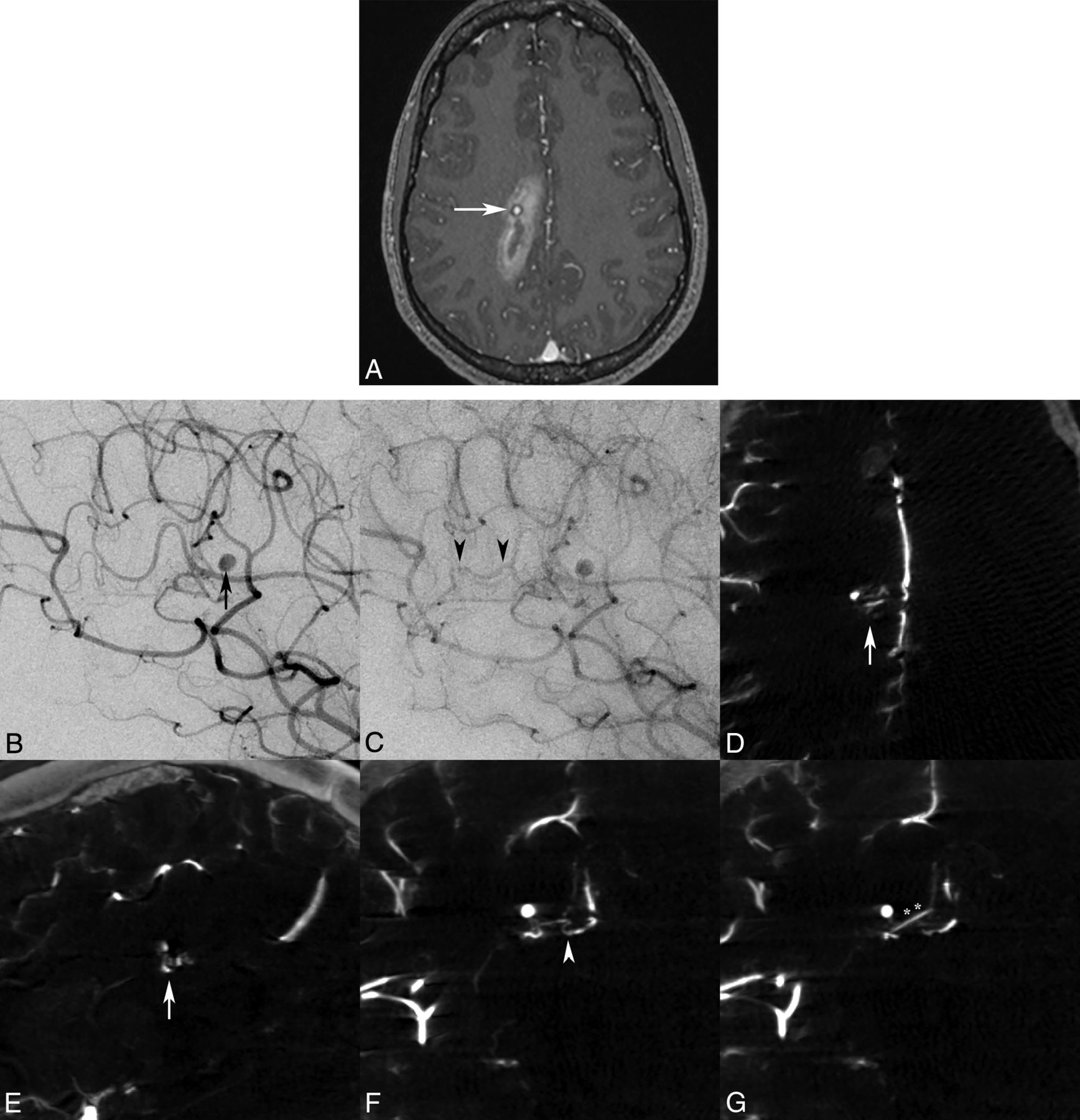
    Axial MR imaging MPRAGE postgadolinium (A) image demonstrates a right parasagittal frontoparietal intraparenchymal hemorrhage with a contrast-enhancing pseudoaneurysm (white arrow), consistent with the rupture site. Lateral oblique DSA images in the early arterial phase confirm a pericallosal anterior cerebral artery aneurysm/pseudoaneurysm (B, black arrow), with a subtle early draining vein in the capillary phase (C, black arrowheads), but no distinct vascular nidus was identified by either observer. Only IA-CBCTA multiplanar reconstructions clearly delineate a <5-mm micronidus on axial and sagittal reconstructions (D and E, white arrows). Coronal multiplanar reconstructions also assist in identification of the small arterial feeder from the pericallosal-splenial artery branch (F, white arrowhead) and single draining vein (G, double asterisks) directly associated with the micronidus and flow-induced pseudoaneurysm.

  • Fig 3.
    • Download figure
    • Open in new tab
    • Download powerpoint
    Fig 3.

    Lateral DSA (A) and coronal IA-CBCTA reconstruction (B) images both demonstrate a small <1-cm micronidus (arrows), supplied by tortuous parietal branches of the pericallosal anterior cerebral artery and inferior division of the MCA (arrowheads), with early venous drainage into bifurcating cortical veins (double asterisks). Although both observers did not report the improved diagnostic value of IA-CBCTA in this case, both neurosurgeons reported increased confidence in treatment planning, and IA-CBCTA was incorporated into the neuronavigation system for microsurgical resection. 3D IA-CBCTA and MR imaging datasets were merged with sagittal overlay (C) delineating the micronidus (arrow) and draining cortical vein (double asterisks) complex in relation to the adjacent hemorrhage. Both datasets were imported into the intraoperative neuronavigation system (D, BrainLAB), allowing anatomic localization of the micronidus within a specific sulcus guiding the surgical approach as well as the presumed deep nidal rupture site abutting the hematoma (black arrow).

Tables

  • Figures
    • View popup
    Table 1:

    Qualitative diagnostic scoring of DSA and IA-CBCTA imaging

    Observer 1Observer 2
    Arterial FeederNidusVenous DrainageOverallRelative CBCTA Diagnostic ValueaArterial FeederNidusVenous DrainageOverallRelative CBCTA Diagnostic Valuea
    DSACBCTADSACBCTADSACBCTADSACBCTADSACBCTADSACBCTADSACBCTADSACBCTA
    1120222363020222264
    2021222264021222363
    3121222462021222363
    4120212264120212264
    5121222462120222363
    6220222462221222561
    7121222462221222561
    8222201451122201352
    922222266021222164−2
    10220222462121222462
    P.02.009.157.007.03.009.564.016
    • τ coefficient = 0.66, P = .018.

    • View popup
    Table 2:

    Treatment-planning scoring of IA-CBCTA relative to DSAa

    PatientNeurosurgeon 1Relative CBCTA Treatment ValueNeurosurgeon 2Relative CBCTA Treatment Value
    DSACBCTADSACBCTA
    1Not sufficientOperation2Not sufficientOperation2
    2RadiosurgeryRadiosurgery1RadiosurgeryRadiosurgery1
    3RadiosurgeryRadiosurgery1RadiosurgeryRadiosurgery0
    4OperationOperation1OperationOperation1
    5OperationOperation1OperationOperation1
    6RadiosurgeryRadiosurgery1RadiosurgeryRadiosurgery1
    7OperationOperation1OperationOperation1
    8RadiosurgeryRadiosurgery1RadiosurgeryRadiosurgery1
    9OperationOperation1OperationOperation1
    10OperationOperation1OperationOperation1
    • ↵a τ coefficient = 0.73, P = .025.

PreviousNext
Back to top

In this issue

American Journal of Neuroradiology: 39 (9)
American Journal of Neuroradiology
Vol. 39, Issue 9
1 Sep 2018
  • Table of Contents
  • Index by author
  • Complete Issue (PDF)
Advertisement
Print
Download PDF
Email Article

Thank you for your interest in spreading the word on American Journal of Neuroradiology.

NOTE: We only request your email address so that the person you are recommending the page to knows that you wanted them to see it, and that it is not junk mail. We do not capture any email address.

Enter multiple addresses on separate lines or separate them with commas.
Adjunctive Efficacy of Intra-Arterial Conebeam CT Angiography Relative to DSA in the Diagnosis and Surgical Planning of Micro-Arteriovenous Malformations
(Your Name) has sent you a message from American Journal of Neuroradiology
(Your Name) thought you would like to see the American Journal of Neuroradiology web site.
CAPTCHA
This question is for testing whether or not you are a human visitor and to prevent automated spam submissions.
Cite this article
A.S. Al-Smadi, A. Elmokadem, A. Shaibani, M.C. Hurley, M.B. Potts, B.S. Jahromi, S.A. Ansari
Adjunctive Efficacy of Intra-Arterial Conebeam CT Angiography Relative to DSA in the Diagnosis and Surgical Planning of Micro-Arteriovenous Malformations
American Journal of Neuroradiology Sep 2018, 39 (9) 1689-1695; DOI: 10.3174/ajnr.A5745

Citation Manager Formats

  • BibTeX
  • Bookends
  • EasyBib
  • EndNote (tagged)
  • EndNote 8 (xml)
  • Medlars
  • Mendeley
  • Papers
  • RefWorks Tagged
  • Ref Manager
  • RIS
  • Zotero
0 Responses
Respond to this article
Share
Bookmark this article
Adjunctive Efficacy of Intra-Arterial Conebeam CT Angiography Relative to DSA in the Diagnosis and Surgical Planning of Micro-Arteriovenous Malformations
A.S. Al-Smadi, A. Elmokadem, A. Shaibani, M.C. Hurley, M.B. Potts, B.S. Jahromi, S.A. Ansari
American Journal of Neuroradiology Sep 2018, 39 (9) 1689-1695; DOI: 10.3174/ajnr.A5745
del.icio.us logo Twitter logo Facebook logo Mendeley logo
  • Tweet Widget
  • Facebook Like
  • Google Plus One
Purchase

Jump to section

  • Article
    • Abstract
    • ABBREVIATIONS:
    • Materials and Methods
    • Results
    • Discussion
    • Conclusions
    • Acknowledgments
    • Footnotes
    • References
  • Figures & Data
  • Supplemental
  • Info & Metrics
  • Responses
  • References
  • PDF

Related Articles

  • PubMed
  • Google Scholar

Cited By...

  • No citing articles found.
  • Crossref (10)
  • Google Scholar

This article has been cited by the following articles in journals that are participating in Crossref Cited-by Linking.

  • Impact of Cone-Beam Computed Tomography Angiography on Visualization of Sylvian Veins
    Yukishige Hashimoto, Toshinori Matsushige, Taichi Ogawa, Hiroshi Sakuragouchi, Koji Shimonaga, Hiroki Takahashi, Michitsura Yoshiyama, Chiaki Ono, Shigeyuki Sakamoto
    World Neurosurgery 2020 143
  • Added Value of Selective Intra-arterial Cone-Beam CT Angiography in the Management of Visceral Artery Aneurysms
    Marton Berczeli, Ponraj Chinnadurai, Daniel S. Veress, Orlando Diaz, Charudatta S. Bavare, Alan B. Lumsden
    Journal of Endovascular Therapy 2024 31 2
  • Aplicaciones de la tomografía computarizada de haz cónico en procedimientos mínimamente invasivos guiados por imagen
    E. Serrano, J. Valcárcel José, A. Páez-Carpio, M. Matute-González, M.F. Werner, A. López-Rueda
    Radiología 2025 67 1
  • Endovascular treatment of pancreaticoduodenal aneurysm with braided stent-assisted coil embolization using intraoperative cone-beam computed tomography guidance
    Marton Berczeli, Ponraj Chinnadurai, Ross G. McFall, Orlando Diaz, Alan B. Lumsden
    Journal of Vascular Surgery Cases, Innovations and Techniques 2022 8 2
  • Adjunctive efficacy of intra-arterial cone-beam computed tomography angiography in the endovascular treatment of vertebra–venous fistula
    Yu-Chun Lu, Chung-Yu Wen, Shun-Tai Yang, Yueh-Hsun Lu, I-Chang Su
    Journal of Surgical Case Reports 2021 2021 8
  • Application of Three-dimensional Cerebral Angiography for Presurgical Simulation of Cerebral Aneurysm Clipping
    Yoshiro ITO, Masayuki SATO, Yuji MATSUMARU, Aiki MARUSHIMA, Shinya MINAMIMOTO, Tenyu HINO, Mikito HAYAKAWA, Eiichi ISHIKAWA, Akira MATSUMURA
    Surgery for Cerebral Stroke 2022 50 3
  • Comparison of computerized tomography angiography and digital subtraction angiography in aneurysmal subarachnoid hemorrhage
    Şeyho Cem YÜCETAŞ, Hakan KAYA, Safiye KAFADAR, Süleyman KILINÇ, İrem KARATAŞ, Hüseyin KAFADAR
    Journal of Surgery and Medicine 2021 5 3
  • Cone Beam computed tomography (CBCT) applications in image-guided minimally invasive procedures
    E. Serrano, J. Valcárcel José, A. Páez-Carpio, M. Matute-González, M.F. Werner, A. López-Rueda
    Radiología (English Edition) 2025 67 1
  • Gross Total Resection of a Ruptured Micro-arteriovenous Malformation within the Cerebellar Peduncle: A Case Report and Qualitative Review of the Literature
    M. Omar Iqbal, Nicholas Lariccia, Christopher Markosian, Catherine Christie, Luke D. Tomycz
    Journal of Neurological Surgery Part A: Central European Neurosurgery 2023 84 06
  • Interobserver reliability of computed tomography angiography in the assessment of ruptured intracranial aneurysm and impact on patient management
    Ali H Elmokadem, Basma Abdelmonaem Elged, Ahmed Abdel Razek, Lamiaa Galal El-Serougy, Mohamed Ali Kasem, Mohamed Ali EL-Adalany
    World Journal of Radiology 2023 15 6

More in this TOC Section

  • SAVE vs. Solumbra Techniques for Thrombectomy
  • Contrast-Induced Encephalopathy after NeuroIR
  • CT Perfusion&Reperfusion in Acute Ischemic Stroke
Show more Interventional

Similar Articles

Advertisement

Indexed Content

  • Current Issue
  • Accepted Manuscripts
  • Article Preview
  • Past Issues
  • Editorials
  • Editor's Choice
  • Fellows' Journal Club
  • Letters to the Editor
  • Video Articles

Cases

  • Case Collection
  • Archive - Case of the Week
  • Archive - Case of the Month
  • Archive - Classic Case

Special Collections

  • AJNR Awards
  • ASNR Foundation Special Collection
  • Most Impactful AJNR Articles
  • Photon-Counting CT
  • Spinal CSF Leak Articles (Jan 2020-June 2024)

More from AJNR

  • Trainee Corner
  • Imaging Protocols
  • MRI Safety Corner

Multimedia

  • AJNR Podcasts
  • AJNR Scantastics

Resources

  • Turnaround Time
  • Submit a Manuscript
  • Submit a Video Article
  • Submit an eLetter to the Editor/Response
  • Manuscript Submission Guidelines
  • Statistical Tips
  • Fast Publishing of Accepted Manuscripts
  • Graphical Abstract Preparation
  • Imaging Protocol Submission
  • Evidence-Based Medicine Level Guide
  • Publishing Checklists
  • Author Policies
  • Become a Reviewer/Academy of Reviewers
  • News and Updates

About Us

  • About AJNR
  • Editorial Board
  • Editorial Board Alumni
  • Alerts
  • Permissions
  • Not an AJNR Subscriber? Join Now
  • Advertise with Us
  • Librarian Resources
  • Feedback
  • Terms and Conditions
  • AJNR Editorial Board Alumni

American Society of Neuroradiology

  • Not an ASNR Member? Join Now

© 2025 by the American Society of Neuroradiology All rights, including for text and data mining, AI training, and similar technologies, are reserved.
Print ISSN: 0195-6108 Online ISSN: 1936-959X

Powered by HighWire