Advertisement

Main menu

  • Home
  • Content
    • Current Issue
    • Accepted Manuscripts
    • Article Preview
    • Past Issue Archive
    • Video Articles
    • AJNR Case Collection
    • Case of the Week Archive
    • Case of the Month Archive
    • Classic Case Archive
  • Special Collections
    • AJNR Awards
    • ASNR Foundation Special Collection
    • Most Impactful AJNR Articles
    • Photon-Counting CT
    • Spinal CSF Leak Articles (Jan 2020-June 2024)
  • Multimedia
    • AJNR Podcasts
    • AJNR SCANtastic
    • Trainee Corner
    • MRI Safety Corner
    • Imaging Protocols
  • For Authors
    • Submit a Manuscript
    • Submit a Video Article
    • Submit an eLetter to the Editor/Response
    • Manuscript Submission Guidelines
    • Statistical Tips
    • Fast Publishing of Accepted Manuscripts
    • Graphical Abstract Preparation
    • Imaging Protocol Submission
    • Author Policies
  • About Us
    • About AJNR
    • Editorial Board
    • Editorial Board Alumni
  • More
    • Become a Reviewer/Academy of Reviewers
    • Subscribers
    • Permissions
    • Alerts
    • Feedback
    • Advertisers
    • ASNR Home

User menu

  • Alerts
  • Log in

Search

  • Advanced search
American Journal of Neuroradiology

American Journal of Neuroradiology

ASHNR American Society of Functional Neuroradiology ASHNR American Society of Pediatric Neuroradiology ASSR
  • Alerts
  • Log in

Advanced Search

  • Home
  • Content
    • Current Issue
    • Accepted Manuscripts
    • Article Preview
    • Past Issue Archive
    • Video Articles
    • AJNR Case Collection
    • Case of the Week Archive
    • Case of the Month Archive
    • Classic Case Archive
  • Special Collections
    • AJNR Awards
    • ASNR Foundation Special Collection
    • Most Impactful AJNR Articles
    • Photon-Counting CT
    • Spinal CSF Leak Articles (Jan 2020-June 2024)
  • Multimedia
    • AJNR Podcasts
    • AJNR SCANtastic
    • Trainee Corner
    • MRI Safety Corner
    • Imaging Protocols
  • For Authors
    • Submit a Manuscript
    • Submit a Video Article
    • Submit an eLetter to the Editor/Response
    • Manuscript Submission Guidelines
    • Statistical Tips
    • Fast Publishing of Accepted Manuscripts
    • Graphical Abstract Preparation
    • Imaging Protocol Submission
    • Author Policies
  • About Us
    • About AJNR
    • Editorial Board
    • Editorial Board Alumni
  • More
    • Become a Reviewer/Academy of Reviewers
    • Subscribers
    • Permissions
    • Alerts
    • Feedback
    • Advertisers
    • ASNR Home
  • Follow AJNR on Twitter
  • Visit AJNR on Facebook
  • Follow AJNR on Instagram
  • Join AJNR on LinkedIn
  • RSS Feeds

AJNR is seeking candidates for the AJNR Podcast Editor. Read the position description.

Research ArticleInterventional
Open Access

Application of High-Resolution C-Arm CT Combined with Streak Metal Artifact Removal Technology for the Stent-Assisted Embolization of Intracranial Aneurysms

T.-F. Li, J. Ma, X.-W. Han, P.-J. Fu, R.-N. Niu, W.-Z. Luo and J.-Z. Ren
American Journal of Neuroradiology October 2019, 40 (10) 1752-1758; DOI: https://doi.org/10.3174/ajnr.A6190
T.-F. Li
aFrom the Departments of Interventional Radiology (T.-F.L., J.M., X.-W.H., P.-J.F., J.-Z.R.)
dInterventional Institute of Zhengzhou University (T.-F.L., J.M., X.-W.H., P.-J.F., J.-Z.R), Zhengzhou, China.
  • Find this author on Google Scholar
  • Find this author on PubMed
  • Search for this author on this site
  • ORCID record for T.-F. Li
J. Ma
aFrom the Departments of Interventional Radiology (T.-F.L., J.M., X.-W.H., P.-J.F., J.-Z.R.)
dInterventional Institute of Zhengzhou University (T.-F.L., J.M., X.-W.H., P.-J.F., J.-Z.R), Zhengzhou, China.
  • Find this author on Google Scholar
  • Find this author on PubMed
  • Search for this author on this site
  • ORCID record for J. Ma
X.-W. Han
aFrom the Departments of Interventional Radiology (T.-F.L., J.M., X.-W.H., P.-J.F., J.-Z.R.)
dInterventional Institute of Zhengzhou University (T.-F.L., J.M., X.-W.H., P.-J.F., J.-Z.R), Zhengzhou, China.
  • Find this author on Google Scholar
  • Find this author on PubMed
  • Search for this author on this site
  • ORCID record for X.-W. Han
P.-J. Fu
aFrom the Departments of Interventional Radiology (T.-F.L., J.M., X.-W.H., P.-J.F., J.-Z.R.)
dInterventional Institute of Zhengzhou University (T.-F.L., J.M., X.-W.H., P.-J.F., J.-Z.R), Zhengzhou, China.
  • Find this author on Google Scholar
  • Find this author on PubMed
  • Search for this author on this site
  • ORCID record for P.-J. Fu
R.-N. Niu
bNeurology (R.-N.N.)
  • Find this author on Google Scholar
  • Find this author on PubMed
  • Search for this author on this site
  • ORCID record for R.-N. Niu
W.-Z. Luo
cNeurosurgery (W.-Z.L.), The First Affiliated Hospital of Zhengzhou University, Zhengzhou, China
  • Find this author on Google Scholar
  • Find this author on PubMed
  • Search for this author on this site
  • ORCID record for W.-Z. Luo
J.-Z. Ren
aFrom the Departments of Interventional Radiology (T.-F.L., J.M., X.-W.H., P.-J.F., J.-Z.R.)
dInterventional Institute of Zhengzhou University (T.-F.L., J.M., X.-W.H., P.-J.F., J.-Z.R), Zhengzhou, China.
  • Find this author on Google Scholar
  • Find this author on PubMed
  • Search for this author on this site
  • ORCID record for J.-Z. Ren
  • Article
  • Figures & Data
  • Info & Metrics
  • Responses
  • References
  • PDF
Loading

Article Figures & Data

Figures

  • Tables
  • Fig 1.
    • Download figure
    • Open in new tab
    • Download powerpoint
    Fig 1.

    Different types of stents and coils in fluoroscopy, DynaCT, and DynaCT Micro combined with SMART image. A–C, Enterprise stent. D–F, Neuroform EZ stent. G–I, LVIS stent. J–L, Solitarie AB stent.

  • Fig 2.
    • Download figure
    • Open in new tab
    • Download powerpoint
    Fig 2.

    LVIS stent-assisted embolization of an unruptured aneurysm in the right ocular artery segments. A and B, DSA and 3D images show a wide-neck aneurysm in the right ocular artery segments (1.6 × 2.1 mm). C, LVIS stent-assisted complete embolization (4.5 × 20 mm) of an aneurysm classified as Raymond embolism class I. D, Normal DynaCT reconstructed images immediately after intervention show that the double helical marker wires of the stent are well developed, the nickel-titanium wires are poorly developed, and the coil metal artifacts are large. E, Immediate postoperative DynaCT Micro reconstructed images show that the double helical marker of the stent is well developed, but the nickel-titanium wire is still poorly developed. However, the appearance of metal wire and coil metal artifacts is significantly improved relative to normal DynaCT. F, Immediately after the operation, DynaCT Micro combined with SMART-reconstructed images shows that the double helix marker wires and nickel-titanium wire are well developed, the metal artifacts of the nickel-titanium wire and coils are significantly reduced, and the overall image quality is significantly improved.
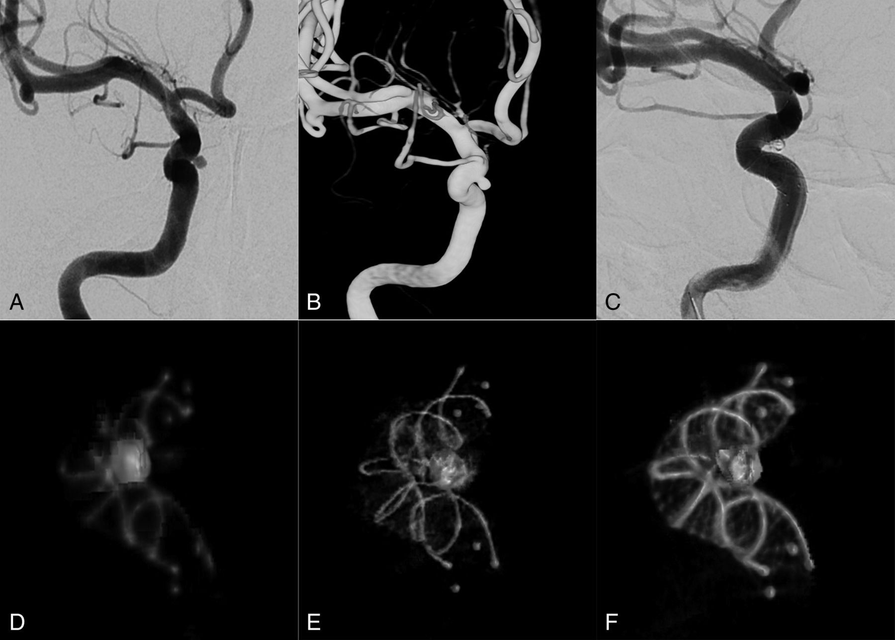
  • Fig 3.
    • Download figure
    • Open in new tab
    • Download powerpoint
    Fig 3.

    Neuroform EZ stent-assisted embolization of a ruptured aneurysm in the right posterior communicating artery. A and B, DSA and 3D reconstructed images show a right posterior communicating wide-neck aneurysm with an irregular petal shape (tumor neck: 3.4 mm; 2 daughter sacs of 2.8 × 3.6 and 2.4 × 3.1 mm). C, Complete aneurysm embolization with the Neuroform EZ stent (3.5 × 30 mm) while maintaining artery patency. D, Normal DynaCT reconstructed images immediately after the operation show that the stent mark is clearly developed, the stent wire is poorly developed, and the coil metal artifacts are large. E, DynaCT Micro reconstructed images immediately after the operation show that the mark at the 2 ends of the stent is well developed, the stent wire is significantly well developed (relative to normal DynaCT), and the coil metal artifacts are still large; F, Reconstructed images of DynaCT Micro combined with SMART immediately after the operation show that the marks of the stent and the nickel-titanium wire are well developed, the metal artifacts of the stent wire and coils are significantly reduced, and the overall image quality is significantly improved.

Tables

  • Figures
    • View popup
    Table 1:

    Aneurysm measurements

    Mean (mm)
    Dome width5.6 ± 3.7
    Dome-to-neck ratio1.2 ± 0.7
    Proximal parent artery diameter3.6 ± 1.6
    Distal parent artery diameter3.4 ± 1.5
    • View popup
    Table 2:

    Evaluation of images obtained with DynaCT, DynaCT Micro, and DynaCT Micro combined with SMART (Neuroform EZ stent, n = 35)

    MethodsPlatinum MarksNickel-Titanium WireCoil Metal ArtifactsNickel-Titanium Wire Artifacts
    01010101
    DynaCT035350350350
    DynaCT Micro035323278287
    DynaCT Micro combined with SMART035827728629
    • View popup
    Table 3:

    Evaluation of images obtained with DynaCT, DynaCT Micro, and DynaCT Micro combined with SMART (LVIS stent, n = 72)

    MethodsDouble Helix Marker WiresNickel-Titanium WireCoil Metal ArtifactsNickel-Titanium Wire Artifacts
    01010101
    DynaCT072720720720
    DynaCT Micro072666657657
    DynaCT Micro combined with SMART072215126462943
    • View popup
    Table 4:

    Evaluation of images obtained with DynaCT, DynaCT Micro, and DynaCT Micro combined with SMART (Enterprise stent, n = 11)

    MethodsPlatinum MarksNickel-Titanium WireCoil Metal ArtifactsNickel-Titanium Wire Artifacts
    01010101
    DynaCT011110110110
    DynaCT Micro0111018392
    DynaCT Micro combined with SMART011384738
    • View popup
    Table 5:

    Comparison of the image quality in different groups (P values)

    Comparison GroupsNeuroform EZ StentLVIS StentEnterprise Stent
    Nickel-Titanium WireCoil Metal ArtifactsNickel-Titanium Wire ArtifactsNickel-Titanium WireCoil Metal ArtifactsNickel-Titanium Wire ArtifactsNickel-Titanium WireCoil Metal ArtifactsNickel-Titanium Wire Artifacts
    DynaCT vs DynaCT Micro.239.050.050.028.013.0131.214.476
    DynaCT vs DynaCT Micro combined with SMART<.001<.001<.001<.001<.001<.001.001<.004.001
    DynaCT Micro vs DynaCT Micro combined with SMART<.001<.001<.001<.001<.001<.001.008.199.030
PreviousNext
Back to top

In this issue

American Journal of Neuroradiology: 40 (10)
American Journal of Neuroradiology
Vol. 40, Issue 10
1 Oct 2019
  • Table of Contents
  • Index by author
  • Complete Issue (PDF)
Advertisement
Print
Download PDF
Email Article

Thank you for your interest in spreading the word on American Journal of Neuroradiology.

NOTE: We only request your email address so that the person you are recommending the page to knows that you wanted them to see it, and that it is not junk mail. We do not capture any email address.

Enter multiple addresses on separate lines or separate them with commas.
Application of High-Resolution C-Arm CT Combined with Streak Metal Artifact Removal Technology for the Stent-Assisted Embolization of Intracranial Aneurysms
(Your Name) has sent you a message from American Journal of Neuroradiology
(Your Name) thought you would like to see the American Journal of Neuroradiology web site.
CAPTCHA
This question is for testing whether or not you are a human visitor and to prevent automated spam submissions.
Cite this article
T.-F. Li, J. Ma, X.-W. Han, P.-J. Fu, R.-N. Niu, W.-Z. Luo, J.-Z. Ren
Application of High-Resolution C-Arm CT Combined with Streak Metal Artifact Removal Technology for the Stent-Assisted Embolization of Intracranial Aneurysms
American Journal of Neuroradiology Oct 2019, 40 (10) 1752-1758; DOI: 10.3174/ajnr.A6190

Citation Manager Formats

  • BibTeX
  • Bookends
  • EasyBib
  • EndNote (tagged)
  • EndNote 8 (xml)
  • Medlars
  • Mendeley
  • Papers
  • RefWorks Tagged
  • Ref Manager
  • RIS
  • Zotero
0 Responses
Respond to this article
Share
Bookmark this article
Application of High-Resolution C-Arm CT Combined with Streak Metal Artifact Removal Technology for the Stent-Assisted Embolization of Intracranial Aneurysms
T.-F. Li, J. Ma, X.-W. Han, P.-J. Fu, R.-N. Niu, W.-Z. Luo, J.-Z. Ren
American Journal of Neuroradiology Oct 2019, 40 (10) 1752-1758; DOI: 10.3174/ajnr.A6190
del.icio.us logo Twitter logo Facebook logo Mendeley logo
  • Tweet Widget
  • Facebook Like
  • Google Plus One
Purchase

Jump to section

  • Article
    • Abstract
    • ABBREVIATIONS:
    • Materials and Methods
    • Results
    • Discussion
    • Conclusions
    • Footnotes
    • References
  • Figures & Data
  • Info & Metrics
  • Responses
  • References
  • PDF

Related Articles

  • No related articles found.
  • PubMed
  • Google Scholar

Cited By...

  • Use of the Neuroform Atlas Stent or LVIS Jr Stent for Treatment of Unruptured Intracranial Aneurysms in Parent Arteries of <2 mm in Diameter: A Multicenter Experience
  • Correlation of Flow Diverter Malapposition at the Aneurysm Neck with Incomplete Aneurysm Occlusion in Patients with Small Intracranial Aneurysms: A Single-Center Experience
  • Crossref (4)
  • Google Scholar

This article has been cited by the following articles in journals that are participating in Crossref Cited-by Linking.

  • Correlation of Flow Diverter Malapposition at the Aneurysm Neck with Incomplete Aneurysm Occlusion in Patients with Small Intracranial Aneurysms: A Single-Center Experience
    Shuhai Long, Shuailong Shi, Qi Tian, Zhuangzhuang Wei, Ji Ma, Ye Wang, Jie Yang, Xinwei Han, Tengfei Li
    American Journal of Neuroradiology 2024 45 1
  • Clinical Efficiency of an Artificial Intelligence-Based 3D-Angiography for Visualization of Cerebral Aneurysm: Comparison with the Conventional Method
    Kojiro Ishikawa, Takashi Izumi, Masahiro Nishihori, Takahiro Imaizumi, Shunsaku Goto, Keita Suzuki, Kinya Yokoyama, Fumiaki Kanamori, Kenji Uda, Yoshio Araki, Ryuta Saito
    Clinical Neuroradiology 2023 33 4
  • Safety and Efficacy of LVIS Jr Stent-assisted Coiling of Intracranial Aneurysms in Small-diameter Parent Arteries
    Shuailong Shi, Shuhai Long, Fangfang Hui, Qi Tian, Zhuangzhuang Wei, Ji Ma, Jie Yang, Ye Wang, Xinwei Han, Tengfei Li
    Clinical Neuroradiology 2024 34 3
  • Use of the Neuroform Atlas Stent or LVIS Jr Stent for Treatment of Unruptured Intracranial Aneurysms in Parent Arteries of &lt;2 mm in Diameter: A Multicenter Experience
    Tengfei Li, Shuailong Shi, Qingliang Chen, Chao Jiang, Wenxian Feng, Qi Tian, Shuhai Long, Zhuangzhuang Wei, Jie Yang, Ye Wang, Jianzhuang Ren, Xinwei Han, Ji Ma
    American Journal of Neuroradiology 2024 45 7

More in this TOC Section

  • SAVE vs. Solumbra Techniques for Thrombectomy
  • Contrast-Induced Encephalopathy after NeuroIR
  • CT Perfusion&Reperfusion in Acute Ischemic Stroke
Show more Interventional

Similar Articles

Advertisement

Indexed Content

  • Current Issue
  • Accepted Manuscripts
  • Article Preview
  • Past Issues
  • Editorials
  • Editor's Choice
  • Fellows' Journal Club
  • Letters to the Editor
  • Video Articles

Cases

  • Case Collection
  • Archive - Case of the Week
  • Archive - Case of the Month
  • Archive - Classic Case

Special Collections

  • AJNR Awards
  • ASNR Foundation Special Collection
  • Most Impactful AJNR Articles
  • Photon-Counting CT
  • Spinal CSF Leak Articles (Jan 2020-June 2024)

More from AJNR

  • Trainee Corner
  • Imaging Protocols
  • MRI Safety Corner

Multimedia

  • AJNR Podcasts
  • AJNR Scantastics

Resources

  • Turnaround Time
  • Submit a Manuscript
  • Submit a Video Article
  • Submit an eLetter to the Editor/Response
  • Manuscript Submission Guidelines
  • Statistical Tips
  • Fast Publishing of Accepted Manuscripts
  • Graphical Abstract Preparation
  • Imaging Protocol Submission
  • Evidence-Based Medicine Level Guide
  • Publishing Checklists
  • Author Policies
  • Become a Reviewer/Academy of Reviewers
  • News and Updates

About Us

  • About AJNR
  • Editorial Board
  • Editorial Board Alumni
  • Alerts
  • Permissions
  • Not an AJNR Subscriber? Join Now
  • Advertise with Us
  • Librarian Resources
  • Feedback
  • Terms and Conditions
  • AJNR Editorial Board Alumni

American Society of Neuroradiology

  • Not an ASNR Member? Join Now

© 2025 by the American Society of Neuroradiology All rights, including for text and data mining, AI training, and similar technologies, are reserved.
Print ISSN: 0195-6108 Online ISSN: 1936-959X

Powered by HighWire