Advertisement

Main menu

  • Home
  • Content
    • Current Issue
    • Accepted Manuscripts
    • Article Preview
    • Past Issue Archive
    • Video Articles
    • AJNR Case Collection
    • Case of the Week Archive
    • Case of the Month Archive
    • Classic Case Archive
  • Special Collections
    • AJNR Awards
    • ASNR Foundation Special Collection
    • Most Impactful AJNR Articles
    • Photon-Counting CT
    • Spinal CSF Leak Articles (Jan 2020-June 2024)
  • Multimedia
    • AJNR Podcasts
    • AJNR SCANtastic
    • Trainee Corner
    • MRI Safety Corner
    • Imaging Protocols
  • For Authors
    • Submit a Manuscript
    • Submit a Video Article
    • Submit an eLetter to the Editor/Response
    • Manuscript Submission Guidelines
    • Statistical Tips
    • Fast Publishing of Accepted Manuscripts
    • Graphical Abstract Preparation
    • Imaging Protocol Submission
    • Author Policies
  • About Us
    • About AJNR
    • Editorial Board
    • Editorial Board Alumni
  • More
    • Become a Reviewer/Academy of Reviewers
    • Subscribers
    • Permissions
    • Alerts
    • Feedback
    • Advertisers
    • ASNR Home

User menu

  • Alerts
  • Log in

Search

  • Advanced search
American Journal of Neuroradiology

American Journal of Neuroradiology

ASHNR American Society of Functional Neuroradiology ASHNR American Society of Pediatric Neuroradiology ASSR
  • Alerts
  • Log in

Advanced Search

  • Home
  • Content
    • Current Issue
    • Accepted Manuscripts
    • Article Preview
    • Past Issue Archive
    • Video Articles
    • AJNR Case Collection
    • Case of the Week Archive
    • Case of the Month Archive
    • Classic Case Archive
  • Special Collections
    • AJNR Awards
    • ASNR Foundation Special Collection
    • Most Impactful AJNR Articles
    • Photon-Counting CT
    • Spinal CSF Leak Articles (Jan 2020-June 2024)
  • Multimedia
    • AJNR Podcasts
    • AJNR SCANtastic
    • Trainee Corner
    • MRI Safety Corner
    • Imaging Protocols
  • For Authors
    • Submit a Manuscript
    • Submit a Video Article
    • Submit an eLetter to the Editor/Response
    • Manuscript Submission Guidelines
    • Statistical Tips
    • Fast Publishing of Accepted Manuscripts
    • Graphical Abstract Preparation
    • Imaging Protocol Submission
    • Author Policies
  • About Us
    • About AJNR
    • Editorial Board
    • Editorial Board Alumni
  • More
    • Become a Reviewer/Academy of Reviewers
    • Subscribers
    • Permissions
    • Alerts
    • Feedback
    • Advertisers
    • ASNR Home
  • Follow AJNR on Twitter
  • Visit AJNR on Facebook
  • Follow AJNR on Instagram
  • Join AJNR on LinkedIn
  • RSS Feeds

AJNR is seeking candidates for the AJNR Podcast Editor. Read the position description.

Research ArticleInterventional

Optimizing the Quality of 4D-DSA Temporal Information

K.L. Ruedinger, E.C. Harvey, S. Schafer, M.A. Speidel and C.M. Strother
American Journal of Neuroradiology December 2019, 40 (12) 2124-2129; DOI: https://doi.org/10.3174/ajnr.A6290
K.L. Ruedinger
aFrom the Department of Biomedical Engineering (K.L.R.)
  • Find this author on Google Scholar
  • Find this author on PubMed
  • Search for this author on this site
  • ORCID record for K.L. Ruedinger
E.C. Harvey
cDepartment of Medical Physics (E.H., M.A.S.)
  • Find this author on Google Scholar
  • Find this author on PubMed
  • Search for this author on this site
  • ORCID record for E.C. Harvey
S. Schafer
eSiemens Healthineers Forchheim Germany (S.S.), Hoffman Estates, Illinois.
  • Find this author on Google Scholar
  • Find this author on PubMed
  • Search for this author on this site
  • ORCID record for S. Schafer
M.A. Speidel
cDepartment of Medical Physics (E.H., M.A.S.)
  • Find this author on Google Scholar
  • Find this author on PubMed
  • Search for this author on this site
  • ORCID record for M.A. Speidel
C.M. Strother
dDepartment of Radiology (C.M.S.), University of Wisconsin-Madison, Madison, Wisconsin
  • Find this author on Google Scholar
  • Find this author on PubMed
  • Search for this author on this site
  • ORCID record for C.M. Strother
  • Article
  • Figures & Data
  • Info & Metrics
  • Responses
  • References
  • PDF
Loading

Article Figures & Data

Figures

  • Tables
  • Fig 1.
    • Download figure
    • Open in new tab
    • Download powerpoint
    Fig 1.

    Closed-loop flow system. Setup includes a pulsatile pump, flow transducer, power injector, patient-specific vascular model, flow-diverting loop to simulate systemic vasculature, a compliance chamber, and location of the x-ray detector.
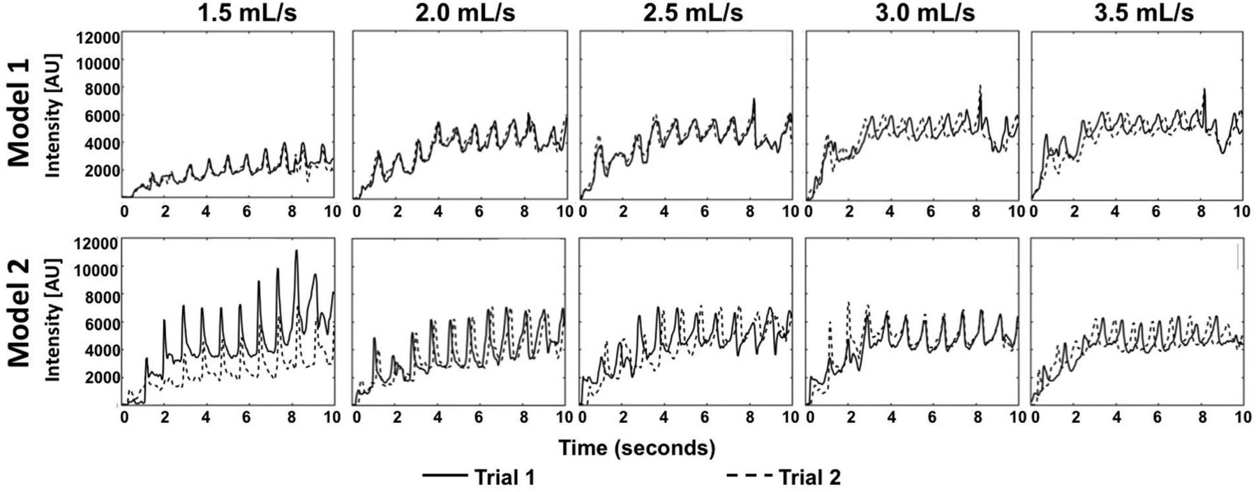
  • Fig 2.
    • Download figure
    • Open in new tab
    • Download powerpoint
    Fig 2.

    TDCs from both trials of the 5 different injection rates, 1.5, 2.0, 2.5, 3.0, 3.5 mL/s. TDCs show differences in contrast intensity from varying injection rates. Variation at the 1.5-mL/s injection rate in model 2 stems from contrast layering.

  • Fig 3.
    • Download figure
    • Open in new tab
    • Download powerpoint
    Fig 3.

    SBR and amplitude of oscillation within the TDC for model 1. The median SBR at different injection rates is presented for the entire inlet section for the combination of trials (lower bar graph). A higher SBR corresponds to stronger contrast pulsatility, with an injection rate of 2.5 mL/s producing the highest SBR. The amplitude of oscillation is studied in 2 cubes that transect the vessel diameter at 2 locations within the inlet of model 1. The median of the oscillation amplitude and median absolute derivation within each cube of voxels are presented for each trial (upper bar graphs). AU indicates Arbitrary Units, and the error bars show SBR variance from the voxels within the 2 cubes.

  • Fig 4.
    • Download figure
    • Open in new tab
    • Download powerpoint
    Fig 4.

    SBR and amplitude of oscillation within the TDC for model 2. The median SBR at different injection rates is presented for the entire inlet section for the combination of trials (lower bar graph). A high SBR represents strong contrast pulsatility; 1.5 mL/s is abnormally high due to contrast layering causing an erroneous signal in the 4D-DSA reconstruction. The amplitude of oscillation is studied in 2 cubes that transect the vessel diameter at 2 locations within the inlet of model 2. The median of the oscillation amplitude and median absolute derivation within each cube of voxels are presented for each trial (upper bar graphs). AU indicates Arbitrary Units, and the error bars show SBR variance from the voxels within the 2 cubes.

  • Fig 5.
    • Download figure
    • Open in new tab
    • Download powerpoint
    Fig 5.

    Left, effect of contrast layering on volumetric reconstruction. The absolute difference (millimeters) between the inlet diameter measurement using the workstation tool and the ground truth inlet diameter from micro-CT measurements is shown in the bar graphs (right). The greatest geometric difference in both models occurs with the 1.5-mL/s injection rate. Error bars represent variance in absolute differences based on multiple measurements.

Tables

  • Figures
  • Hemodynamic parameters characterizing flow profiles achieved from various injection rates within the closed-loop flow setupa

    Injection RateQf (mL/min)Vf (cm/s)Vc (cm/s)RefRecCt
    1.5120.30101.60.69
    2160.40135.50.52
    2.525222.27200.50328.72169.30.41
    3240.60203.20.35
    3.5280.70237.00.30
    1.5120.30101.60.70
    2160.40135.50.52
    2.526022.43200.50335.05169.30.42
    3240.60203.20.35
    3.5280.70237.00.30
    • Note:—Significant figures varied depending on the hemodynamic parameter. Qf indicates flow rate of working fluid; Vf, velocity of working fluid; Vc, velocity contrast; Ref = Reynolds number of working fluid; Rec, Reynolds number of contrast.

    • ↵a Upper half refers to model 1, and bottom half refers to model 2.

PreviousNext
Back to top

In this issue

American Journal of Neuroradiology: 40 (12)
American Journal of Neuroradiology
Vol. 40, Issue 12
1 Dec 2019
  • Table of Contents
  • Index by author
  • Complete Issue (PDF)
Advertisement
Print
Download PDF
Email Article

Thank you for your interest in spreading the word on American Journal of Neuroradiology.

NOTE: We only request your email address so that the person you are recommending the page to knows that you wanted them to see it, and that it is not junk mail. We do not capture any email address.

Enter multiple addresses on separate lines or separate them with commas.
Optimizing the Quality of 4D-DSA Temporal Information
(Your Name) has sent you a message from American Journal of Neuroradiology
(Your Name) thought you would like to see the American Journal of Neuroradiology web site.
CAPTCHA
This question is for testing whether or not you are a human visitor and to prevent automated spam submissions.
Cite this article
K.L. Ruedinger, E.C. Harvey, S. Schafer, M.A. Speidel, C.M. Strother
Optimizing the Quality of 4D-DSA Temporal Information
American Journal of Neuroradiology Dec 2019, 40 (12) 2124-2129; DOI: 10.3174/ajnr.A6290

Citation Manager Formats

  • BibTeX
  • Bookends
  • EasyBib
  • EndNote (tagged)
  • EndNote 8 (xml)
  • Medlars
  • Mendeley
  • Papers
  • RefWorks Tagged
  • Ref Manager
  • RIS
  • Zotero
0 Responses
Respond to this article
Share
Bookmark this article
Optimizing the Quality of 4D-DSA Temporal Information
K.L. Ruedinger, E.C. Harvey, S. Schafer, M.A. Speidel, C.M. Strother
American Journal of Neuroradiology Dec 2019, 40 (12) 2124-2129; DOI: 10.3174/ajnr.A6290
del.icio.us logo Twitter logo Facebook logo Mendeley logo
  • Tweet Widget
  • Facebook Like
  • Google Plus One
Purchase

Jump to section

  • Article
    • Abstract
    • ABBREVIATIONS:
    • MATERIALS AND METHODS
    • RESULTS
    • DISCUSSION
    • CONCLUSIONS
    • Footnotes
    • References
  • Figures & Data
  • Info & Metrics
  • Responses
  • References
  • PDF

Related Articles

  • No related articles found.
  • PubMed
  • Google Scholar

Cited By...

  • 4D DSA: technical addition or big revolution?
  • 4D-DSA: Development and Current Neurovascular Applications
  • Interrater Reliability in the Measurement of Flow Characteristics on Color-Coded Quantitative DSA of Brain AVMs
  • Crossref (6)
  • Google Scholar

This article has been cited by the following articles in journals that are participating in Crossref Cited-by Linking.

  • 4D-DSA: Development and Current Neurovascular Applications
    K.L. Ruedinger, S. Schafer, M.A. Speidel, C.M. Strother
    American Journal of Neuroradiology 2021 42 2
  • Interrater Reliability in the Measurement of Flow Characteristics on Color-Coded Quantitative DSA of Brain AVMs
    K.H Narsinh, K. Mueller, J. Nelson, J. Massachi, D.C. Murph, A.Z. Copelan, S.W. Hetts, V.V. Halbach, R.T. Higashida, A.A. Abla, M.R. Amans, C.F. Dowd, H. Kim, D.L. Cooke
    American Journal of Neuroradiology 2020 41 12
  • Recent progress understanding pathophysiology and genesis of brain AVM—a narrative review
    Hans-Jakob Steiger
    Neurosurgical Review 2021 44 6
  • Machine Learning Application With Quantitative Digital Subtraction Angiography for Detection of Hemorrhagic Brain Arteriovenous Malformations
    Jia-Sheng Hong, Chung-Jung Lin, Yue-Hsin Lin, Cheng-Chia Lee, Huai-Che Yang, Ling-Hsuan Meng, Te-Ming Lin, Yong-Sin Hu, Wan-Yuo Guo, Wei-Fa Chu, Yu-Te Wu
    IEEE Access 2020 8
  • Estimating blood flow velocity using time-resolved 3D angiography and a derived physical law of contrast media
    Ko-Kung Chen, Chung-Jung Lin, Wan-Yuo Guo, Wei-Fa Chu, Yu-Te Wu
    Physiological Measurement 2021 42 2
  • 4D DSA: technical addition or big revolution?
    Isil Saatci, H Saruhan Cekirge
    Journal of NeuroInterventional Surgery 2021 13 11

More in this TOC Section

  • SAVE vs. Solumbra Techniques for Thrombectomy
  • Contrast-Induced Encephalopathy after NeuroIR
  • CT Perfusion&Reperfusion in Acute Ischemic Stroke
Show more Interventional

Similar Articles

Advertisement

Indexed Content

  • Current Issue
  • Accepted Manuscripts
  • Article Preview
  • Past Issues
  • Editorials
  • Editor's Choice
  • Fellows' Journal Club
  • Letters to the Editor
  • Video Articles

Cases

  • Case Collection
  • Archive - Case of the Week
  • Archive - Case of the Month
  • Archive - Classic Case

Special Collections

  • AJNR Awards
  • ASNR Foundation Special Collection
  • Most Impactful AJNR Articles
  • Photon-Counting CT
  • Spinal CSF Leak Articles (Jan 2020-June 2024)

More from AJNR

  • Trainee Corner
  • Imaging Protocols
  • MRI Safety Corner

Multimedia

  • AJNR Podcasts
  • AJNR Scantastics

Resources

  • Turnaround Time
  • Submit a Manuscript
  • Submit a Video Article
  • Submit an eLetter to the Editor/Response
  • Manuscript Submission Guidelines
  • Statistical Tips
  • Fast Publishing of Accepted Manuscripts
  • Graphical Abstract Preparation
  • Imaging Protocol Submission
  • Evidence-Based Medicine Level Guide
  • Publishing Checklists
  • Author Policies
  • Become a Reviewer/Academy of Reviewers
  • News and Updates

About Us

  • About AJNR
  • Editorial Board
  • Editorial Board Alumni
  • Alerts
  • Permissions
  • Not an AJNR Subscriber? Join Now
  • Advertise with Us
  • Librarian Resources
  • Feedback
  • Terms and Conditions
  • AJNR Editorial Board Alumni

American Society of Neuroradiology

  • Not an ASNR Member? Join Now

© 2025 by the American Society of Neuroradiology All rights, including for text and data mining, AI training, and similar technologies, are reserved.
Print ISSN: 0195-6108 Online ISSN: 1936-959X

Powered by HighWire