Advertisement

Main menu

  • Home
  • Content
    • Current Issue
    • Accepted Manuscripts
    • Article Preview
    • Past Issue Archive
    • Video Articles
    • AJNR Case Collection
    • Case of the Week Archive
    • Case of the Month Archive
    • Classic Case Archive
  • Special Collections
    • AJNR Awards
    • ASNR Foundation Special Collection
    • Most Impactful AJNR Articles
    • Photon-Counting CT
    • Spinal CSF Leak Articles (Jan 2020-June 2024)
  • Multimedia
    • AJNR Podcasts
    • AJNR SCANtastic
    • Trainee Corner
    • MRI Safety Corner
    • Imaging Protocols
  • For Authors
    • Submit a Manuscript
    • Submit a Video Article
    • Submit an eLetter to the Editor/Response
    • Manuscript Submission Guidelines
    • Statistical Tips
    • Fast Publishing of Accepted Manuscripts
    • Graphical Abstract Preparation
    • Imaging Protocol Submission
    • Author Policies
  • About Us
    • About AJNR
    • Editorial Board
    • Editorial Board Alumni
  • More
    • Become a Reviewer/Academy of Reviewers
    • Subscribers
    • Permissions
    • Alerts
    • Feedback
    • Advertisers
    • ASNR Home

User menu

  • Alerts
  • Log in

Search

  • Advanced search
American Journal of Neuroradiology

American Journal of Neuroradiology

ASHNR American Society of Functional Neuroradiology ASHNR American Society of Pediatric Neuroradiology ASSR
  • Alerts
  • Log in

Advanced Search

  • Home
  • Content
    • Current Issue
    • Accepted Manuscripts
    • Article Preview
    • Past Issue Archive
    • Video Articles
    • AJNR Case Collection
    • Case of the Week Archive
    • Case of the Month Archive
    • Classic Case Archive
  • Special Collections
    • AJNR Awards
    • ASNR Foundation Special Collection
    • Most Impactful AJNR Articles
    • Photon-Counting CT
    • Spinal CSF Leak Articles (Jan 2020-June 2024)
  • Multimedia
    • AJNR Podcasts
    • AJNR SCANtastic
    • Trainee Corner
    • MRI Safety Corner
    • Imaging Protocols
  • For Authors
    • Submit a Manuscript
    • Submit a Video Article
    • Submit an eLetter to the Editor/Response
    • Manuscript Submission Guidelines
    • Statistical Tips
    • Fast Publishing of Accepted Manuscripts
    • Graphical Abstract Preparation
    • Imaging Protocol Submission
    • Author Policies
  • About Us
    • About AJNR
    • Editorial Board
    • Editorial Board Alumni
  • More
    • Become a Reviewer/Academy of Reviewers
    • Subscribers
    • Permissions
    • Alerts
    • Feedback
    • Advertisers
    • ASNR Home
  • Follow AJNR on Twitter
  • Visit AJNR on Facebook
  • Follow AJNR on Instagram
  • Join AJNR on LinkedIn
  • RSS Feeds

AJNR is seeking candidates for the AJNR Podcast Editor. Read the position description.

Research ArticleInterventional
Open Access

Histologic and Biomolecular Similarities in Healing between Aneurysms and Cutaneous Skin Wounds

J.R. Ayers-Ringler, Z. Khashim, Y.-H. Ding, D.F. Kallmes and R. Kadirvel
American Journal of Neuroradiology June 2019, 40 (6) 1018-1021; DOI: https://doi.org/10.3174/ajnr.A6046
J.R. Ayers-Ringler
aFrom the Department of Radiology, Mayo Clinic, Saint Mary's Hospital, Rochester, Minnesota.
  • Find this author on Google Scholar
  • Find this author on PubMed
  • Search for this author on this site
  • ORCID record for J.R. Ayers-Ringler
Z. Khashim
aFrom the Department of Radiology, Mayo Clinic, Saint Mary's Hospital, Rochester, Minnesota.
  • Find this author on Google Scholar
  • Find this author on PubMed
  • Search for this author on this site
  • ORCID record for Z. Khashim
Y.-H. Ding
aFrom the Department of Radiology, Mayo Clinic, Saint Mary's Hospital, Rochester, Minnesota.
  • Find this author on Google Scholar
  • Find this author on PubMed
  • Search for this author on this site
  • ORCID record for Y.-H. Ding
D.F. Kallmes
aFrom the Department of Radiology, Mayo Clinic, Saint Mary's Hospital, Rochester, Minnesota.
  • Find this author on Google Scholar
  • Find this author on PubMed
  • Search for this author on this site
  • ORCID record for D.F. Kallmes
R. Kadirvel
aFrom the Department of Radiology, Mayo Clinic, Saint Mary's Hospital, Rochester, Minnesota.
  • Find this author on Google Scholar
  • Find this author on PubMed
  • Search for this author on this site
  • ORCID record for R. Kadirvel
  • Article
  • Figures & Data
  • Info & Metrics
  • Responses
  • References
  • PDF
Loading

Abstract

SUMMARY: The poorly understood mechanisms of aneurysm healing contribute substantially to the pressing medical problem of coiled aneurysm recanalization. Using an established saccular aneurysm model, we developed an animal model system in rabbits to study aneurysm and skin wound healing concurrently in the same animal. We found treated aneurysm healing to be similar to skin wound healing both histologically and in biomarker gene and protein expression, but in a delayed fashion.

ABBREVIATIONS:

GAPDH
glyceraldehyde phosphate dehydrogenase
H&E
hematoxylin-eosin
SMA1
α smooth muscle actin
TGFβ1
transforming growth factor β 1
PDGF
platelet derived growth factor
VEGF
vascular endothelial growth factor

Despite wide use and considerable safety of endovascular coiling of intracranial aneurysms, the recurrence rate is high even with second-generation coils,1 partially due to a dearth of knowledge about healing mechanisms involved.2 Conversely, cutaneous wound healing is well-established. The 4 distinct-yet-overlapping stages of cutaneous wound healing (hemostasis, inflammation, proliferation, and remodeling) have been thoroughly described.3 Here we demonstrate that concurrent study, in the same research subjects, of endovascular-versus-cutaneous healing provides insight into deficiencies in the cellular, molecular, and humoral features in aneurysm healing, enabling elucidation of potential targets for improved outcomes.

Materials and Methods

Aneurysm Creation and Coil Embolization

All procedures were approved by the Institutional Animal Care and Use Committee before study initiation. Elastase-induced saccular aneurysms were created at the origin of the right common carotid artery in 12 female New Zealand white rabbits.4 At least 3 weeks later, aneurysms were treated with platinum coils. DSA was performed after embolization.

Cutaneous Ear Wound Creation

Immediately after aneurysm treatment, an ear skin defect was created on the ventral side of 1 ear using a 1-cm stainless steel punch,5 with removal of skin down to the cartilage. Hemostasis was achieved with pressure, followed by occlusive dressing (TegaDerm; 3M, St. Paul, Minnesota). Ear wounds were assessed visually daily for healing and wound closure rate.

Follow-Up Imaging and Tissue Harvest

At days 1, 5, 14, and 30 (groups 1–4) following treatment, DSA was performed from a left femoral approach, and the animals were euthanized. The aneurysm, ear wound, and contralateral control tissues were harvested for histology or cut in 2 for biomolecular studies.

Angiographic Analysis

Aneurysm occlusions at follow-up were rated either stable or progressive, or recanalizated compared with posttreatment angiograms using the modified Raymond scale.6

Histologic Analysis

Formalin-fixed aneurysms were evaluated for the degree of neck tissue coverage.7 Following a modified histologic technique, aneurysms and ear wounds were sectioned and stained with hematoxylin-eosin (H&E) or Masson Trichrome for collagen. A semiquantitative scoring for thrombus organization, neoepithelialization, inflammation, cellularity, and collagen deposition was performed by a blinded reviewer. In addition, an ordinal grading system was used to evaluate histologic healing of aneurysms between groups.

RNA Extraction and Expression Analysis by Real-Time Polymerase Chain Reaction

Total RNA was isolated and converted to complementary DNA followed by real-time polymerase chain reaction with SYBR greenER (www.thermofisher.com). Target gene expression (Table) was normalized to glyceraldehyde phosphate dehydrogenase (GAPDH) and reported as a fold change relative to control tissues (contralateral tissues) using the 2-ΔΔCt method.

View this table:
  • View inline
  • View popup

Genes important in cutaneous wound healing

Protein Extraction and Analysis by Sodium Dodecyl Sulfate Polyacrylamide Gel Electrophoresis and Western Blot

Soluble proteins were extracted from tissue samples with RIPA buffer (https://www.thermofisher.com/order/catalog/product/89900). Protein separation and western analysis were performed using a Wes system (ProteinSimple, San Jose, California). Protein expression data were normalized to control proteins (GAPDH or β-actin) using Compass Software (http://www.compass-software.de/en-us) and reported as fold change relative to control tissues.

Statistical Analysis

Continuous variables were compared using a Student t test, and categoric variables were compared using the Fisher exact test.

Results

Aneurysm Angiography

Among the first 3 groups, mean aneurysm neck, width, and height were 3.3 ± 1.8 mm, 4.0 ± 1.8 mm, and 9.8 ± 1.6 mm, respectively. The aneurysms were completely occluded immediately after coil embolization in 11 of 12 cases (92%), the other being incompletely occluded (8%). Two of the 3 aneurysms in the 24-hour group, and 1 in the 14-day group showed recanalization at sacrifice. All the other aneurysms (9 of 12) remained completely occluded (75%) (Fig 1).

Fig 1.
  • Download figure
  • Open in new tab
  • Download powerpoint
Fig 1.

Angiograms with H&E-stained tissue sections. Elastase-induced aneurysm (A) on the day of platinum coil embolization and ear wound creation. Angiogram of coiled aneurysm at 14 days (B) with a corresponding ear wound (C, H&E, magnification ×100), macro image of the ear wound on day of sacrifice (D, ruler markings are in centimeters, the smallest ticks in millimeters). Angiogram of 30-day coiled aneurysm (E) with corresponding aneurysm (F, H&E, magnification ×200). Note diffuse, dense myofibroblast (long, spindle-shaped) proliferation in both H&E photomicrographs.

Histology

Histologic analysis revealed necrosis and inflammatory cell infiltration in both epidermal and dermal layers of ear wounds at both 24 hours and 5 days, whereas scab formation in the epidermis and diffuse, dense cellular proliferation of myofibroblasts within the dermal layer with minimal inflammatory cells was found by 14 days. Aneurysm features were consistent with previous findings,8 with sparse myofibroblasts at the periphery of the aneurysm dome by 14 days, increasing dramatically by 30 days (Fig 1D, -F).

Biomolecules

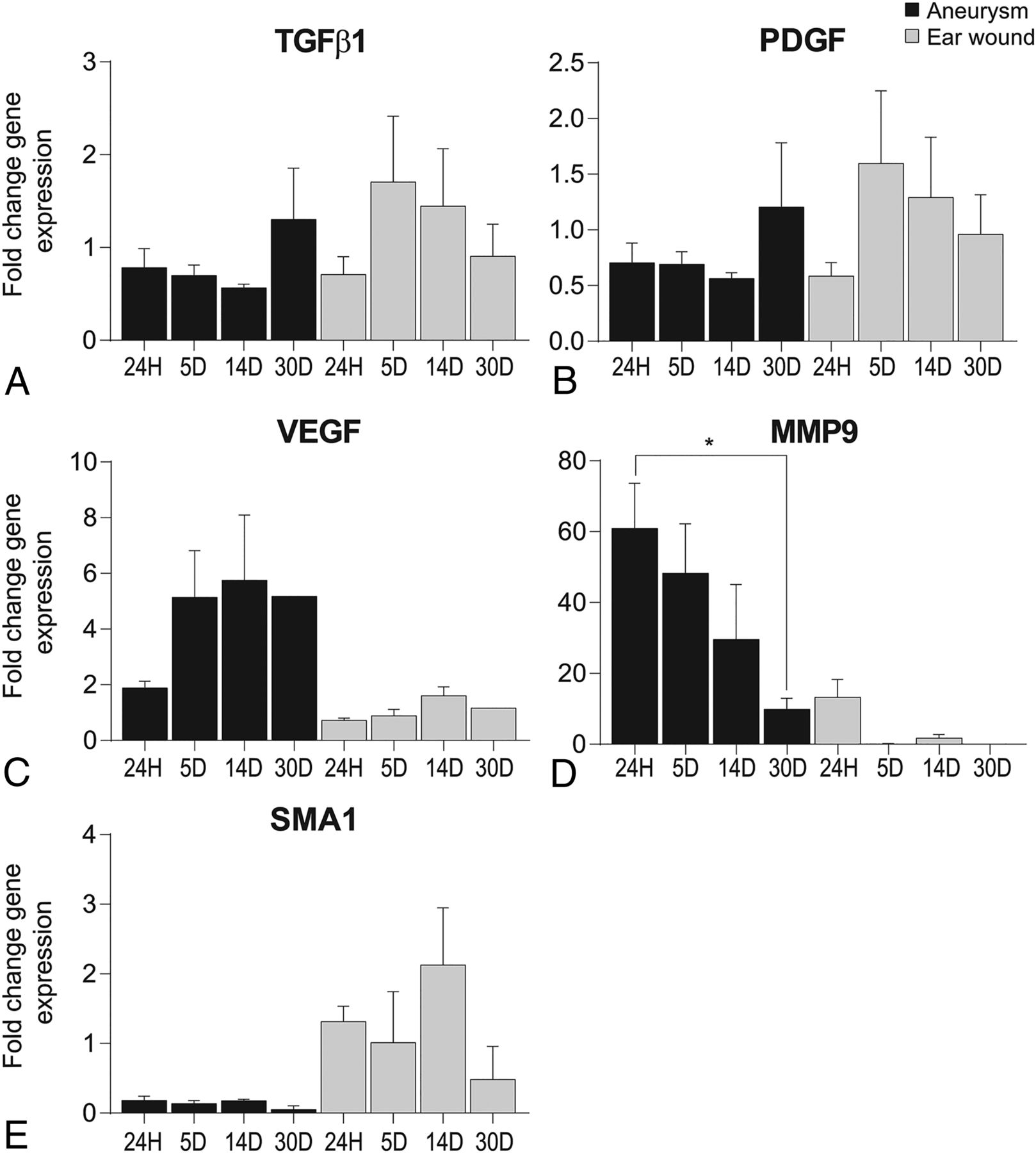
Inflammatory marker gene expression was elevated at 5 days in ear wounds but reduced/delayed until 30 days in aneurysms (Fig 2A, -B). Protein expression, however, was only 2-fold for transforming growth factor β1 (TGFβ1) at 24 hours and 30 days in ear wounds (data not shown). Proliferation marker expressions in aneurysms were higher compared with ear wounds (Fig 2C, -D) and with inflammatory markers, overall. α smooth-muscle actin (SMA1) remodeling markerexpressions were relatively unremarkable, with a small increase at 14 days in ear wounds (Fig 2E). Macrophage analysis showed an overall M1 polarization, with peak CD86 levels at 24 hours in aneurysms and 5 days in ear wounds (data not shown).

Fig 2.
  • Download figure
  • Open in new tab
  • Download powerpoint
Fig 2.

Gene expression data from aneurysms and cutaneous ear wounds after 24 hours (24H), 5 days (5D), and 14 days (14D). Data were normalized to GAPDH and displayed as a fold change relative to normal tissues (left carotid artery or contralateral ear, respectively) from the same animal (P = .0422 in D) (n = 3 animals, 3 experimental replicates). PDGF indicates platelet-derived growth factor; VEGF, vascular endothelial growth factor; MMP9, matrix metalloproteinase 9; 30D, 30 days.

Discussion

Our results reveal similarities and differences in wound-healing mechanisms for these 2 disparate environments. Histologically, skin wounds at 2 weeks resemble aneurysms at 1 month following platinum coil treatment,8 suggesting a roughly 2-week healing delay in coiled aneurysms relative to skin wounds. Previous studies8 showed similar findings, supporting this issue of deficient immune cell infiltration and immune-driven healing mechanisms in platinum-coiled aneurysms.2 The biomolecular profiles reflect this healing delay and confirm an M1 polarization in the macrophage profile, indicating a deficient prohealing phenotype.9

Conclusions

This novel animal model enabling longitudinal study of platinum-coiled aneurysm healing in parallel with cutaneous wound healing provides an exceptional level of control and will help determine key signaling events necessary for proper arterial wound healing, promising development of improved treatments.

Acknowledgments

We thank Chunfeng Zhao, MD, Praveen Kolumam Parameswaran, and Daiying Dai for essential work done for this article.

Footnotes

  • Disclosures: David F. Kallmes—UNRELATED: Stock/Stock Options: Superior Medical Experts, Comments: founder/shareholder. Ram Kadirvel—RELATED: Grant: National Institutes of Health, Comments: grant No. NS076491.* *Money paid to the institution.

  • This work was supported by Mayo Clinic Radiology Research and R01 grant number NS076491.

Indicates open access to non-subscribers at www.ajnr.org

REFERENCES

  1. 1.↵
    1. Broeders JA,
    2. Ahmed Ali U,
    3. Molyneux AJ, et al
    . Bioactive versus bare platinum coils for the endovascular treatment of intracranial aneurysms: systematic review and meta-analysis of randomized clinical trials. J Neurointerv Surg 2016;8:898–908 doi:10.1136/neurintsurg-2015-011881 pmid:26359214
    Abstract/FREE Full Text
  2. 2.↵
    1. Brinjikji W,
    2. Kallmes DF,
    3. Kadirvel R
    . Mechanisms of healing in coiled intracranial aneurysms: a review of the literature. AJNR Am J Neuroradiol 2015;36:1216–22 doi:10.3174/ajnr.A4175
    Abstract/FREE Full Text
  3. 3.↵
    1. Baum CL,
    2. Arpey C. J
    . Normal cutaneous wound healing: clinical correlation with cellular and molecular events. Dermatol Surg 2005;31:674–86; discussion 686 doi:10.1097/00042728-200506000-00011 pmid:15996419
    CrossRefPubMed
  4. 4.↵
    1. Altes TA,
    2. Cloft HJ,
    3. Short JG, et al
    . 1999 ARRS Executive Council Award: creation of saccular aneurysms in the rabbit—a model suitable for testing endovascular devices. American Roentgen Ray Society. AJR Am J Roentgenol 2000;174:349–54 doi:10.2214/ajr.174.2.1740349 pmid:10658703
    CrossRefPubMed
  5. 5.↵
    1. Houdek MT,
    2. Wyles CC,
    3. Stalboerger PG, et al
    . Collagen and fractionated platelet-rich plasma scaffold for dermal regeneration. Plast Reconstr Surg 2016;137:1498–1506 doi:10.1097/PRS.0000000000002094 pmid:27119924
    CrossRefPubMed
  6. 6.↵
    1. Raymond J,
    2. Salazkin I,
    3. Georganos S, et al
    . Endovascular treatment of experimental wide neck aneurysms: comparison of results using coils or cyanoacrylate with the assistance of an aneurysm neck bridge device. AJNR Am J Neuroradiol 2002;23:1710–16 pmid:12427629
    Abstract/FREE Full Text
  7. 7.↵
    1. Dai D,
    2. Ding H,
    3. Lewis DA, et al
    . A proposed ordinal scale for grading histology in elastase-induced, saccular aneurysms. AJNR Am J Neuroradiol 2006;27:132–38 pmid:16418372
    Abstract/FREE Full Text
  8. 8.↵
    1. Dai D,
    2. Ding YH,
    3. Kadirvel R, et al
    . A longitudinal immunohistochemical study of the healing of experimental aneurysms after embolization with platinum coils. AJNR Am J Neuroradiol 2006;27:736–41 pmid:16611755
    Abstract/FREE Full Text
  9. 9.↵
    1. Hoh BL,
    2. Hosaka K,
    3. Downes DP, et al
    . Monocyte chemotactic protein-1 promotes inflammatory vascular repair of murine carotid aneurysms via a macrophage inflammatory protein-1α and macrophage inflammatory protein-2-dependent pathway. Circulation 2011;124:2243–52 doi:10.1161/circulationaha.111.036061 pmid:22007074
    Abstract/FREE Full Text
  • Received March 13, 2019.
  • Accepted after revision March 19, 2019.
  • © 2019 by American Journal of Neuroradiology
View Abstract
PreviousNext
Back to top

In this issue

American Journal of Neuroradiology: 40 (6)
American Journal of Neuroradiology
Vol. 40, Issue 6
1 Jun 2019
  • Table of Contents
  • Index by author
  • Complete Issue (PDF)
Advertisement
Print
Download PDF
Email Article

Thank you for your interest in spreading the word on American Journal of Neuroradiology.

NOTE: We only request your email address so that the person you are recommending the page to knows that you wanted them to see it, and that it is not junk mail. We do not capture any email address.

Enter multiple addresses on separate lines or separate them with commas.
Histologic and Biomolecular Similarities in Healing between Aneurysms and Cutaneous Skin Wounds
(Your Name) has sent you a message from American Journal of Neuroradiology
(Your Name) thought you would like to see the American Journal of Neuroradiology web site.
CAPTCHA
This question is for testing whether or not you are a human visitor and to prevent automated spam submissions.
Cite this article
J.R. Ayers-Ringler, Z. Khashim, Y.-H. Ding, D.F. Kallmes, R. Kadirvel
Histologic and Biomolecular Similarities in Healing between Aneurysms and Cutaneous Skin Wounds
American Journal of Neuroradiology Jun 2019, 40 (6) 1018-1021; DOI: 10.3174/ajnr.A6046

Citation Manager Formats

  • BibTeX
  • Bookends
  • EasyBib
  • EndNote (tagged)
  • EndNote 8 (xml)
  • Medlars
  • Mendeley
  • Papers
  • RefWorks Tagged
  • Ref Manager
  • RIS
  • Zotero
0 Responses
Respond to this article
Share
Bookmark this article
Histologic and Biomolecular Similarities in Healing between Aneurysms and Cutaneous Skin Wounds
J.R. Ayers-Ringler, Z. Khashim, Y.-H. Ding, D.F. Kallmes, R. Kadirvel
American Journal of Neuroradiology Jun 2019, 40 (6) 1018-1021; DOI: 10.3174/ajnr.A6046
del.icio.us logo Twitter logo Facebook logo Mendeley logo
  • Tweet Widget
  • Facebook Like
  • Google Plus One
Purchase

Jump to section

  • Article
    • Abstract
    • ABBREVIATIONS:
    • Materials and Methods
    • Results
    • Discussion
    • Conclusions
    • Acknowledgments
    • Footnotes
    • REFERENCES
  • Figures & Data
  • Info & Metrics
  • Responses
  • References
  • PDF

Related Articles

  • No related articles found.
  • PubMed
  • Google Scholar

Cited By...

  • No citing articles found.
  • Crossref
  • Google Scholar

This article has not yet been cited by articles in journals that are participating in Crossref Cited-by Linking.

More in this TOC Section

  • SAVE vs. Solumbra Techniques for Thrombectomy
  • Contrast-Induced Encephalopathy after NeuroIR
  • CT Perfusion&Reperfusion in Acute Ischemic Stroke
Show more Interventional

Similar Articles

Advertisement

Indexed Content

  • Current Issue
  • Accepted Manuscripts
  • Article Preview
  • Past Issues
  • Editorials
  • Editor's Choice
  • Fellows' Journal Club
  • Letters to the Editor
  • Video Articles

Cases

  • Case Collection
  • Archive - Case of the Week
  • Archive - Case of the Month
  • Archive - Classic Case

Special Collections

  • AJNR Awards
  • ASNR Foundation Special Collection
  • Most Impactful AJNR Articles
  • Photon-Counting CT
  • Spinal CSF Leak Articles (Jan 2020-June 2024)

More from AJNR

  • Trainee Corner
  • Imaging Protocols
  • MRI Safety Corner

Multimedia

  • AJNR Podcasts
  • AJNR Scantastics

Resources

  • Turnaround Time
  • Submit a Manuscript
  • Submit a Video Article
  • Submit an eLetter to the Editor/Response
  • Manuscript Submission Guidelines
  • Statistical Tips
  • Fast Publishing of Accepted Manuscripts
  • Graphical Abstract Preparation
  • Imaging Protocol Submission
  • Evidence-Based Medicine Level Guide
  • Publishing Checklists
  • Author Policies
  • Become a Reviewer/Academy of Reviewers
  • News and Updates

About Us

  • About AJNR
  • Editorial Board
  • Editorial Board Alumni
  • Alerts
  • Permissions
  • Not an AJNR Subscriber? Join Now
  • Advertise with Us
  • Librarian Resources
  • Feedback
  • Terms and Conditions
  • AJNR Editorial Board Alumni

American Society of Neuroradiology

  • Not an ASNR Member? Join Now

© 2025 by the American Society of Neuroradiology All rights, including for text and data mining, AI training, and similar technologies, are reserved.
Print ISSN: 0195-6108 Online ISSN: 1936-959X

Powered by HighWire