
Article Figures & Data
Figures
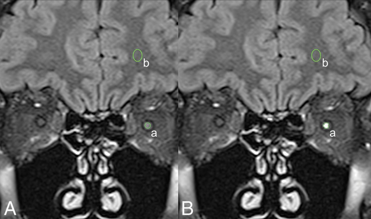
- Fig 1.
Measurement of the CNR between the optic nerve and ipsilateral white matter. Baseline (A) and processed (B) coronal FLAIR images show equivalent placement of ROIs to measure signal intensities of the optic nerve (a) and the ipsilateral white matter (b). The SD of the intensity in air around the head (c, not shown) was used as a measure of noise. CNR was defined as (a − b) / c. In this patient with left optic neuritis, processing resulted in an increase of the left optic nerve signal intensity from 355 at baseline to 754 after processing. Other measurements were unaffected by processing.
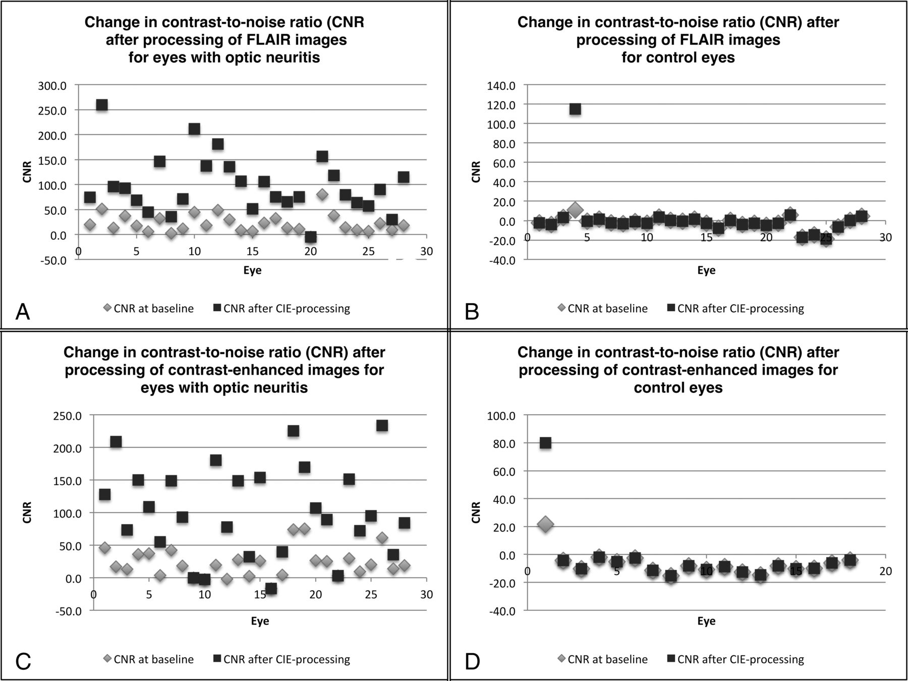
- Fig 2.
The effect of image processing on the CNR for optic nerves with and without optic neuritis. Scatterplots show the contrast-to-noise ratio of the optic nerve for the baseline images and the postprocessed images in both eyes with optic neuritis and control eyes.
- Fig 3.
Examples highlighting the effect of processing on FLAIR and contrast-enhanced T1WI in patients with optic neuritis. A, Baseline FLAIR image of a patient with left-sided optic neuritis. B, Processed version of the same FLAIR image (from A) of a patient with left-sided optic neuritis. C, Baseline FLAIR image of a patient with right-sided optic neuritis. D, Processed version of the same FLAIR image (from C) of a patient with right-sided optic neuritis. E, Baseline contrast-enhanced image of a patient with right-sided optic neuritis. F, Processed version of the same contrast-enhanced image (from E) of a patient with right-sided optic neuritis. G, Baseline contrast-enhanced image of a patient with left-sided optic neuritis. H, Processed version of the same contrast-enhanced image (from G) of a patient with left-sided optic neuritis.


Tables
- Table 1:
Technical parameters for the coronal FLAIR and coronal contrast-enhanced T1WI used in the study
Parameter FLAIR Contrast-Enhanced T1WI Slice thickness (mm) 3–4 3–4 TR (ms) 9000–10,000 400–800 TE (ms) 74–102 10–20 TI (ms) 2500 NA FOV (mm) 200–220 × 166–186 180–220 × 170–200 Matrix 256–512 × 192–384 226–512 × 288–512 Fat saturation Yes Yes Contrast material used NA Omniscan,a MultiHance,b Dotaremc - Table 2:
Effect of image processing on sensitivity and specificity for detection of optic neuritis on FLAIR and contrast-enhanced T1WI by 6 masked readers including 2 radiology residents (readers 1, 2), 2 neuroradiology fellows (readers 3, 4), and 2 attending neuroradiologists (readers 5, 6)
Reader 1 2 3 4 5 6 Average Sensitivity for baseline FLAIR image 18/28 16/28 19/28 27/28 19/28 21/28 71.4 ± 13.6 64% 57% 68% 96% 68% 75% Sensitivity for processed FLAIR image 26/28 26/28 26/28 27/28 27/28 27/28 94.6 ± 2.0 93% 93% 93% 96% 96% 96% Specificity for baseline FLAIR image 28/28 28/28 26/28 20/28 26/28 27/28 92.3 ± 10.7 100% 100% 93% 71% 93% 96% Specificity for processed FLAIR image 26/28 25/28 26/28 24/28 26/28 27/28 91.7 ± 3.7 93% 89% 93% 86% 93% 96% P valuea <.001 <.001 .02 .02 .01 .01 Sensitivity for baseline contrast-enhanced image 17/28 18/28 20/28 25/28 19/28 22/28 72.0 ± 10.5 61% 64% 71% 89% 68% 79% Sensitivity for processed contrast-enhanced image 22/28 20/28 21/28 23/28 24/28 22/28 78.6 ± 5.1 79% 71% 75% 82% 86% 79% Specificity for baseline contrast-enhanced image 18/18 18/18 18/18 13/18 17/18 18/18 94.4 ± 11.1 100% 100% 100% 72% 94% 100% Specificity for processed contrast-enhanced image 18/18 18/18 18/18 17/18 17/18 18/18 98.1 ± 2.9 100% 100% 100% 94% 94% 100% P valuea <.001 <.001 .002 .12 .01 .003 ↵a P value for improvement of diagnostic performance in the optic neuritis eyes compared with the control eyes.


