
Article Figures & Data
Figures
- FIG 1.
Flow chart.
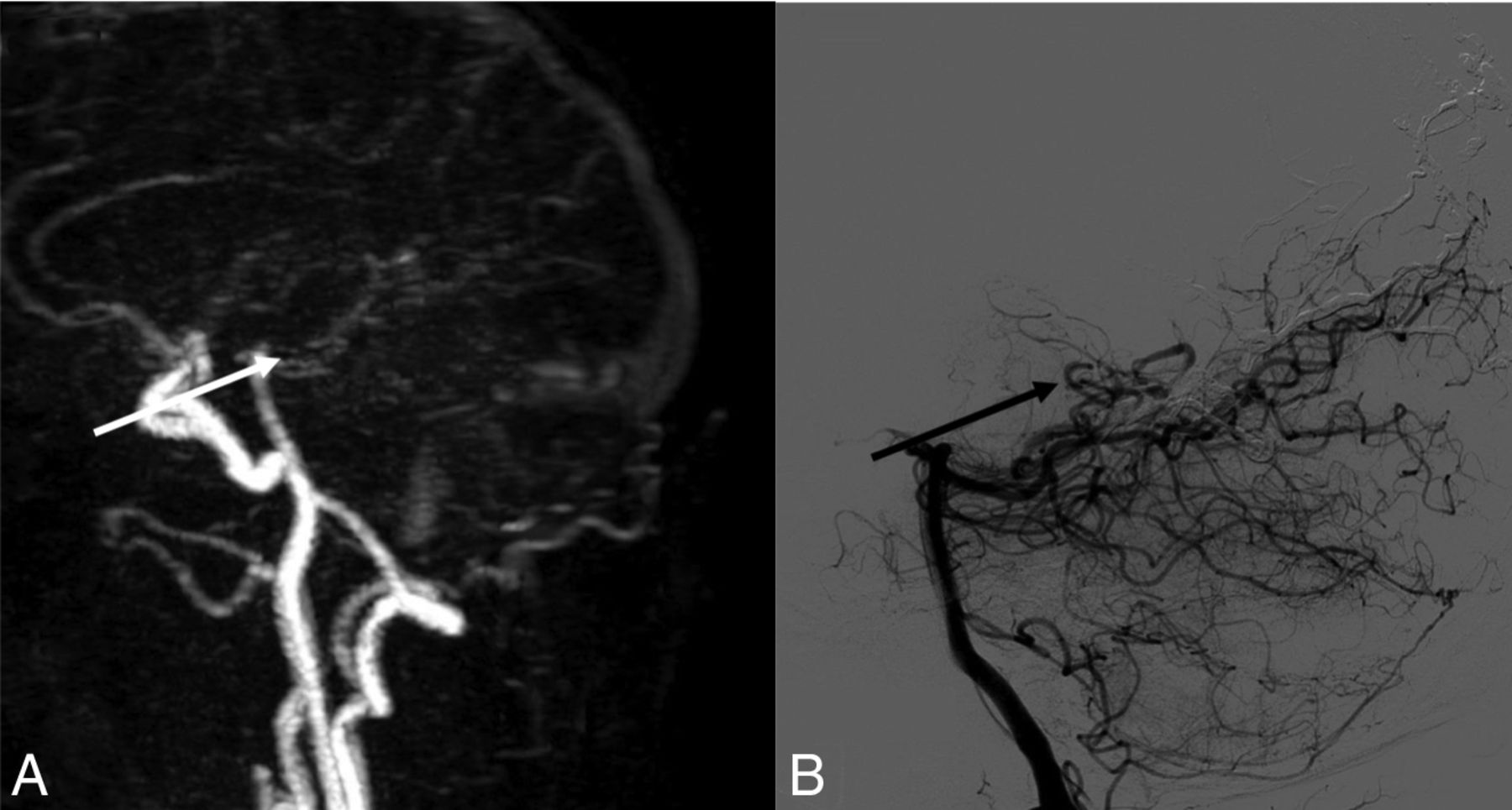
- FIG 2.
A, Sagittal MIP of 4D-MRA at arterial phase. B, Cerebral arteriography through the left vertebral artery in a sagittal view. The white arrow in A and the black arrow in B show early opacification of an epiphyseal vein, before the superior sagittal sinus, confirming an arteriovenous shunt. This examination was rated type III on both imaging modalities.
- FIG 3.
A, Sagittal MIP of 4D-MRA at late arterial phase. B, Cerebral arteriography through the left external carotid artery in a sagittal view. The black arrow in B shows early opacification of an occipital vein, confirming an arteriovenous shunt not found in A.


Tables
Dural Fistula Location 4D-MRA Classification Reader 1 4D-MRA Classification Reader 2 4D-MRA Classification Consensus DSA Classification 4D-MRA Consensus Quality Score Hemispheric right 0 III III III 3 Parasagittal right 0 III 0 0 3 SSS 0 IIa 0 0 1 Epiphyseal 0 III III III 3 Sinus lateralis left I 0 I I 3 Medulla III 0 0 V 3 Note:—SSS indicates superior sagittal sinus.
- Table 2:
Contingency table of residual/recurrent DAVF grading according to technique (4D-MRA and DSA)a
DSA 4D-MRA No Shunt Type Ib Type IIab Type IIbc Type IIa+bc Type IIIc Type IVc Type Vc No shunt 28 0 0 0 1 0 0 0 Type Ib 1 3 0 0 0 0 0 0 Type IIab 1 0 3 0 0 0 0 0 Type IIbc 0 0 1 0 0 0 0 0 Type IIa + bc 0 0 0 0 0 0 0 0 Type IIIc 4 0 0 0 0 4 3 0 Type IVc 1 0 0 0 0 0 0 0 Type Vc 1 0 0 0 0 0 0 0 - Table 3:
Intermodality discrepancy in residual/recurrent dural arteriovenous fistula detection and classificationa
Location Pretreatment Grading 4D-MRA/DSA Interval (Days) DSA Grading 4D-MRA Consensus Grading 4D-MRA Reader Disagreement (Yes/No) Year 4D-MRA Consensus Quality Score Possible Explanation Parasagittal right III 65 IV 0 No 2010 3 Confusion between occipital artery and a cortical vein Cavernous sinus right III 59 III 0 No 2015 2 Cortical vein visible but missed by 4D-MRA readers Transverse sinus left IIb 112 I 0 No 2017 2 Artery and vein overlay SSS IIb 129 0 IIa + b No 2012 3 Early drainage due to a meningioma Cavernous sinus left III 0 III 0 No 2015 3 Cortical vein visible but missed by 4D-MRA readers Posterior fossa right IV 43 III 0 No 2009 3 Early opacification of all dural sinuses; shunt missed due to misinterpretation of time sequence in 4D-MRA Sigmoid sinus left III 2 IIa 0 No 2011 2 Cortical vein visible but missed by 4D-MRA readers Medulla V 3 V 0 Yes (III/0) 2011 3 Not visible in the FOV Parieto-occipital left III 85 III 0 No 2018 2 Cortical vein visible but missed by 4D-MRA readers Note:—SSS indicates superior sagittal sinus.
↵a Grading according to Cognard et al.5


