Advertisement

Main menu

  • Home
  • Content
    • Current Issue
    • Accepted Manuscripts
    • Article Preview
    • Past Issue Archive
    • Video Articles
    • AJNR Case Collection
    • Case of the Week Archive
    • Case of the Month Archive
    • Classic Case Archive
  • Special Collections
    • AJNR Awards
    • ASNR Foundation Special Collection
    • Most Impactful AJNR Articles
    • Photon-Counting CT
    • Spinal CSF Leak Articles (Jan 2020-June 2024)
  • Multimedia
    • AJNR Podcasts
    • AJNR SCANtastic
    • Trainee Corner
    • MRI Safety Corner
    • Imaging Protocols
  • For Authors
    • Submit a Manuscript
    • Submit a Video Article
    • Submit an eLetter to the Editor/Response
    • Manuscript Submission Guidelines
    • Statistical Tips
    • Fast Publishing of Accepted Manuscripts
    • Graphical Abstract Preparation
    • Imaging Protocol Submission
    • Author Policies
  • About Us
    • About AJNR
    • Editorial Board
    • Editorial Board Alumni
  • More
    • Become a Reviewer/Academy of Reviewers
    • Subscribers
    • Permissions
    • Alerts
    • Feedback
    • Advertisers
    • ASNR Home

User menu

  • Alerts
  • Log in

Search

  • Advanced search
American Journal of Neuroradiology

American Journal of Neuroradiology

ASHNR American Society of Functional Neuroradiology ASHNR American Society of Pediatric Neuroradiology ASSR
  • Alerts
  • Log in

Advanced Search

  • Home
  • Content
    • Current Issue
    • Accepted Manuscripts
    • Article Preview
    • Past Issue Archive
    • Video Articles
    • AJNR Case Collection
    • Case of the Week Archive
    • Case of the Month Archive
    • Classic Case Archive
  • Special Collections
    • AJNR Awards
    • ASNR Foundation Special Collection
    • Most Impactful AJNR Articles
    • Photon-Counting CT
    • Spinal CSF Leak Articles (Jan 2020-June 2024)
  • Multimedia
    • AJNR Podcasts
    • AJNR SCANtastic
    • Trainee Corner
    • MRI Safety Corner
    • Imaging Protocols
  • For Authors
    • Submit a Manuscript
    • Submit a Video Article
    • Submit an eLetter to the Editor/Response
    • Manuscript Submission Guidelines
    • Statistical Tips
    • Fast Publishing of Accepted Manuscripts
    • Graphical Abstract Preparation
    • Imaging Protocol Submission
    • Author Policies
  • About Us
    • About AJNR
    • Editorial Board
    • Editorial Board Alumni
  • More
    • Become a Reviewer/Academy of Reviewers
    • Subscribers
    • Permissions
    • Alerts
    • Feedback
    • Advertisers
    • ASNR Home
  • Follow AJNR on Twitter
  • Visit AJNR on Facebook
  • Follow AJNR on Instagram
  • Join AJNR on LinkedIn
  • RSS Feeds

AJNR is seeking candidates for the AJNR Podcast Editor. Read the position description.

Review ArticleHead & Neck
Open Access

Retinoblastoma: What the Neuroradiologist Needs to Know

V.M. Silvera, J.B. Guerin, W. Brinjikji and L.A. Dalvin
American Journal of Neuroradiology April 2021, 42 (4) 618-626; DOI: https://doi.org/10.3174/ajnr.A6949
V.M. Silvera
aFrom the Departments of Neuroradiology (V.M.S., J.B.G., W.B.)
  • Find this author on Google Scholar
  • Find this author on PubMed
  • Search for this author on this site
  • ORCID record for V.M. Silvera
J.B. Guerin
aFrom the Departments of Neuroradiology (V.M.S., J.B.G., W.B.)
  • Find this author on Google Scholar
  • Find this author on PubMed
  • Search for this author on this site
  • ORCID record for J.B. Guerin
W. Brinjikji
aFrom the Departments of Neuroradiology (V.M.S., J.B.G., W.B.)
bNeurosurgery (W.B.)
  • Find this author on Google Scholar
  • Find this author on PubMed
  • Search for this author on this site
  • ORCID record for W. Brinjikji
L.A. Dalvin
cOphthalmology (L.A.D.), Mayo Clinic, Rochester, Minnesota
  • Find this author on Google Scholar
  • Find this author on PubMed
  • Search for this author on this site
  • ORCID record for L.A. Dalvin
  • Article
  • Figures & Data
  • Info & Metrics
  • Responses
  • References
  • PDF
Loading

Abstract

SUMMARY: Retinoblastoma is the most common primary intraocular tumor of childhood. Accurate diagnosis at an early stage is important to maximize patient survival, globe salvage, and visual acuity. Management of retinoblastoma is individualized based on the presenting clinical and imaging features of the tumor, and a multidisciplinary team is required to optimize patient outcomes. The neuroradiologist is a key member of the retinoblastoma care team and should be familiar with characteristic diagnostic and prognostic imaging features of this disease. Furthermore, with the adoption of intra-arterial chemotherapy as a standard of care option for globe salvage therapy in many centers, the interventional neuroradiologist may play an active role in retinoblastoma treatment. In this review, we discuss the clinical presentation of retinoblastoma, ophthalmic imaging modalities, neuroradiology imaging features, and current treatment options.

ABBREVIATIONS:

IAC
intra-arterial chemotherapy
IVC
IV chemotherapy
IvitC
intravitreal chemotherapy
EBRT
external-beam radiation therapy
OA
ophthalmic artery

Retinoblastoma is the most common primary intraocular malignancy in children. Prompt diagnosis is essential to preserving life, eye, and sight. Neuroradiologists play an important role in diagnosis, staging, and treatment of patients with retinoblastoma. In this review, we aim to educate neuroradiologists regarding retinoblastoma imaging features and basic principles of treatment.

EPIDEMIOLOGY

Retinoblastoma affects 1 in 16,000 births,1 with 8000–10,000 children diagnosed annually. There are no known geographic, racial, or sex predilections. Heritable retinoblastoma is diagnosed at a median age of 12 months and nonheritable disease at 24 months, with 80% diagnosed before 4 years of age.

CLINICAL PRESENTATION AND DIAGNOSIS

Retinoblastoma is diagnosed by physical examination and classified as International Classification of Retinoblastoma groups A through E according to increasing severity. The most common presenting feature is a white pupillary reflex called leukocoria, often recognized first by parents. Strabismus and decreased vision are also common. Patients with advanced disease can present with iris color changes, an enlarged cornea and globe, orbital inflammation, and exophthalmos. Biopsy is avoided because of a risk of tumor seeding, though aqueous biopsy is under investigation.

PATHOPHYSIOLOGY

Retinoblastoma results from a biallelic mutation of the retinoblastoma gene (RB-1) in developing retinal cells, following the 2-hit model of tumor suppressor gene inactivation.2 In the heritable form, seen in up to 50% of patients, a germline mutation is followed by a second acquired somatic mutation. In the nonheritable form, 2 somatic mutations must occur in the same gene in a single cell.

Retinoblastoma originates in the retina and can display endophytic growth into the vitreous chamber; exophytic growth into the subretinal space; or diffuse, infiltrative growth along the retina. Most tumors demonstrate both endophytic and exophytic growth and can cause retinal detachment as well as vitreous and subretinal tumor seeding.

OPHTHALMIC IMAGING

Color Fundus Photography

The first step in diagnosing retinoblastoma is a complete dilated eye examination, often performed under anesthesia. Widefield color fundus, anterior segment, and external photography facilitate serial monitoring.

Ultrasonography

Ocular ultrasonography is a critical diagnostic test with resolution approaching 150 microns using 10-MHz frequency. On B-scan ultrasonography, retinoblastoma is an attenuated, round tumor with focal areas of high internal reflectivity, corresponding to calcification, which create shadowing that persists with low sonography gain.3 Sonography biomicroscopy uses higher frequencies (50–100 MHz) to obtain anterior segment images and detect anterior segment, iris, ciliary body, and anterior chamber angle invasion.4

Fluorescein Angiography

Fluorescein angiography uses IV sodium fluorescein dye to allow direct visualization of blood flow through retinal vasculature. The dye emits green fluorescent light (520–530 nm) after excitation by blue light (465–490 nm). Key imaging features that this technique may highlight include iris neovascularization, dilated tumor-associated retinal vessels, intrinsic tumor vasculature, venous leakage, and microvasculature changes.5 Compared with completely perfused tumors, partially perfused tumors may require fewer cycles of intra-arterial chemotherapy (IAC) to achieve regression.6 Fluorescein angiography can also detect treatment-related ischemic side effects.7

Optical Coherence Tomography

Optical coherence tomography uses low-coherence light to provide a high-resolution cross-sectional image of the retina, showing details as small as 10 microns and permitting detection of clinically invisible tumors.8,9 Newer optical coherence tomography angiography can image retinal vasculature without injection of IV dye and may prove useful for future diagnosis and monitoring.10

Neuroradiology Imaging

High-resolution contrast-enhanced MR imaging is important for optimal assessment of key features that impact treatment decisions in children with retinoblastoma, such as optic nerve invasion, extraocular tumor extension, and intracranial disease. Imaging approaches differ per institution and individual preferences with variations regarding the use of small surface coils or multichannel head coils and imaging at 1.5T or 3T magnet strength. In this regard, the European Retinoblastoma Imaging Collaboration has published imaging guidelines for diagnostic evaluation of retinoblastoma that specify hardware considerations, optimal pulse sequences, and acceptable minimum spatial resolution.11 Carefully performed MR imaging can help confirm retinoblastoma diagnosis, exclude alternative diagnoses, assess tumor extent and stage, and provide long-term monitoring. Poor prognostic features of choroidal invasion and optic nerve extension cannot be reliably detected on clinical examination, so MR imaging detection of extraocular disease and intracranial tumor is crucial. MR imaging can also provide intraocular tumor detail in patients with tumor-related media opacity and can detect congenital brain anomalies in patients with retinoblastoma and 13q-deletion syndrome.12

On imaging, retinoblastoma is typically irregular in contour. On CT, retinoblastoma has intermediate attenuation with variable calcification and mild to moderate enhancement. On MR imaging, tumor is slightly hyperintense relative to vitreous on T1-weighted images, is dark on T2-weighted images, shows reduced diffusivity on diffusion-weighted images, and demonstrates heterogeneous enhancement with gadolinium. Tumor calcification may be evident as signal voids on susceptibility-weighted sequences.

Calcification is a key diagnostic feature present in approximately 95% of cases. Although CT has been used historically to detect tumor calcification, ultrasonography has good sensitivity, and because of potential increased risk for second cancers with ionizing radiation exposure, especially in germline retinoblastoma, sonography for detection of calcification has replaced CT, and the role of CT in the evaluation of retinoblastoma is now considered obsolete.13⇓⇓-16

IMAGING APPROACH TO DISEASE

Intraocular Tumor

The smallest intraocular retinoblastomas are well-circumscribed and confined to the retina. These are typically best visualized on heavily T2-weighted 3D images, such as 3D CISS/FIESTA (Fig 1A). Larger endophytic tumors are associated with vitreous seeding and exophytic tumors with subretinal seeding. Diffusely infiltrating retinoblastoma is uncommon, accounts for approximately 2% of cases, presents at an older mean age of 6 years with inflammatory features, and demonstrates linear growth without a discrete mass or calcification.17,18

FIG 1.
  • Download figure
  • Open in new tab
  • Download powerpoint
FIG 1.

A 21-month-old boy with right group B and left group E retinoblastoma. Coronal reformat of 3D FIESTA (A) shows a hypointense calcified mass in the left globe with serous retinal detachment. Two of 3 small retinoblastoma tumors in the right globe are shown (black solid arrows). Axial post-gadolinium T1-weighted image with fat saturation (B) shows enhancing tumor abutting the normally enhancing choroid (white arrow) making focal tumor invasion challenging to assess. Tiny foci of enhancement surrounding the left optic nerve cuff represent normal small vessels (black and white arrow). The left globe was larger than the right consistent with buphthalmos. Gross pathology specimen of the enucleated left eye (C) shows the posteriorly located intraocular neoplasm (arrow). Histopathologic examination showed moderately differentiated retinoblastoma without choroidal invasion and with tumor involving the optic nerve head and lamina cribrosa, but without postlaminar tumor.

Anterior Chamber Involvement

Tumor extension into the anterior chamber is uncommon, may be evident by enhancement on MR imaging, and is associated with iris neovascularization, hyperemia, and inflammation.19,20 Anterior chamber, ciliary body, or iris invasion may necessitate enucleation.21,22

Vitreous Seeding

Vitreous involvement can range from small conglomerates of tumor cells to scattered, discrete tumor foci to diffuse involvement. High-resolution MR imaging can identify vitreous seeding as small foci of T2 signal shortening within the vitreous chamber, but clinical examination is more sensitive with a potential role for optical coherence tomography monitoring.23,24

Vitreous Hemorrhage

Vitreous hemorrhage complicates tumor assessment in children with retinoblastoma, obscuring tumor margins on clinical and MR imaging examination, and is present in fewer than 3% of cases.

Subretinal Tumor Invasion

Retinoblastoma that proliferates within the subretinal space between the retina and the choroid is termed subretinal tumor seeding. Subretinal tumor can be complicated by secondary serous retinal detachment and less frequently retinal hemorrhage (Figs 2 and 3).25

FIG 2.
  • Download figure
  • Open in new tab
  • Download powerpoint
FIG 2.

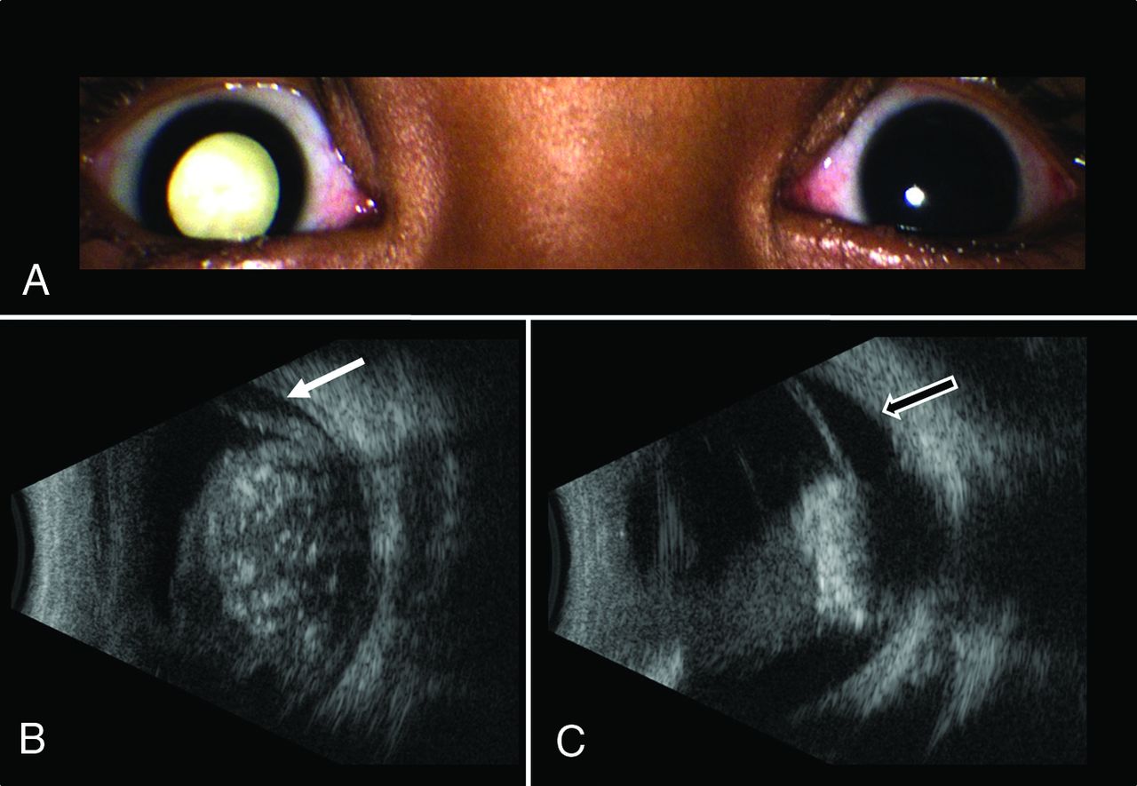
Group E retinoblastoma managed with IAC and IvitC. A 2-year-old boy presented with leukocoria in the right eye (A). Ultrasonography (B) revealed a large, round, hyperechoic retinal mass with intrinsic calcifications seen as foci of high reflectivity and serous retinal detachment (white arrow). After 3 cycles of IAC and IvitC using melphalan and topotecan, a follow-up sonography image (C) shows regression of tumor to a calcified hyperechoic scar with persistent serous retinal detachment (black arrow).

FIG 3.
  • Download figure
  • Open in new tab
  • Download powerpoint
FIG 3.

A 21-month-old boy with group E retinoblastoma of the right eye. Axial fat-saturated T2-weighted image (A) shows a large hypointense intraocular tumor (short white arrow) in the right globe with retinal detachment and hemorrhage with a fluid–fluid level (long white arrow). Abnormal thickening of the right optic nerve (arrowhead) is consistent with optic nerve invasion. Sagittal postgadolinium T1-weighted image with fat saturation (B) demonstrates enhancing tumor contacting the lens (short arrow) and tumor invading the optic nerve and extending posteriorly along the optic nerve sheath (long arrow) to the level of the optic canal (open arrow). Histopathologic examination showed poorly differentiated retinoblastoma with choroidal and extensive postlaminar optic nerve invasion.

Choroidal Invasion

Exophytic growth, breaching the Bruch membrane, can result in tumor invasion of the choroid between the retina and sclera. On MR imaging, the choroid normally appears as a linear, uniformly enhancing layer deep to the hypointense sclera. Choroidal invasion is suggested by a focal region of choroidal thickening, either enhancing or nonenhancing, or a focal region of choroidal thinning with decreased enhancement (Fig 1B).25⇓⇓-28 Subtle choroidal invasion remains challenging to discern when enhancing tumor abuts the normally enhancing choroid with accuracies estimated at 57%–72%.25,29 Massive choroidal invasion ≥3 mm is associated with increased metastatic risk, warranting enucleation and adjuvant systemic chemotherapy.30,31

Additional Intraocular Complications

Additional tumor-related complications include a subluxed or dislocated lens, an enlarged globe from secondary glaucoma (Fig 1B), a shallow anterior chamber with increased intraocular pressure, intraocular hemorrhage, and rarely phthisis bulbi.32 Advanced retinoblastoma can cause exophthalmos and periorbital soft tissue swelling from tumor necrosis that can mimic periorbital cellulitis.33

Extraocular Retinoblastoma

Extraocular tumor can manifest by extension of tumor through the sclera into the retrobulbar fat and by growth along the optic nerve and meningeal sheath (Figs 1, 3, and 4).34

FIG 4.
  • Download figure
  • Open in new tab
  • Download powerpoint
FIG 4.

A 2-year-old girl with group E retinoblastoma. Axial fat-saturated T2-weighted image (A) shows a hypointense intraocular tumor centered within the posterior right globe with retinal detachment. Axial trace DWI shows reduced diffusivity of the tumor (B). Axial postgadolinium T1-weighted image with fat saturation (C) shows enhancement of the distal optic nerve in continuity with tumor over 1.2 mm (arrow) and enhancing tumor abutting the enhancing choroid, limiting the assessment for focal choroidal invasion. Histopathologic examination showed poorly differentiated retinoblastoma (grade IV of IV) largely confined to the posterior eye with postlaminar optic nerve invasion and no choroidal invasion.

Scleral and Extrascleral Involvement

Tumor infiltration of the sclera may be apparent on MR imaging by interruption of its normally thin, linear hypointense appearance on both T1-weighted and T2-weighted images and by extrascleral extension of tumor into the retro-ocular fat. The normal discontinuity of the sclera where the optic nerve inserts into the globe should not be mistaken for pathology, though tumor can extend into the optic nerve through the lamina cribrosa at this site. Scleral invasion and extraocular tumor extension are rare in developed countries and signify a poor prognosis and increased likelihood of metastasis.35 Occasionally, enhancement of retrobulbar fat without a tumor mass is observed, which can represent peritumoral inflammation rather than extraocular tumor extension.

Optic Nerve Involvement

Assessment of the optic nerve is essential for accurate staging. Prelaminar and intralaminar invasion of the optic nerve are not considered high-risk features.34 Postlaminar optic nerve invasion occurs in approximately 8% of patients36 and typically requires enucleation followed by systemic chemotherapy because of increased risk for metastatic disease and mortality.28,34,37,38 On MR imaging, the location of the lamina can be estimated at the junction between the optic disc and the midpoint between the enhancing choroid and the low intensity sclera. A definitive diagnosis of postlaminar optic nerve invasion is based on histopathology, which may not be available if eye-preserving treatment is considered. Thickening and enhancement of the postlaminar optic nerve on MR imaging usually indicates tumor extension (Fig 4). However, more subtle abnormalities at the globe–nerve junction can be challenging to accurately classify. Enhancement of the optic nerve at the globe–nerve junction can represent postlaminar tumor, pre- or intralaminar tumor with a posteriorly displaced lamina cribrosa, central retinal vessels, or inflammation (Fig 1).20,36,39⇓⇓⇓-43 Postlaminar optic nerve invasion can be diagnosed based on visual assessment of optic nerve enhancement and tumor size with sensitivity estimated at 59% and specificity estimated at 94%.39,44,45 Lower accuracy may be related to differences in the level of suspicion, imaging technique, and the experience of the interpreting radiologist.46⇓-48 The most reliable direct MR imaging criteria to rule out advanced optic nerve invasion are normal optic nerve size, normal optic nerve signal on T2-weighted images, and optic nerve enhancement of ≤3 mm on postcontrast imaging.36,39 Normal enhancement of the central retinal vessels in the optic nerve and “double-dot” enhancement, seen as punctate foci of enhancement straddling the central retinal vessels at the globe–nerve junction, should not be mistaken for tumor.39 In addition, increased intraocular pressure can cause posterior bulging of the lamina cribrosa that can simulate optic nerve invasion and result in a false-positive interpretation.36,41 MR imaging remains limited in its ability to detect early stage optic nerve invasion with pathology remaining the criterion standard for assessing high-risk features.28,39

TREATMENT MODALITIES

Retinoblastoma treatment is individualized depending on International Classification of Retinoblastoma group staging, laterality of involvement, germline testing results, institutional resources, and psychosocial factors.49 Current treatment modalities include IV chemotherapy (IVC), IAC, intravitreal chemotherapy (IvitC), intracameral chemotherapy, consolidation therapies (cryotherapy and transpupillary thermotherapy), radiation-based therapies (external-beam radiation therapy [EBRT] and plaque radiation therapy), and enucleation.

Intravenous Chemotherapy

Systemic IVC, introduced in the 1990s, remains a key treatment tool, especially for bilateral and germline disease. The most frequently used regimen consists of vincristine, etoposide, and carboplatin (VEC), administered through a central or peripheral catheter monthly for 6 cycles.50 Tumor control is best for less advanced disease.51 Systemic chemotherapy may provide protection against second cancers, metastasis, and pineal embryonal tumor in patients with germline disease.52,53 Intensified chemotherapy regimens are used to treat high-risk disease after enucleation. Neoadjuvant chemotherapy can be used in advanced disease with choroidal or postlaminar optic nerve invasion before enucleation and may prevent exenteration in orbital retinoblastoma.54,55

Intra-arterial Chemotherapy

Introduced in 2004, IAC is now a standard of care first-line therapy, especially for advanced, unilateral tumors, and carries a lower risk of side effects such as ototoxicity and neurotoxicity associated with systemic chemotherapy.56⇓⇓-59 Melphalan, topotecan, and carboplatin are the most commonly used drugs, administered as combinations of 1, 2, or 3 agents depending on tumor severity.50 Treatment is usually administered monthly for 3 or 4 cycles. IAC can be used as a primary or secondary treatment and has higher success rates for globe salvage in advanced tumors compared with IVC (Fig 5).49,60

FIG 5.
  • Download figure
  • Open in new tab
  • Download powerpoint
FIG 5.

Group D retinoblastoma managed with IAC and IvitC. A 7-year-old boy presented with a large, white inferiorly located orbital tumor shown in the color fundus photograph (A). Ultrasonography revealed a round, hyperechoic retinal mass (B). After 4 cycles of IAC and IvitC using melphalan and topotecan, the tumor regressed to a small, calcified scar seen on fundoscopy (C) and confirmed by ultrasonography (D) (white arrow).

An experienced interventional neurosurgeon or neuroradiologist is re-quired to perform this procedure and this approach achieves superselective delivery of 10 times the chemotherapy dose to the eye compared with IVC.61 The target artery into which the chemotherapy is administered is the central retinal artery, a branch of the ophthalmic artery (OA). The central retinal artery is too small to visualize on conventional angiography, so the posterior ciliary arteries, which cause the choroid blush, are often used as the surrogate (Fig 6). The OA is the first intradural branch of the internal carotid artery and it is the dominant arterial supply to the retina in more than 98% of adults. However, during fetal life, there are robust connections between the OA and branches of the middle meningeal artery. These connections persist into early childhood, and compared with adults, there may be a higher prevalence of dual supply to the retina from both the middle meningeal artery (ie, the artery of the superior orbital fissure) and the OA (Fig 6).62 Preoperative MRA can detect anatomic variations.

FIG 6.
  • Download figure
  • Open in new tab
  • Download powerpoint
FIG 6.

Competitive flow to the choroid from the OA and middle meningeal artery (MMA). A lateral angiographic image provides reference for OA supply to the globe (A); the posterior ciliary artery is the vascular supply to the choroid and produces the choroid blush. This is the imaging surrogate for the origin of the central retinal artery. Right ICA injection (B) shows the normal OA origin (black arrow) and expected choroid blush (white arrow). Right ECA injection (C) shows the artery of the superior orbital fissure (Art SOF; black and white arrows), which anastomoses with distal branches of the OA and then fills the OA in a retrograde fashion (black arrows) indicating competitive flow between the MMA and OA for supply to the retina. MR angiogram (D) shows supply to the orbit through the artery of the superior orbital fissure (white arrow), a branch of the MMA (white and black arrow).

Regarding technical aspects, IAC delivery is a relatively straightforward procedure. At our center, we typically place a 4F Berenstein catheter (Boston Scientific) in the femoral artery and advance it to the carotid artery on the side of the lesion. Internal and external carotid artery injections are performed to identify the source of the choroid blush. After this, a microcatheter (Headway Duo, MicroVention; or Magic, Balt; 1.2F) is advanced in a flow-directed technique (ie, wireless) to the ostium of the OA, and after confirmation of positioning, the chemotherapeutic agents are administered over the course of 1 hour.

Complications can occur. Catheterization and contrast injection into the OA can trigger an oculocardiac reflex and severe bradycardia. Ocular side effects can include transient periorbital edema, cutaneous hyperemia, madarosis, blepharoptosis, scalp hair loss, and extraocular motility abnormalities.63⇓-65 More lasting vision-threatening ophthalmic vascular events include vitreous hemorrhage, retinal and choroidal ischemia with resulting atrophy, branch or central retinal artery occlusion, and OA spasm or occlusion.7,60,63,66⇓-68 Retinal detachment occurs in up to 25% of treated eyes presumably because of rapid tumor regression. The rate of serious ophthalmic vascular events that result in chorioretinal atrophy and vision loss has decreased over time and correlates with increased experience of the treating physician.68 Risks are similar when IAC is used as primary or secondary therapy.69 Complications related to chemotherapy administration include neutropenia, fever, and intraoperative bronchospasm.

Intraocular Chemotherapy

IvitC is used in combination with IAC to treat vitreous seeding. The most common drugs are melphalan and topotecan, used alone or in combination. Treatment is administered every 2–4 weeks and tailored to tumor response. Globe salvage for group E eyes has improved to >50% by combining IAC and IvitC (Fig 5).70,71 Small chemotherapy doses can be delivered to the anterior chamber (intracameral chemotherapy) for aqueous seeding.72

Focal Therapies

Focal therapies may be the primary treatment for small retinoblastomas but are typically used for consolidation in combination with IVC.73 Whereas cryotherapy with a triple-freeze–thaw technique is used for peripheral tumors, transpupillary thermotherapy with a diode laser is typically used for posteriorly located tumors that cannot be reached with the cryotherapy probe.

External-Beam Radiation Therapy

EBRT is generally avoided because of associated side effects, the most serious of which is increased risk for second primary malignancies, especially in patients with germline retinoblastoma.16,74,75 EBRT maintains a role in treating extraocular tumor extension, orbital recurrence, and a positive optic nerve margin following enucleation.76

Plaque Radiation Therapy

Plaque radiation therapy, also known as brachytherapy, delivers localized, low-dose radiation, often as secondary treatment for small- to medium-sized tumors that are chemoresistant or for localized recurrence.77 Unlike EBRT, plaque radiation therapy is not associated with increased risk for second cancers.78,79

Enucleation

Enucleation is sometimes necessary to protect the life of patients with advanced disease. Enucleation is typically reserved for advanced group E tumors, especially if there is poor visualization of the fundus or concern for optic nerve or extraocular extension.21,80⇓⇓-83 Tumors that have failed other therapies may also require enucleation. If there are high-risk features on histopathology of the enucleated globe, additional systemic chemotherapy is required for prevention of metastatic disease.84

Advanced Disease

Trilateral Retinoblastoma.

Trilateral retinoblastoma refers to the presence of intracranial tumor histologically similar to retinoblastoma in the pineal gland or suprasellar cistern (supratentorial CNS embryonal tumor) in addition to bilateral ocular involvement.85 This occurs in 10%–15% of patients with heritable retinoblastoma.86 The presence of CNS tumor in both midline locations is sometimes referred to as quadrilateral retinoblastoma (Fig 7). The incidence of trilateral retinoblastoma is 3.2%. These intracranial tumors can be synchronous with intraocular retinoblastoma at diagnosis but are more frequently found on surveillance MR imaging. The lead time from asymptomatic to symptomatic pineal trilateral tumor is approximately 1 year. The prognosis for trilateral retinoblastoma is poor.87⇓-89

FIG 7.
  • Download figure
  • Open in new tab
  • Download powerpoint
FIG 7.

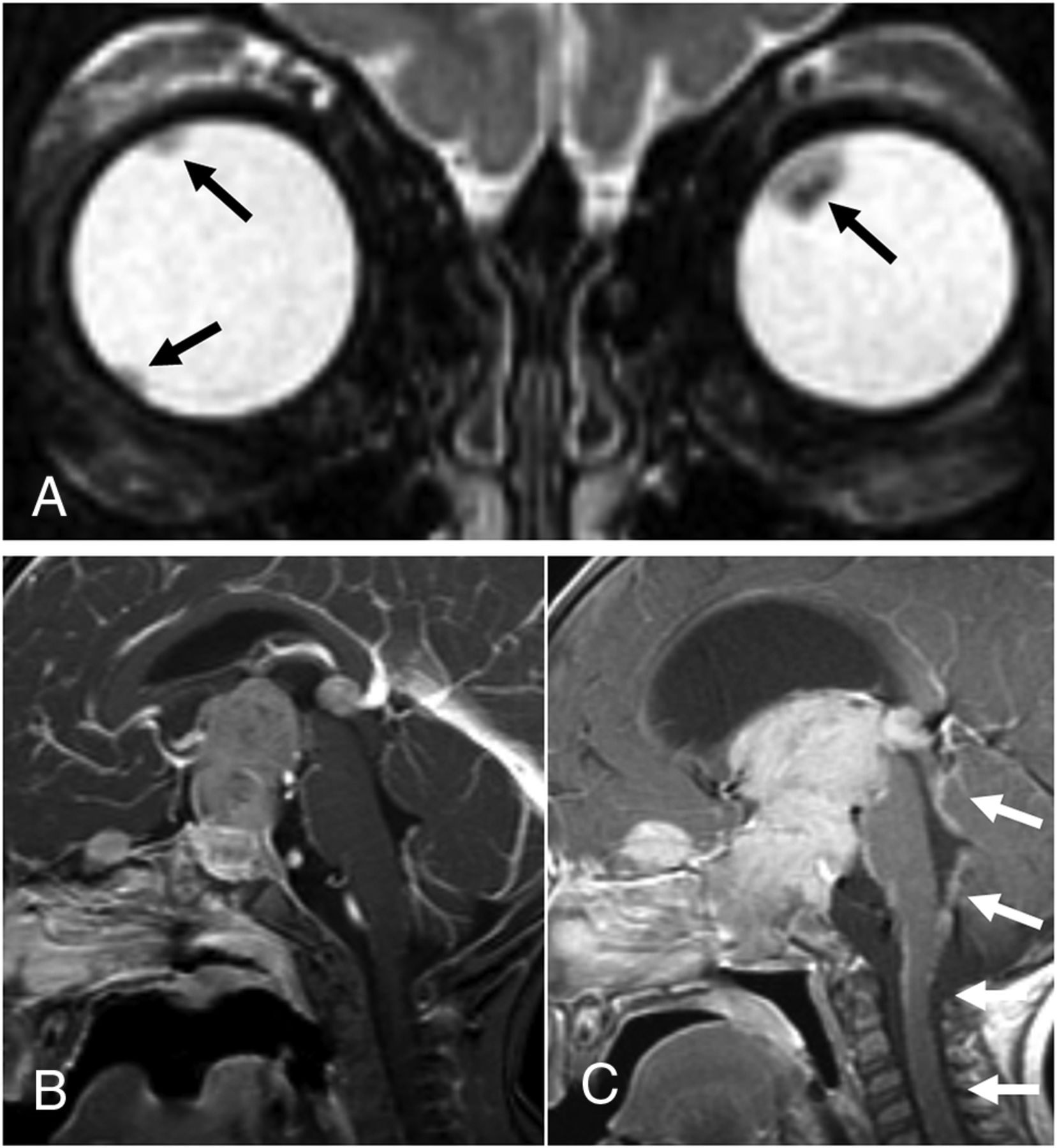
A 16-month-old boy presented with extensive metastatic disseminated quadrilateral retinoblastoma. Coronal T2-weighted image from the initial MR imaging (A) showed hypointense retinal tumors in both globes (black arrows). Sagittal postgadolinium T1-weighted image from the initial MR imaging (B) showed a large enhancing mass occupying the sella, suprasellar cistern, and third ventricle, with separate tumors involving the pineal gland and extending along the planum sphenoidale. Sagittal postgadolinium T1-weighted image from an MR imaging 5 weeks later (C) shows tumor growth, obstructive hydrocephalus, and leptomeningeal dissemination of tumor within the fourth ventricle and along the brain stem and upper cervical cord (white arrows).

Scrutiny of the pineal gland at presentation and on surveillance MR imaging is important for detecting developing pineal region tumor. It is important to not mistake pineal cysts for tumor; pineal cysts are a common incidental finding in the general pediatric population, particularly at 3T. Incidental pineal cysts may grow, have proteinaceous content, and may show enhancement of the cyst wall with mild central enhancement.90,91 Embryonal tumors of the pineal gland, on the other hand, have imaging characteristics similar to retinoblastoma and are predominantly solid and dark on T2-weighted images, enhance, and show reduced diffusivity (Fig 7).

CNS Dissemination and Metastatic Disease.

Tumor spread into the intracranial space is uncommon and carries a poor prognosis. Retinoblastoma can spread through direct extension via the optic nerve and its meningeal sheath into the intracranial compartment and can metastasize systemically via hematogenous or lymphatic routes (Fig 7).28,34 The most commonly affected lymph nodes are in the preauricular and cervical regions. Systemic metastases are most frequently observed in the lung, liver, bone, and brain.

Tumor Surveillance and Treatment-Related Findings

Posttreatment imaging includes tumor surveillance to identify local or intracranial recurrence, new intracranial or spinal CSF seeding, and development of trilateral or quadrilateral retinoblastoma. The contralateral globe must be monitored to identify metachronous tumor. Treatment-related complications include cataract, vitreous hemorrhage from radiation retinopathy, enophthalmos, phthisis bulbi, myositis, nonspecific inflammatory changes, enhancement of orbital fat and optic nerve, and orbital and midface hypoplasia from EBRT.92⇓-94 Secondary tumors are seen in heritable forms of retinoblastoma, especially after radiation. Patients with germline disease are at increased risk for osteosarcoma and soft tissue sarcoma, melanoma, and malignancies of the brain and nasal cavities. The cumulative incidence for new cancer at 50 years after retinoblastoma diagnosis is 36% for those with hereditary retinoblastoma and 5.7% for those with nonhereditary retinoblastoma.75

CONCLUSIONS

Retinoblastoma is a complex disease that requires careful work-up for accurate diagnosis and an expert, multidisciplinary team for appropriate treatment, optimized vision-sparing survival, and long-term surveillance. The neuroradiologist is a critical part of the retinoblastoma care team and should be familiar with key diagnostic and prognostic features of this disease.

Indicates open access to non-subscribers at www.ajnr.org

References

  1. 1.↵
    1. Dimaras H,
    2. Kimani K,
    3. Dimba EA, et al
    . Retinoblastoma. Lancet 2012;379:1436–46 doi:10.1016/S0140-6736(11)61137-9 pmid:22414599
    CrossRefPubMed
  2. 2.↵
    1. Dimaras H,
    2. Corson TW,
    3. Cobrinik D, et al
    . Retinoblastoma. Nat Rev Dis Primers 2015;1:15021 doi:10.1038/nrdp.2015.21 pmid:27189421
    CrossRefPubMed
  3. 3.↵
    1. Shields JA,
    2. Leonard BC,
    3. Michelson JB, et al
    . B-scan ultrasonography in the diagnosis of atypical retinoblastomas. Can J Ophthalmol 1976;11:42–51 pmid:1247938
    PubMed
  4. 4.↵
    1. Chawla B,
    2. Bhaskaran K,
    3. Dada T, et al
    . Evaluation of the role of ultrasound biomicroscopy in advanced retinoblastoma: a prospective study on Asian Indian children. Ophthalmic Genet 2020;1–6 doi:10.1080/13816810.2020.1737946 pmid:32176559
    CrossRefPubMed
  5. 5.↵
    1. Kim JW,
    2. Ngai LK,
    3. Sadda S, et al
    . Retcam fluorescein angiography findings in eyes with advanced retinoblastoma. Br J Ophthalmol 2014;98:1666–71 doi:10.1136/bjophthalmol-2014-305180 pmid:25053763
    Abstract/FREE Full Text
  6. 6.↵
    1. Lim LS,
    2. Dalvin LA,
    3. Ancona-Lezama D, et al
    . Retinoblastoma vascular perfusion and intra-arterial chemotherapy cycle requirements. Clin Exp Ophthalmol 2019;47:1164–72 doi:10.1111/ceo.13616 pmid:31432625
    CrossRefPubMed
  7. 7.↵
    1. Bianciotto C,
    2. Shields CL,
    3. Iturralde JC, et al
    . Fluorescein angiographic findings after intra-arterial chemotherapy for retinoblastoma. Ophthalmology 2012;119:843–49 doi:10.1016/j.ophtha.2011.09.040 pmid:22137042
    CrossRefPubMed
  8. 8.↵
    1. Seider MI,
    2. Grewal DS,
    3. Mruthyunjaya P
    . Portable optical coherence tomography detection or confirmation of ophthalmoscopically invisible or indeterminate active retinoblastoma. Ophthalmic Surg Lasers Imaging Retina 2016;47:965–68 doi:10.3928/23258160-20161004-12 pmid:27759865
    CrossRefPubMed
  9. 9.↵
    1. Gonzalez-Montpetit ME,
    2. Samara WA,
    3. Magrath GN, et al
    . Detection of minimally visible recurrent retinoblastoma by hand-held spectral-domain optical coherence tomography. J Pediatr Ophthalmol Strabismus 2017;54:e6–e8 doi:10.3928/01913913-20170201-03 pmid:28196267
    CrossRefPubMed
  10. 10.↵
    1. Nadiarnykh O,
    2. Davidoiu V,
    3. Grafe MGO, et al
    . Phase-based OCT angiography in diagnostic imaging of pediatric retinoblastoma patients: abnormal blood vessels in post-treatment regression patterns. Biomed Opt Express 2019;10:2213–26 doi:10.1364/BOE.10.002213 pmid:31143490
    CrossRefPubMed
  11. 11.↵
    1. de Graaf P,
    2. Göricke S,
    3. Rodjan F, et al
    . Guidelines for imaging retinoblastoma: imaging principles and MRI standardization. Pediatr Radiol 2012;42:2–14 doi:10.1007/s00247-011-2201-5 pmid:21850471
    CrossRefPubMed
  12. 12.↵
    1. Rodjan F,
    2. de Graaf P,
    3. Moll AC, et al
    . Brain abnormalities on MR imaging in patients with retinoblastoma. AJNR Am J Neuroradiol 2010;31:1385–89 doi:10.3174/ajnr.A2102 pmid:20413604
    Abstract/FREE Full Text
  13. 13.↵
    1. Galluzzi P,
    2. Hadjistilianou T,
    3. Cerase A, et al
    . Is CT still useful in the study protocol of retinoblastoma? AJNR Am J Neuroradiol 2009;30:1760–65 doi:10.3174/ajnr.A1716 pmid:19617447
    Abstract/FREE Full Text
  14. 14.↵
    1. Marees T,
    2. Moll AC,
    3. Imhof SM, et al
    . Risk of second malignancies in survivors of retinoblastoma: more than 40 years of follow-up. J Natl Cancer Inst 2008;100:1771–79 doi:10.1093/jnci/djn394 pmid:19066271
    CrossRefPubMed
  15. 15.↵
    1. Murray T
    . Cancer incidence after retinoblastoma: radiation dose and sarcoma risk. Surv Ophthalmol 1998;43:288–89 doi:10.1016/s0039-6257(98)00043-5 pmid:9862317
    CrossRefPubMed
  16. 16.↵
    1. Wong FL,
    2. Boice JD Jr.,
    3. Abramson DH, et al
    . Cancer incidence after retinoblastoma. Radiation dose and sarcoma risk. JAMA 1997;278:1262–67 doi:10.1001/jama.1997.03550150066037 pmid:9333268
    CrossRefPubMed
  17. 17.↵
    1. Brisse HJ,
    2. Lumbroso L,
    3. Freneaux PC, et al
    . Sonographic, CT, and MR imaging findings in diffuse infiltrative retinoblastoma: report of two cases with histologic comparison. AJNR Am J Neuroradiol 2001;22:499–504 pmid:11237973
    Abstract/FREE Full Text
  18. 18.↵
    1. Shields CL,
    2. Ghassemi F,
    3. Tuncer S, et al
    . Clinical spectrum of diffuse infiltrating retinoblastoma in 34 consecutive eyes. Ophthalmology 2008;115:2253–58 doi:10.1016/j.ophtha.2008.07.003 pmid:18962920
    CrossRefPubMed
  19. 19.↵
    1. de Graaf P,
    2. van der Valk P,
    3. Moll AC, et al
    . Contrast-enhancement of the anterior eye segment in patients with retinoblastoma: correlation between clinical, MR imaging, and histopathologic findings. AJNR Am J Neuroradiol 2010;31:237–45 doi:10.3174/ajnr.A1825 pmid:19833805
    Abstract/FREE Full Text
  20. 20.↵
    1. Galluzzi P,
    2. Cerase A,
    3. Hadjistilianou T, et al
    . Retinoblastoma: abnormal gadolinium enhancement of anterior segment of eyes at MR imaging with clinical and histopathologic correlation. Radiology 2003;228:683–90 doi:10.1148/radiol.2283020466 pmid:12881579
    CrossRefPubMed
  21. 21.↵
    1. Chintagumpala M,
    2. Chevez-Barrios P,
    3. Paysse EA, et al
    . Retinoblastoma: review of current management. Oncologist 2007;12:1237–46 doi:10.1634/theoncologist.12-10-1237 pmid:17962617
    Abstract/FREE Full Text
  22. 22.↵
    1. Kopelman JE,
    2. McLean IW,
    3. Rosenberg SH
    . Multivariate analysis of risk factors for metastasis in retinoblastoma treated by enucleation. Ophthalmology 1987;94:371–77 doi:10.1016/S0161-6420(87)33436-0 pmid:3587919
    CrossRefPubMed
  23. 23.↵
    1. Barkhof F,
    2. Smeets M,
    3. van der Valk P, et al
    . MR imaging in retinoblastoma. Eur Radiology 1997;7:726–31 doi:10.1007/BF02742934 pmid:9166573
    CrossRefPubMed
  24. 24.↵
    1. Schueler AO,
    2. Hosten N,
    3. Bechrakis NE, et al
    . High resolution magnetic resonance imaging of retinoblastoma. Br J Ophthalmol 2003;87:330–35 doi:10.1136/bjo.87.3.330 pmid:12598449
    Abstract/FREE Full Text
  25. 25.↵
    1. de Graaf P,
    2. Barkhof F,
    3. Moll AC, et al
    . Retinoblastoma: MR imaging parameters in detection of tumor extent. Radiology 2005;235:197–207 doi:10.1148/radiol.2351031301 pmid:15695620
    CrossRefPubMed
  26. 26.↵
    1. Rauschecker AM,
    2. Patel CV,
    3. Yeom KW, et al
    . High-resolution MR imaging of the orbit in patients with retinoblastoma. Radiographics 2012;32:1307–26 doi:10.1148/rg.325115176 pmid:22977020
    CrossRefPubMed
  27. 27.↵
    1. Sirin S,
    2. Schlamann M,
    3. Metz KA, et al
    . High-resolution MRI using orbit surface coils for the evaluation of metastatic risk factors in 143 children with retinoblastoma: part 1: MRI vs. histopathology. Neuroradiology 2015;57:805–14 doi:10.1007/s00234-015-1544-2 pmid:26041632
    CrossRefPubMed
  28. 28.↵
    1. Khelfaoui F,
    2. Validire P,
    3. Auperin A, et al
    . Histopathologic risk factors in retinoblastoma: a retrospective study of 172 patients treated in a single institution. Cancer 1996;77:1206–13 doi:10.1002/(SICI)1097-0142(19960315)77:6<1206::AID-CNCR30>3.0.CO;2-3 pmid:8635145
    CrossRefPubMed
  29. 29.↵
    1. Chawla B,
    2. Sharma S,
    3. Sen S, et al
    . Correlation between clinical features, magnetic resonance imaging, and histopathologic findings in retinoblastoma: a prospective study. Ophthalmology 2012;119:850–56 doi:10.1016/j.ophtha.2011.09.037 pmid:22218144
    CrossRefPubMed
  30. 30.↵
    1. Novetsky DE,
    2. Abramson DH,
    3. Kim JW, et al
    . Published International Classification of Retinoblastoma (ICRB) definitions contain inconsistencies—an analysis of impact. Ophthalmic Genet 2009;30:40–44 doi:10.1080/13816810802452168 pmid:19172510
    CrossRefPubMed
  31. 31.↵
    1. Chévez-Barrios P,
    2. Eagle RC Jr.,
    3. Krailo M, et al
    . Study of unilateral retinoblastoma with and without histopathologic high-risk features and the role of adjuvant chemotherapy: a Children's Oncology Group study. J Clin Oncol 2019;37:2883–91 doi:10.1200/JCO.18.01808 pmid:31539297
    CrossRefPubMed
  32. 32.↵
    1. Kashyap S,
    2. Meel R,
    3. Pushker N, et al
    . Phthisis bulbi in retinoblastoma. Clin Exp Ophthalmol 2011;39:105–10 doi:10.1111/j.1442-9071.2010.02426.x pmid:21040312
    CrossRefPubMed
  33. 33.↵
    1. Shields JA,
    2. Shields CL,
    3. Suvarnamani C, et al
    . Retinoblastoma manifesting as orbital cellulitis. Am J Ophthalmol 1991;112:442–49 doi:10.1016/s0002-9394(14)76255-5 pmid:1928248
    CrossRefPubMed
  34. 34.↵
    1. Shields CL,
    2. Shields JA,
    3. Baez K, et al
    . Optic nerve invasion of retinoblastoma. Metastatic potential and clinical risk factors. Cancer 1994;73:692–98 doi:10.1002/1097-0142(19940201)73:3<692::AID-CNCR2820730331>3.0.CO;2-8 pmid:8299091
    CrossRefPubMed
  35. 35.↵
    1. Cuenca A,
    2. Giron F,
    3. Castro D, et al
    . Microscopic scleral invasion in retinoblastoma: clinicopathological features and outcome. Arch Ophthalmol 2009;127:1006–10 doi:10.1001/archophthalmol.2009.174 pmid:19667337
    CrossRefPubMed
  36. 36.↵
    1. Brisse HJ,
    2. Guesmi M,
    3. Aerts I, et al
    . Relevance of CT and MRI in retinoblastoma for the diagnosis of postlaminar invasion with normal-size optic nerve: a retrospective study of 150 patients with histological comparison. Pediatr Radiol 2007;37:649–56 doi:10.1007/s00247-007-0491-4 pmid:17479257
    CrossRefPubMed
  37. 37.↵
    1. Li Z,
    2. Guo J,
    3. Xu X, et al
    . Diagnosis of postlaminar optic nerve invasion in retinoblastoma with MRI features. J Magn Reson Imaging 2020;51:1045–52 doi:10.1002/jmri.26961 pmid:31617290
    CrossRefPubMed
  38. 38.↵
    1. Magramm I,
    2. Abramson DH,
    3. Ellsworth RM
    . Optic nerve involvement in retinoblastoma. Ophthalmology 1989;96:217–22 doi:10.1016/S0161-6420(89)32910-1 pmid:2704542
    CrossRefPubMed
  39. 39.↵
    1. Brisse HJ,
    2. de Graaf P,
    3. Galluzzi P
    , et al. Assessment of early-stage optic nerve invasion in retinoblastoma using high-resolution 1.5 Tesla MRI with surface coils: a multicentre, prospective accuracy study with histopathological correlation. Eur Radio 2015;25:1443–52 doi:10.1007/s00330-014-3514-1 pmid:25433413
    CrossRefPubMed
  40. 40.↵
    1. Cui Y,
    2. Luo R,
    3. Wang R, et al
    . Correlation between conventional MR imaging combined with diffusion-weighted imaging and histopathologic findings in eyes primarily enucleated for advanced retinoblastoma: a retrospective study. Eur Radiol 2018;28:620–29 doi:10.1007/s00330-017-4993-7 pmid:28786011
    CrossRefPubMed
  41. 41.↵
    1. Lee BJ,
    2. Kim JH,
    3. Kim DH, et al
    . The validity of routine brain MRI in detecting post-laminar optic nerve involvement in retinoblastoma. Br J Ophthalmol 2012;96:1237–41 doi:10.1136/bjophthalmol-2012-301502
    Abstract/FREE Full Text
  42. 42.↵
    1. Khurana A,
    2. Eisenhut CA,
    3. Wan W, et al
    . Comparison of the diagnostic value of MR imaging and ophthalmoscopy for the staging of retinoblastoma. Eur Radiol 2013;23:1271–80 doi:10.1007/s00330-012-2707-8 pmid:23160663
    CrossRefPubMed
  43. 43.↵
    1. de Graaf P,
    2. Moll AC,
    3. Imhof SM, et al
    . Retinoblastoma and optic nerve enhancement on MRI: not always extraocular tumour extension. Br J Ophthalmol 2006;90:800–01 doi:10.1136/bjo.2005.089383 pmid:16714275
    FREE Full Text
  44. 44.↵
    1. Lemke AJ,
    2. Kazi I,
    3. Mergner U, et al
    . Retinoblastoma—MR appearance using a surface coil in comparison with histopathological results. Eur Radiol 2007;17:49–60 doi:10.1007/s00330-006-0197-2 pmid:16636801
    CrossRefPubMed
  45. 45.↵
    1. Song KD,
    2. Eo H,
    3. Kim JH, et al
    . Can preoperative MR imaging predict optic nerve invasion of retinoblastoma? Eur J Radiol 2012;81:4041–45 doi:10.1016/j.ejrad.2012.03.034 pmid:23017191
    CrossRefPubMed
  46. 46.↵
    1. Wilson MW,
    2. Rodriguez-Galindo C,
    3. Billups C, et al
    . Lack of correlation between the histologic and magnetic resonance imaging results of optic nerve involvement in eyes primarily enucleated for retinoblastoma. Ophthalmology 2009;116:1558–63 doi:10.1016/j.ophtha.2009.02.018 pmid:19545904
    CrossRefPubMed
  47. 47.↵
    1. Hiasat JG,
    2. Saleh A,
    3. Al-Hussaini M, et al
    . The predictive value of magnetic resonance imaging of retinoblastoma for the likelihood of high-risk pathologic features. Eur J Ophthalmol 2019;29:262–68 doi:10.1177/1120672118781200 pmid:29890860
    CrossRefPubMed
  48. 48.↵
    1. de Jong MC,
    2. de Graaf P,
    3. Noij DP, et al
    . Diagnostic performance of magnetic resonance imaging and computed tomography for advanced retinoblastoma: a systematic review and meta-analysis. Ophthalmology 2014;121:1109–18 doi:10.1016/j.ophtha.2013.11.021 pmid:24589388
    CrossRefPubMed
  49. 49.↵
    1. Shields CL,
    2. Mashayekhi A,
    3. Au AK, et al
    . The International Classification of Retinoblastoma predicts chemoreduction success. Ophthalmology 2006;113:2276–80 doi:10.1016/j.ophtha.2006.06.018 pmid:16996605
    CrossRefPubMed
  50. 50.↵
    1. Shields CL,
    2. Lally SE,
    3. Leahey AM, et al
    . Targeted retinoblastoma management: when to use intravenous, intra-arterial, periocular, and intravitreal chemotherapy. Curr Opin Ophthalmol 2014;25:374–85 doi:10.1097/ICU.0000000000000091 pmid:25014750
    CrossRefPubMed
  51. 51.↵
    1. Shields CL,
    2. Bas Z,
    3. Tadepalli S, et al
    . Long-term (20-year) real-world outcomes of intravenous chemotherapy (chemoreduction) for retinoblastoma in 964 eyes of 554 patients at a single centre. Br J Ophthalmol 2020;104:1548–55 doi:10.1136/bjophthalmol-2019-315572 pmid:32051141
    Abstract/FREE Full Text
  52. 52.↵
    1. Shields CL,
    2. Meadows AT,
    3. Shields JA, et al
    . Chemoreduction for retinoblastoma may prevent intracranial neuroblastic malignancy (trilateral retinoblastoma). Arch Ophthalmol 2001;119:1269–72 doi:10.1001/archopht.119.9.1269 pmid:11545631
    CrossRefPubMed
  53. 53.↵
    1. Turaka K,
    2. Shields CL,
    3. Meadows AT, et al
    . Second malignant neoplasms following chemoreduction with carboplatin, etoposide, and vincristine in 245 patients with intraocular retinoblastoma. Pediatr Blood Cancer 2012;59:121–25 doi:10.1002/pbc.23278 pmid:21826785
    CrossRefPubMed
  54. 54.↵
    1. Luna-Fineman S,
    2. Chantada G,
    3. Alejos A, et al
    . Delayed enucleation with neoadjuvant chemotherapy in advanced intraocular unilateral retinoblastoma: AHOPCA II, a prospective, multi-institutional protocol in Central America. J Clin Oncol 2019;37:2875–82 doi:10.1200/JCO.18.00141 pmid:31536438
    CrossRefPubMed
  55. 55.↵
    1. Radhakrishnan V,
    2. Kashyap S,
    3. Pushker N, et al
    . Outcome, pathologic findings, and compliance in orbital retinoblastoma (International Retinoblastoma Staging System stage III) treated with neoadjuvant chemotherapy: a prospective study. Ophthalmology 2012;119:1470–77 doi:10.1016/j.ophtha.2012.01.029 pmid:22463820
    CrossRefPubMed
  56. 56.↵
    1. Yamane T,
    2. Kaneko A,
    3. Mohri M
    . The technique of ophthalmic arterial infusion therapy for patients with intraocular retinoblastoma. Int J Clin Oncol 2004;9:69–73 doi:10.1007/s10147-004-0392-6 pmid:15108036
    CrossRefPubMed
  57. 57.↵
    1. Abramson DH,
    2. Dunkel IJ,
    3. Brodie SE, et al
    . A phase I/II study of direct intraarterial (ophthalmic artery) chemotherapy with melphalan for intraocular retinoblastoma initial results. Ophthalmology 2008;115:1398–404, 404.e1 doi:10.1016/j.ophtha.2007.12.014 pmid:18342944
    CrossRefPubMed
  58. 58.↵
    1. Gobin YP,
    2. Dunkel IJ,
    3. Marr BP, et al
    . Intra-arterial chemotherapy for the management of retinoblastoma: four-year experience. Arch Ophthalmol 2011;129:732–37 doi:10.1001/archophthalmol.2011.5 pmid:21320950
    CrossRefPubMed
  59. 59.↵
    1. Shields CL,
    2. Bianciotto CG,
    3. Jabbour P, et al
    . Intra-arterial chemotherapy for retinoblastoma: report No. 1, control of retinal tumors, subretinal seeds, and vitreous seeds. Arch Ophthalmol 2011;129:1399–406 doi:10.1001/archophthalmol.2011.150 pmid:21670328
    CrossRefPubMed
  60. 60.↵
    1. Shields CL,
    2. Manjandavida FP,
    3. Lally SE, et al
    . Intra-arterial chemotherapy for retinoblastoma in 70 eyes: outcomes based on the International Classification of Retinoblastoma. Ophthalmology 2014;121:1453–60 doi:10.1016/j.ophtha.2014.01.026 pmid:24656794
    CrossRefPubMed
  61. 61.↵
    1. Jabbour P,
    2. Chalouhi N,
    3. Tjoumakaris S, et al
    . Pearls and pitfalls of intraarterial chemotherapy for retinoblastoma. J Neurosurg Pediatr 2012;10:175–81 doi:10.3171/2012.5.PEDS1277 pmid:22793160
    CrossRefPubMed
  62. 62.↵
    1. Bonasia S,
    2. Bojanowski M,
    3. Robert T
    . Embryology and anatomical variations of the ophthalmic artery. Neuroradiology 2020;62:139–52 doi:10.1007/s00234-019-02336-4 pmid:31863143
    CrossRefPubMed
  63. 63.↵
    1. Shields CL,
    2. Bianciotto CG,
    3. Jabbour P, et al
    . Intra-arterial chemotherapy for retinoblastoma: report No. 2, treatment complications. Arch Ophthalmol 2011;129:1407–15 doi:10.1001/archophthalmol.2011.151 pmid:21670326
    CrossRefPubMed
  64. 64.↵
    1. Marr B,
    2. Gobin PY,
    3. Dunkel IJ, et al
    . Spontaneously resolving periocular erythema and ciliary madarosis following intra-arterial chemotherapy for retinoblastoma. Middle East Afr J Ophthalmol 2010;17:207–09 doi:10.4103/0974-9233.65492 pmid:20844675
    CrossRefPubMed
  65. 65.↵
    1. Vajzovic LM,
    2. Murray TG,
    3. Aziz-Sultan MA, et al
    . Supraselective intra-arterial chemotherapy: evaluation of treatment-related complications in advanced retinoblastoma. Clin Ophthalmol 2011;5:171–76 doi:10.2147/OPTH.S12665 pmid:21383945
    CrossRefPubMed
  66. 66.↵
    1. Zanaty M,
    2. Barros G,
    3. Chalouhi N, et al
    . Update on intra-arterial chemotherapy for retinoblastoma. ScientificWorldJournal 2014;2014:869604 doi:10.1155/2014/869604 pmid:25405236
    CrossRefPubMed
  67. 67.↵
    1. Monroy JE,
    2. Orbach DB,
    3. VanderVeen D
    . Complications of intra-arterial chemotherapy for retinoblastoma. Semin Ophthalmol 2014;29:429–33 doi:10.3109/08820538.2014.959188 pmid:25325870
    CrossRefPubMed
  68. 68.↵
    1. Dalvin LA,
    2. Ancona-Lezama D,
    3. Lucio-Alvarez JA, et al
    . Ophthalmic vascular events after primary unilateral intra-arterial chemotherapy for retinoblastoma in early and recent eras. Ophthalmology 2018;125:1803–11 doi:10.1016/j.ophtha.2018.05.013 pmid:29921454
    CrossRefPubMed
  69. 69.↵
    1. Ancona-Lezama D,
    2. Dalvin LA,
    3. Lucio-Alvarez JA, et al
    . Ophthalmic vascular events following intra-arterial chemotherapy for retinoblastoma. Real-world comparison between primary and secondary treatments. Retina 2019;39:2264–72 doi:10.1097/IAE.0000000000002315 pmid:30204728
    CrossRefPubMed
  70. 70.↵
    1. Shields CL,
    2. Alset AE,
    3. Say EA, et al
    . Retinoblastoma control with primary intra-arterial chemotherapy: outcomes before and during the intravitreal chemotherapy era. J Pediatr Ophthalmol Strabismus 2016;53:275–84 doi:10.3928/01913913-20160719-04 pmid:27486728
    CrossRefPubMed
  71. 71.↵
    1. Dalvin LA,
    2. Kumari M,
    3. Essuman VA, et al
    . Primary intra-arterial chemotherapy for retinoblastoma in the intravitreal chemotherapy era: five years of experience. Ocul Oncol Pathol 2019;5:139–46 doi:10.1159/000491580 pmid:30976594
    CrossRefPubMed
  72. 72.↵
    1. Munier FL,
    2. Gaillard MC,
    3. Decembrini S, et al
    . Intracameral chemotherapy (melphalan) for aqueous seeding in retinoblastoma: bicameral injection technique and related toxicity in a pilot case study. Ocul Oncol Pathol 2017;3:149–55 doi:10.1159/000453617 pmid:28868287
    CrossRefPubMed
  73. 73.↵
    1. Ramasubramanian A,
    2. Shields CL
    1. Lumbroso-Le Rouic L,
    2. Levy-Gabriel C,
    3. Desjardins L
    . Local adjuvant therapy. In: Ramasubramanian A, Shields CL, eds. Retinoblastoma. New Delhi: Jaypee Brothers Medical Publishers Ltd; 2012
  74. 74.↵
    1. Abramson DH,
    2. Frank CM
    . Second nonocular tumors in survivors of bilateral retinoblastoma: a possible age effect on radiation-related risk. Ophthalmology 1998;105:573–79; discussion 57 9–80 doi:10.1016/S0161-6420(98)94006-4 pmid:9544627
    CrossRefPubMed
  75. 75.↵
    1. Kleinerman RA,
    2. Tucker MA,
    3. Tarone RE, et al
    . Risk of new cancers after radiotherapy in long-term survivors of retinoblastoma: an extended follow-up. J Clin Oncol 2005;23:2272–79 doi:10.1200/JCO.2005.05.054 pmid:15800318
    Abstract/FREE Full Text
  76. 76.↵
    1. Kim JY,
    2. Park Y
    . Treatment of retinoblastoma: the role of external beam radiotherapy. Yonsei Med J 2015;56:1478–91 doi:10.3349/ymj.2015.56.6.1478 pmid:26446627
    CrossRefPubMed
  77. 77.↵
    1. Shields CL,
    2. Mashayekhi A,
    3. Sun H, et al
    . Iodine 125 plaque radiotherapy as salvage treatment for retinoblastoma recurrence after chemoreduction in 84 tumors. Ophthalmology 2006;113:2087–92 doi:10.1016/j.ophtha.2006.04.032 pmid:16949158
    CrossRefPubMed
  78. 78.↵
    1. Shields CL,
    2. Shields JA,
    3. Minelli S, et al
    . Regression of retinoblastoma after plaque radiotherapy. Am J Ophthalmol 1993;115:181–87 doi:10.1016/s0002-9394(14)73922-4 pmid:8430727
    CrossRefPubMed
  79. 79.↵
    1. Ramasubramanian A,
    2. Shields CL
    1. Paramo DM,
    2. Firestone B,
    3. Mahajan A, et al
    . External beam radiation therapy. In: Ramasubramanian A, Shields CL, eds. Retinoblastoma. New Delhi: Jaypee Brothers Medical Publishers Ltd; 2012
  80. 80.↵
    1. Ramasubramanian A,
    2. Shields CL
    1. Tsimpida M,
    2. Reddy MA.
    3. Sagoo M
    . Enucleation. In: Ramasubramanian A, Shields CL, eds. Retinoblastoma. New Delhi: Jaypee Brothers Medical Publishers Ltd; 2012
  81. 81.↵
    1. de Potter P
    . Current treatment of retinoblastoma. Curr Opin Ophthalmol 2002;13:331–36 doi:10.1097/00055735-200210000-00007 pmid:12218465
    CrossRefPubMed
  82. 82.↵
    1. Shields JA,
    2. Shields CL,
    3. Sivalingam V
    . Decreasing frequency of enucleation in patients with retinoblastoma. Am J Ophthalmol 1989;108:185–88 doi:10.1016/0002-9394(89)90015-9 pmid:2757099
    CrossRefPubMed
  83. 83.↵
    1. Shields CL,
    2. Uysal Y,
    3. Marr BP, et al
    . Experience with the polymer-coated hydroxyapatite implant after enucleation in 126 patients. Ophthalmology 2007;114:367–73 doi:10.1016/j.ophtha.2006.08.030 pmid:17270685
    CrossRefPubMed
  84. 84.↵
    1. Kaliki S,
    2. Shields CL,
    3. Shah SU, et al
    . Postenucleation adjuvant chemotherapy with vincristine, etoposide, and carboplatin for the treatment of high-risk retinoblastoma. Arch Ophthalmol 2011;129:1422–27 doi:10.1001/archophthalmol.2011.289 pmid:22084213
    CrossRefPubMed
  85. 85.↵
    1. Li MH,
    2. Bouffet E,
    3. Hawkins CE, et al
    . Molecular genetics of supratentorial primitive neuroectodermal tumors and pineoblastoma. Neurosurg Focus 2005;19:E3 doi:10.3171/foc.2005.19.5.4 pmid:16398467
    CrossRefPubMed
  86. 86.↵
    1. de Jong MC,
    2. Kors WA,
    3. Moll AC, et al
    . Screening for pineal trilateral retinoblastoma revisited: a meta-analysis. Ophthalmology 2020;127:601–07 doi:10.1016/j.ophtha.2019.10.040 pmid:32061409
    CrossRefPubMed
  87. 87.↵
    1. Rodjan F,
    2. de Graaf P,
    3. Brisse HJ, et al
    . Trilateral retinoblastoma: neuroimaging characteristics and value of routine brain screening on admission. J Neurooncol 2012;109:535–44 doi:10.1007/s11060-012-0922-4 pmid:22802019
    CrossRefPubMed
  88. 88.↵
    1. de Jong MC,
    2. Kors WA,
    3. de Graaf P, et al
    . The incidence of trilateral retinoblastoma: a systematic review and meta-analysis. Am J Ophthalmol 2015;160:1116–26.e5 doi:10.1016/j.ajo.2015.09.009 pmid:26374932
    CrossRefPubMed
  89. 89.↵
    1. Sirin S,
    2. de Jong MC,
    3. Galluzzi P, et al
    . MRI-based assessment of the pineal gland in a large population of children aged 0-5 years and comparison with pineoblastoma: part II, the cystic gland. Neuroradiology 2016;58:713–21 doi:10.1007/s00234-016-1683-0 pmid:27130617
    CrossRefPubMed
  90. 90.↵
    1. Ramasubramanian A,
    2. Kytasty C,
    3. Meadows AT, et al
    . Incidence of pineal gland cyst and pineoblastoma in children with retinoblastoma during the chemoreduction era. Am J Ophthalmol 2013;156:825–29 doi:10.1016/j.ajo.2013.05.023 pmid:23876864
    CrossRefPubMed
  91. 91.↵
    1. Gaillard F,
    2. Jones J
    . Masses of the pineal region: clinical presentation and radiographic features. Postgrad Med J 2010;86:597–607 doi:10.1136/pgmj.2009.087460 pmid:20971711
    Abstract/FREE Full Text
  92. 92.↵
    1. Imhof SM,
    2. Mourits MP,
    3. Hofman P, et al
    . Quantification of orbital and mid-facial growth retardation after megavoltage external beam irradiation in children with retinoblastoma. Ophthalmology 1996;103:263–68 doi:10.1016/S0161-6420(96)30706-9 pmid:8594512
    CrossRefPubMed
  93. 93.↵
    1. Tawansy KA,
    2. Samuel MA,
    3. Shammas M, et al
    . Vitreoretinal complications of retinoblastoma treatment. Retina 2006;26:S47–52 doi:10.1097/01.iae.0000225350.83931.f6 pmid:16946679
    CrossRefPubMed
  94. 94.↵
    1. Abramson DH,
    2. Beaverson KL,
    3. Chang ST, et al
    . Outcome following initial external beam radiotherapy in patients with Reese-Ellsworth group Vb retinoblastoma. Arch Ophthalmol 2004;122:1316–23 doi:10.1001/archopht.122.9.1316 pmid:15364710
    CrossRefPubMed
  • Received June 9, 2020.
  • Accepted after revision October 14, 2020.
  • © 2021 by American Journal of Neuroradiology
PreviousNext
Back to top

In this issue

American Journal of Neuroradiology: 42 (4)
American Journal of Neuroradiology
Vol. 42, Issue 4
1 Apr 2021
  • Table of Contents
  • Index by author
  • Complete Issue (PDF)
Advertisement
Print
Download PDF
Email Article

Thank you for your interest in spreading the word on American Journal of Neuroradiology.

NOTE: We only request your email address so that the person you are recommending the page to knows that you wanted them to see it, and that it is not junk mail. We do not capture any email address.

Enter multiple addresses on separate lines or separate them with commas.
Retinoblastoma: What the Neuroradiologist Needs to Know
(Your Name) has sent you a message from American Journal of Neuroradiology
(Your Name) thought you would like to see the American Journal of Neuroradiology web site.
CAPTCHA
This question is for testing whether or not you are a human visitor and to prevent automated spam submissions.
Cite this article
V.M. Silvera, J.B. Guerin, W. Brinjikji, L.A. Dalvin
Retinoblastoma: What the Neuroradiologist Needs to Know
American Journal of Neuroradiology Apr 2021, 42 (4) 618-626; DOI: 10.3174/ajnr.A6949

Citation Manager Formats

  • BibTeX
  • Bookends
  • EasyBib
  • EndNote (tagged)
  • EndNote 8 (xml)
  • Medlars
  • Mendeley
  • Papers
  • RefWorks Tagged
  • Ref Manager
  • RIS
  • Zotero
0 Responses
Respond to this article
Share
Bookmark this article
Retinoblastoma: What the Neuroradiologist Needs to Know
V.M. Silvera, J.B. Guerin, W. Brinjikji, L.A. Dalvin
American Journal of Neuroradiology Apr 2021, 42 (4) 618-626; DOI: 10.3174/ajnr.A6949
del.icio.us logo Twitter logo Facebook logo Mendeley logo
  • Tweet Widget
  • Facebook Like
  • Google Plus One
Purchase

Jump to section

  • Article
    • Abstract
    • ABBREVIATIONS:
    • EPIDEMIOLOGY
    • CLINICAL PRESENTATION AND DIAGNOSIS
    • PATHOPHYSIOLOGY
    • OPHTHALMIC IMAGING
    • IMAGING APPROACH TO DISEASE
    • TREATMENT MODALITIES
    • CONCLUSIONS
    • References
  • Figures & Data
  • Info & Metrics
  • Responses
  • References
  • PDF

Related Articles

  • No related articles found.
  • PubMed
  • Google Scholar

Cited By...

  • Comparison of HASTE versus EPI-Based DWI for Retinoblastoma and Correlation with Prognostic Histopathologic Parameters
  • Association of Lower Age and Cardiorespiratory Events during Intra-Arterial Chemotherapy for Retinoblastoma: A Prospective Observational Study
  • Crossref (31)
  • Google Scholar

This article has been cited by the following articles in journals that are participating in Crossref Cited-by Linking.

  • Imaging Findings of Pediatric Orbital Masses and Tumor Mimics
    Annie K. Joseph, Julie B. Guerin, Laurence J. Eckel, Lauren A. Dalvin, Gesina F. Keating, Greta B. Liebo, John C. Benson, Waleed Brinjikji, Nadia N. Laack, V. Michelle Silvera
    RadioGraphics 2022 42 3
  • Magnetic resonance imaging of the orbit, Part 2: Characterization of orbital pathologies
    Chinmay P Nagesh, Raksha Rao, Shivaprakash B Hiremath, Santosh G Honavar
    Indian Journal of Ophthalmology 2021 69 10
  • The Optic Nerve: Anatomy and Pathology
    Tomás de Andrade Lourenção Freddi, Ana Carolina Ottaiano
    Seminars in Ultrasound, CT and MRI 2022 43 5
  • Diagnostic Imaging for Retinoblastoma Cancer Staging: Guide for Providing Essential Insights for Ophthalmologists and Oncologists
    Vivek Pai, Prakash Muthusami, Birgit Ertl-Wagner, Manohar M. Shroff, Carmen Parra-Fariñas, Kanchan Sainani, Stephanie Kletke, Marie-Anne Brundler, Ashwin Mallipatna
    RadioGraphics 2024 44 4
  • CTSB Nuclear Translocation Facilitates DNA Damage and Lysosomal Stress to Promote Retinoblastoma Cell Death
    Cairui Li, Shuguang Sun, Yanmei Zhuang, Zhaokui Luo, Guangquan Ji, Zhong Liu
    Molecular Biotechnology 2024 66 9
  • Neuroradiology 2040: A Glimpse into the Future
    Yoshimi Anzai, Birgit Ertl-Wagner
    Radiology 2023 308 3
  • Retinoblastoma: An update on genetic origin, classification, conventional to next-generation treatment strategies
    Ashutosh Pareek, Deepanjali Kumar, Aaushi Pareek, Madan Mohan Gupta, Philippe Jeandet, Yashumati Ratan, Vivek Jain, Mohammad Amjad Kamal, Muhammad Saboor, Ghulam Md Ashraf, Anil Chuturgoon
    Heliyon 2024 10 12
  • METTL14-Mediated m6a Modification of CDKN2A Promotes the Development of Retinoblastoma by Inhibiting the p53 Pathway
    Jing Chen, Bo Zeng
    Critical Reviews in Immunology 2024 44 3
  • Apparent Diffusion Coefficient (ADC) Differentiates Retinoblastoma from Coats Disease on MRI
    WENDY X. ZHANG, JOSHUA S. SHIMONY, GREGG T. LUEDER, MARGARET M. REYNOLDS
    American Journal of Ophthalmology 2024 267
  • Association between MRI Findings and Causative Variations in Unilateral Retinoblastoma
    Nancy K. Rollins
    Radiology 2023 307 5

More in this TOC Section

  • Chondrosarcoma vs Synovial Chondromatosis: Imaging
  • WHO Classification Update: Nasal&Skull Base Tumors
  • Peritumoral Signal in Vestibular Schwannomas
Show more HEAD & NECK

Similar Articles

Advertisement

Indexed Content

  • Current Issue
  • Accepted Manuscripts
  • Article Preview
  • Past Issues
  • Editorials
  • Editor's Choice
  • Fellows' Journal Club
  • Letters to the Editor
  • Video Articles

Cases

  • Case Collection
  • Archive - Case of the Week
  • Archive - Case of the Month
  • Archive - Classic Case

Special Collections

  • AJNR Awards
  • ASNR Foundation Special Collection
  • Most Impactful AJNR Articles
  • Photon-Counting CT
  • Spinal CSF Leak Articles (Jan 2020-June 2024)

More from AJNR

  • Trainee Corner
  • Imaging Protocols
  • MRI Safety Corner

Multimedia

  • AJNR Podcasts
  • AJNR Scantastics

Resources

  • Turnaround Time
  • Submit a Manuscript
  • Submit a Video Article
  • Submit an eLetter to the Editor/Response
  • Manuscript Submission Guidelines
  • Statistical Tips
  • Fast Publishing of Accepted Manuscripts
  • Graphical Abstract Preparation
  • Imaging Protocol Submission
  • Evidence-Based Medicine Level Guide
  • Publishing Checklists
  • Author Policies
  • Become a Reviewer/Academy of Reviewers
  • News and Updates

About Us

  • About AJNR
  • Editorial Board
  • Editorial Board Alumni
  • Alerts
  • Permissions
  • Not an AJNR Subscriber? Join Now
  • Advertise with Us
  • Librarian Resources
  • Feedback
  • Terms and Conditions
  • AJNR Editorial Board Alumni

American Society of Neuroradiology

  • Not an ASNR Member? Join Now

© 2025 by the American Society of Neuroradiology All rights, including for text and data mining, AI training, and similar technologies, are reserved.
Print ISSN: 0195-6108 Online ISSN: 1936-959X

Powered by HighWire