Advertisement

Main menu

  • Home
  • Content
    • Current Issue
    • Accepted Manuscripts
    • Article Preview
    • Past Issue Archive
    • Video Articles
    • AJNR Case Collection
    • Case of the Week Archive
    • Case of the Month Archive
    • Classic Case Archive
  • Special Collections
    • AJNR Awards
    • ASNR Foundation Special Collection
    • Most Impactful AJNR Articles
    • Photon-Counting CT
    • Spinal CSF Leak Articles (Jan 2020-June 2024)
  • Multimedia
    • AJNR Podcasts
    • AJNR SCANtastic
    • Trainee Corner
    • MRI Safety Corner
    • Imaging Protocols
  • For Authors
    • Submit a Manuscript
    • Submit a Video Article
    • Submit an eLetter to the Editor/Response
    • Manuscript Submission Guidelines
    • Statistical Tips
    • Fast Publishing of Accepted Manuscripts
    • Graphical Abstract Preparation
    • Imaging Protocol Submission
    • Author Policies
  • About Us
    • About AJNR
    • Editorial Board
    • Editorial Board Alumni
  • More
    • Become a Reviewer/Academy of Reviewers
    • Subscribers
    • Permissions
    • Alerts
    • Feedback
    • Advertisers
    • ASNR Home

User menu

  • Alerts
  • Log in

Search

  • Advanced search
American Journal of Neuroradiology

American Journal of Neuroradiology

ASHNR American Society of Functional Neuroradiology ASHNR American Society of Pediatric Neuroradiology ASSR
  • Alerts
  • Log in

Advanced Search

  • Home
  • Content
    • Current Issue
    • Accepted Manuscripts
    • Article Preview
    • Past Issue Archive
    • Video Articles
    • AJNR Case Collection
    • Case of the Week Archive
    • Case of the Month Archive
    • Classic Case Archive
  • Special Collections
    • AJNR Awards
    • ASNR Foundation Special Collection
    • Most Impactful AJNR Articles
    • Photon-Counting CT
    • Spinal CSF Leak Articles (Jan 2020-June 2024)
  • Multimedia
    • AJNR Podcasts
    • AJNR SCANtastic
    • Trainee Corner
    • MRI Safety Corner
    • Imaging Protocols
  • For Authors
    • Submit a Manuscript
    • Submit a Video Article
    • Submit an eLetter to the Editor/Response
    • Manuscript Submission Guidelines
    • Statistical Tips
    • Fast Publishing of Accepted Manuscripts
    • Graphical Abstract Preparation
    • Imaging Protocol Submission
    • Author Policies
  • About Us
    • About AJNR
    • Editorial Board
    • Editorial Board Alumni
  • More
    • Become a Reviewer/Academy of Reviewers
    • Subscribers
    • Permissions
    • Alerts
    • Feedback
    • Advertisers
    • ASNR Home
  • Follow AJNR on Twitter
  • Visit AJNR on Facebook
  • Follow AJNR on Instagram
  • Join AJNR on LinkedIn
  • RSS Feeds

AJNR is seeking candidates for the AJNR Podcast Editor. Read the position description.

Research ArticleInterventional

Predictors of the Effects of Flow Diversion in Very Large and Giant Aneurysms

H.J. Bae, Y.K. Park, D.Y. Cho, J.H. Choi, B.S. Kim and Y.S. Shin
American Journal of Neuroradiology June 2021, 42 (6) 1099-1103; DOI: https://doi.org/10.3174/ajnr.A7085
H.J. Bae
aFrom the Department of Neurosurgery (H.J.B.), Yeouido St. Mary's Hospital, The Catholic University of Korea College of Medicine, Seoul, Korea
  • Find this author on Google Scholar
  • Find this author on PubMed
  • Search for this author on this site
  • ORCID record for H.J. Bae
Y.K. Park
bDepartment of Neurosurgery (Y.K.P.), Inje University Ilsan Paik Hospital, Goyang, Korea
  • Find this author on Google Scholar
  • Find this author on PubMed
  • Search for this author on this site
  • ORCID record for Y.K. Park
D.Y. Cho
cDepartment of Neurosurgery (D.Y.C.), Ewha Womans University, Seoul, Korea
  • Find this author on Google Scholar
  • Find this author on PubMed
  • Search for this author on this site
  • ORCID record for D.Y. Cho
J.H. Choi
dDepartment of Neurosurgery (J.H.C., Y.S.S.), Seoul St. Mary's Hospital, The Catholic University of Korea College of Medicine. Seoul, Republic of Korea
  • Find this author on Google Scholar
  • Find this author on PubMed
  • Search for this author on this site
  • ORCID record for J.H. Choi
B.S. Kim
eDepartment of Radiology (B.S.K.), Seoul St. Mary's Hospital, The Catholic University of Korea College of Medicine, Seoul, Korea
  • Find this author on Google Scholar
  • Find this author on PubMed
  • Search for this author on this site
  • ORCID record for B.S. Kim
Y.S. Shin
dDepartment of Neurosurgery (J.H.C., Y.S.S.), Seoul St. Mary's Hospital, The Catholic University of Korea College of Medicine. Seoul, Republic of Korea
  • Find this author on Google Scholar
  • Find this author on PubMed
  • Search for this author on this site
  • ORCID record for Y.S. Shin
  • Article
  • Figures & Data
  • Info & Metrics
  • Responses
  • References
  • PDF
Loading

Abstract

BACKGROUND AND PURPOSE: The treatment paradigm for very large and giant aneurysms has recently changed to flow diversion, in light of the results of the Pipeline for Uncoilable or Failed Aneurysms trial. However, the effects of flow diversion were definitely unknown. We explored this topic and identified the predictors of such effects.

MATERIALS AND METHODS: We retrospectively reviewed 51 patients with unruptured aneurysms admitted to our institution for flow diversion between February 2014 and August 2019. Patients were categorized into an effect group (no filling or remnant entry) and a no-effect group (subtotal or total filling). We evaluated the aneurysm size and shape, incorporation vessel, parent artery stenosis and curvature, stagnation of contrast medium within the aneurysm, use of balloon angioplasty, and intra-aneurysm thrombus as potential predictors of the effects of flow diversion.

RESULTS: The effect group comprised 34 patients (66.7%, 34/51; no filling, 35.3%, 18/51; and remnant entry, 31.4%, 16/51). The no-effect group comprised 17 patients (33.3%, 17/51; subtotal filling, 29.4%, 15/51; and total filling, 3.9%, 2/51). An incorporation vessel and balloon angioplasty were independent risk factors for the no-effect group in multivariate logistic regression analyses (OR = 0.13 and 0.05; 95% confidence intervals, 0.02–0.62 and 0.00–0.32; P values, .021 and .004, respectively).

CONCLUSIONS: Flow diversion is effective for very large and giant aneurysms, but the outcomes require further improvement. The results of this study show that an incorporated vessel and excessive balloon angioplasty might compromise flow diversion. This finding can help improve the outcomes of flow diversion.

ABBREVIATIONS:

FD
flow diversion
FDs
flow-diverter stent

Very large (15–25 mm) and giant (>25 mm) cerebral aneurysms are at high risk for fatal rupture.1,2 Conventional surgical treatment is associated with a low rate of complete ligation and a high incidence of surgical complications.3,4 Given the development of new endovascular techniques and devices and the results of the International Subarachnoid Aneurysm Trial (ISAT),5 coil embolization is now the preferred treatment for many kinds of cerebral aneurysms. However, many studies reported that coil embolization for very large and giant aneurysms has a high recurrence rate.6⇓-8 Thus, considering the results of the Pipeline for Uncoilable or Failed Aneurysms (PUFS) trial,9 the preferred treatment has recently been changed to flow diversion (FD). However, the effects of FD remain definitely unknown. We explored this topic and identified the predictors of the effects of FD.

MATERIALS AND METHODS

Patient Selection

We retrospectively reviewed the records of patients with unruptured aneurysms consecutively admitted to our institution for FD by a single surgeon (Y.S. Shin) from February 2014 to August 2019. According to the approval criteria of the Korean government (Health Insurance Review and Assessment Service) for flow-diverter stents, these were placed in patients with unruptured intracranial aneurysms of ≥15 mm in maximum diameter without coil embolization. A total of 64 patients underwent FD; catheter-based angiographic follow-up data were available for 51 patients. We retrospectively analyzed their outcomes and sought their predictors. The study was approved by the institutional review board of Seoul St. Mary's Hospital.

Baseline and Follow-up Assessments

The records included patient sex and age; the presence of diabetes mellitus, hypertension, and hyperlipidemia; current smoking status; aneurysm size, location, neck diameter, and shape (saccular or fusiform); intra-aneurysm thrombus and incorporated vessel status; stenosis status and curvature of the parent artery in the aneurysm neck region; contrast medium stagnation during angiography; balloon angioplasty in the procedure; and angiographic and clinical outcomes. All angiographic outcomes were blindly evaluated by 1 neurointerventionalist and 1 neuroradiologist using the O'Kelly-Marotta scale based on the degree of filling in catheter-based angiographic follow-up (no filling, remnant entry, subtotal filling, and total filling).10 MR angiography and catheter-based angiography were performed 6 months after treatment. If incomplete occlusion was evident in the first catheter-based angiography, follow-up was performed at 12 and 24 months. Patients were categorized into an effect group (no filling or remnant entry) and a no-effect group (subtotal or total filling) based on the catheter-based angiographic data. The 5-year results of the PUFS trial11 indicated that aneurysms evidencing remnant entry were likely to completely occlude across time without further treatment.

Predictors of the Effects of FD

The parameters evaluated were aneurysm shape (saccular or fusiform), size, and neck diameter; intra-aneurysmal thrombus and incorporated vessel status; stenosis and curvature of the parent artery in the region of the aneurysm neck; balloon angioplasty status; and contrast medium stagnation during angiography as potential predictors of the effects of FD.9,12 The curvature of the parent artery involving the aneurysm neck region was classified as outer or non-outer, depending on the location of the aneurysm neck (Fig 1). Because the pore density of flow-diverter stents is greatly affected by curvature, metal coverage is relatively higher over the inner curve than over the outer curve.13 If the aneurysm neck lies on the outer curve, any effects of flow-diverter stents are likely to be reduced. The stagnation grade of contrast medium was categorized as arterial, capillary, or venous on pre- and post-FD cerebral angiography. Immediately after flow-diverter stent (FDs) deployment, it was determined whether the stagnation grade increased. In addition, prolonged stagnation (for >1 minute) was recorded after FDs deployment. We assume that balloon angioplasty changes the stent pore density because the flow-diverter stents are braided and the aneurysm neck lies in the unconstrained zone.

FIG 1.
  • Download figure
  • Open in new tab
  • Download powerpoint
FIG 1.

This image shows the curvature type of the parent artery involving the neck region of the aneurysm. A, Non-outer type. B, Outer type.

Procedure

All procedures were performed with the patient under general anesthesia following systemic intravenous heparinization and premedication with antiplatelet drugs (aspirin, 100 mg, and clopidogrel, 75 mg, daily for at least 7 days). After the dual-antiplatelet therapy for 6 months, only aspirin, 100 mg, was maintained continuously. The platelet function test was routinely performed on the day of the procedure. The antiplatelet medication was not modified in patients with resistance because our other study showed that antiplatelet drug resistance did not increase the thromboembolic events after stent-assisted coiling.14 Because we encountered delayed rupture of aneurysms treated via FD, dexamethasone was prescribed for 3 weeks after FDs deployment as prophylaxis for intradural aneurysms. The Pipeline Embolization Device (PED; Medtronic) and the Flow-Redirection Endoluminal Device (FRED; MicroVention) were used for flow-diverter stents. When parent artery stenosis or poor stent apposition to the vessel wall was evident in conebeam CT performed immediately after FDs deployment, balloon angioplasty was performed using the Scepter device (MicroVention) along with the FDs.

Statistical Analyses

Categoric and continuous variables are reported as means (SD) and ranges and as frequencies with percentages, respectively. Demographic, clinical, and radiologic variables were compared between the 2 groups using the Student t test, the Mann-Whitney U test, the Fisher exact test, or the χ2 test, as appropriate. Predictors with P < .20 in univariate analyses were included in the multivariate logistic regression model in a backward stepwise method to identify the effects of FD.12 All data were analyzed using R statistical and computing software, Version 3.3.2 (https://www.r-project.org/).

RESULTS

The clinical characteristics of the 51 patients who underwent follow-up angiography after FD are summarized in the Online Supplemental Data. There were 34 patients in the effect group (66.7%, 34/51; no filling, 35.3%, 18/51; remnant entry, 31.4%, 16/5). The no-effect group comprised 17 patients (33.3%, 17/51; subtotal filling, 29.4%, 15/51; total filling, 3.9% 2/51). The mean age of the effect group was lower than that of the no-effect group (51.2 [SD, 14.9] years versus 57.8 [SD, 12.8] years; P = .126). The mean aneurysm size and neck diameter were 21.9 (SD, 4.3) mm and 10.1 (SD, 4.8) mm, respectively (effect group: 21.4 [SD, 4.1] mm, 9.8 [SD, 4.4] mm; no-effect group: 25.4 [SD, 12.7] mm, 13.2 [SD, 15.4] mm; P = .231, P = .385, respectively). The mean aneurysm size and neck diameters were larger in the no-effect group, but these differences did not reach statistical significance. The effect group was observed in 11/19 (57.8%) aneurysms involving the infraclinoid internal carotid artery, 14/20 (70.0%) aneurysms involving the supraclinoid internal carotid artery, 3/4 (75.0%) aneurysms involving the anterior and middle cerebral arteries, and 6/8 (75.0%) aneurysms involving the vertebrobasilar artery. The factors such as aneurysm size, vessel incorporation into the aneurysm, aneurysm neck located on an outer curve of the parent artery, and intraprocedural balloon angioplasty yielded P values <.200 in univariate analyses; thus, these were included as predictors of the effect in multivariate analyses (Table 1). Vessel incorporation into the aneurysm and intraprocedural balloon angioplasty (OR = 0.13 and 0.05; 95% CI, 0.02–0.62 and 0.00–0.32; P value, .021 and .004, respectively) were significant risk factors for the no-effect group in multivariate logistic regression analyses (Table 2). Among all 51 patients, complications occurred in 7, including 4 deaths (3 delayed ruptures and 1 thromboembolic event) and 3 major complications (1 each of rupture, thromboembolic event, and aneurysm mass effect). The overall morbidity and mortality rates were 3.9% (2/51) and 7.8% (4/51), respectively.

View this table:
  • View inline
  • View popup
Table 1:

Results of univariate analysis for predictors of effect group

View this table:
  • View inline
  • View popup
Table 2:

Results of multivariate logistic regression analysis for predictors of effect group

DISCUSSION

Despite recent improvement in endovascular devices, the treatment outcomes of very large and giant aneurysms remain unsatisfactory.6,7,15,16 Sluzewski et al16 reported a 41% (12/29) incomplete occlusion rate after initial and repeat coiling for very large and giant aneurysms. In addition, recurrence after coil embolization for very large and giant aneurysms was more common than after coil embolization for simple aneurysms. Chalouhi et al15 reported recurrence and retreatment rates of 46.8% (29/62) and 37.1% (23/62) for very large aneurysms and 52% (11/21) and 47.6% (10/21) for giant aneurysms, respectively, after coil embolization. After the PUFS trial, the introduction of FD induced a paradigm shift in endovascular treatment of such aneurysms.11,17⇓-19 For large and giant aneurysms after FD, Peschillo et al17 and Oishi et al19 reported 61.5% (16/26) and 63% (63/100) no-filling rates, respectively. In our study, the no-filling and remnant entry rates were 35.3% (18/51) and 31.4% (16/51), respectively. When we considered remnant entry to be equivalent to no filling, our occlusion rate was similar to those reported in other studies.17,19

However, the actual no-filling rate in this study was lower than those reported in other studies9,12 for several reasons: First, the size of the aneurysms in our study was at least 15 mm, and the mean aneurysm size was 21.9 (SD, 4.3) mm, which is larger than the 18.2 mm reported in the PUFS trial. To the best of our knowledge, this is the largest average aneurysm size reported in any study to date. The aneurysm size is known to affect FD.12,18,20⇓-22 Second, only a single FDs was placed in all but 5 patients (in whom a single FDs could not cover the aneurysm neck or it was foreshortened) because the Korean National Insurance scheme does not usually permit using multiple flow-diverter stents or combined with coil embolization.23 Other studies that used multiple flow-diverter stents or coiling combined with FD have reported that these approaches are better than the placement of a single FDs.17,24⇓-26 In addition, other studies with the same treatment inclusion criteria as ours showed similar results. The authors reported an approximately 77% complete or near-complete occlusion rate in a multicenter study.27 Third, the patient inclusion criteria in the PUFS trial were aneurysms involving the internal carotid artery from the petrous segment to the supraclinoid segment. In our study, there were 14/51 (27.4%) aneurysm cases (posterior circulation, 8 cases; internal carotid artery bifurcation, 2; middle cerebral artery, 3; anterior communicating artery, 1) that did not correspond to the PUFS trial. These 14 cases may have more incorporated vessels that were or were not angiographically visible. However, there is a lack of studies analyzing FD outcomes based on the location of aneurysm. Fourth, our study reported short-term results (follow-up duration, 83.0 [SD, 60.5] weeks). The results of the 5-year PUFS trial suggest that the aneurysm healing process following FD occurs progressively. The trial reported that 7 of the 9 remnants identified were occluded on subsequent angiographic studies without retreatment.11 Therefore, we considered remnant entry as the effect group.

In this study, it was confirmed that the presence of incorporated vessels (OR = 0.13, P = .021) was a significant factor in predicting the effects of FD. Among cases with no incorporated vessel, 22/29 (75.8%) showed the effect of FD, whereas in cases with incorporation, only 12/22 (54.6%) cases showed the effect of FD. In patients exhibiting remnant entry or subtotal filling on follow-up angiography, a significant proportion of aneurysms developed thromboses and occlusion, but sometimes remnant aneurysms were apparent close to the incorporated vessels (Fig 2). Bender et al12 studied a single-institution series of 445 cases and reported that vessel incorporation was a risk factor for incomplete occlusion after FD (OR = 2.206, P = .035).

FIG 2.
  • Download figure
  • Open in new tab
  • Download powerpoint
FIG 2.

Remnant aneurysms are apparent close to the incorporated ophthalmic artery on 6-month follow-up angiography.

In-stent balloon angioplasty is performed when the stent-to-wall apposition is poor or stenosis is evident in the parent artery because either condition greatly increases the risk of thromboembolic events and subsequent parent artery occlusion.18,28,29 In addition, high-velocity blood flow into the aneurysm caused by stenosis of the proximal parent artery compromises the effect of FD and might induce delayed aneurysm rupture (Fig 3).20,30,31 However, we found that patients who underwent in-stent balloon angioplasty are more likely to be in the no-effect group (OR = 0.05, P = .004). Of the no-effect group, 6/17 (35.3%) patients underwent in-stent balloon angioplasty, while 3/34 (8.8%) in the effect group underwent angioplasty. To the best of our knowledge, no other study has shown that balloon angioplasty compromises the effect of FD. Because FD efficacy is greatly affected by pore density, interventionists sought to maximize this density using the push-and-pull technique at the unconstrained area of the parent artery during FDs deployment. This balloon angioplasty might increase the pore density in the non-neck area and decrease it in the neck area. However, stenosis of the parent artery confounds the effects of balloon angioplasty on the FDs. As mentioned above, angioplasty was performed when stenosis was apparent. Such stenosis of the parent artery is more likely to occur owing to the mass effect than the size of the aneurysm. However, there was no significant trend toward increased aneurysm size in parent artery stenosis in our study (effect group, 21.9 mm [SD, 8.6 mm] and no-effect group, 25.9 mm [5.4 mm], P = .151). In addition, this information should be interpreted with caution, given the low incidence of balloon angioplasty. Many studies have found that balloon angioplasty is required during FD. Our findings imply that excessive balloon angioplasty, which is performed along the whole stent or when there is a slight suspicion of poor stent-to-wall apposition, should be avoided.

FIG 3.
  • Download figure
  • Open in new tab
  • Download powerpoint
FIG 3.

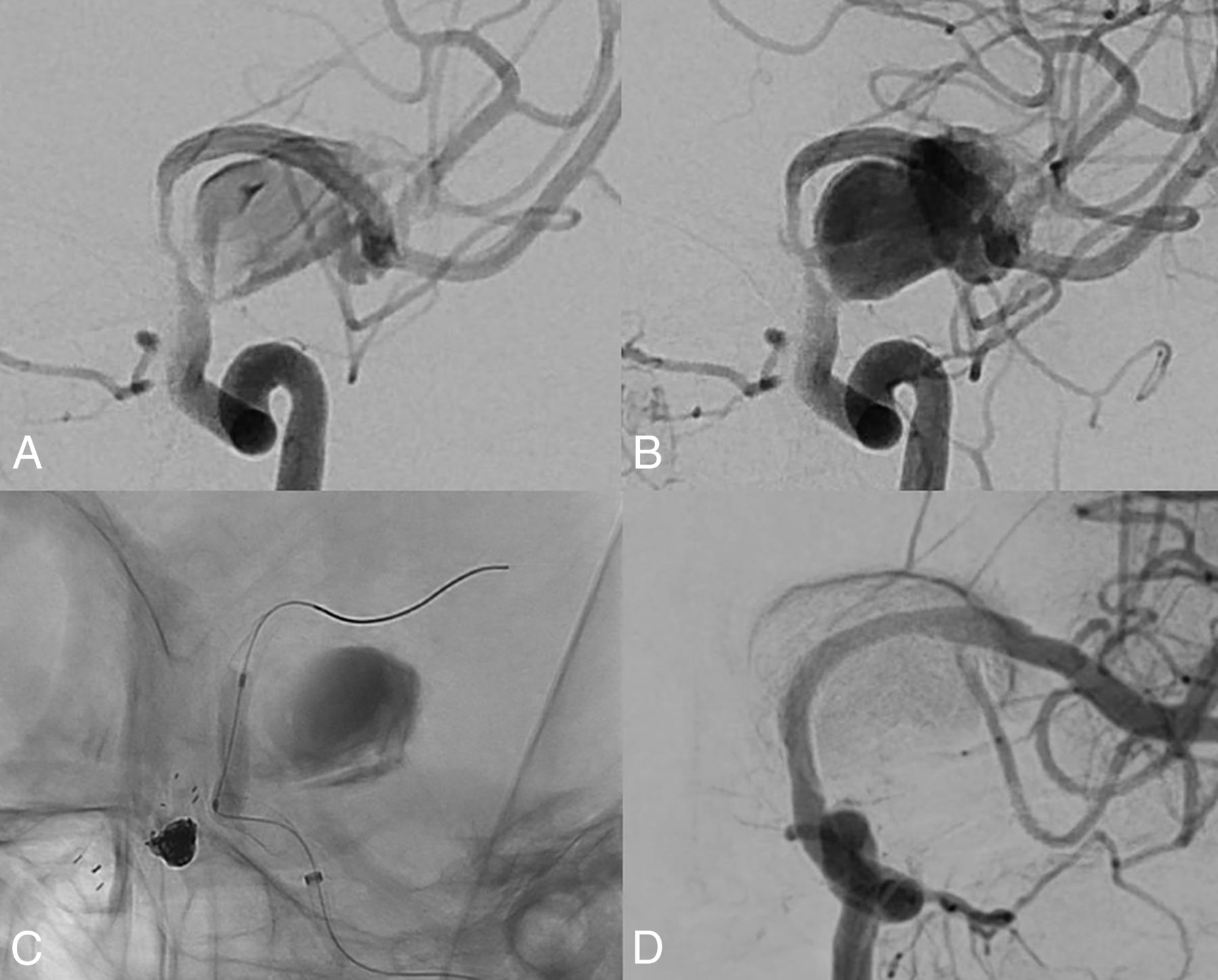
High-velocity blood flow into the aneurysm was caused by proximal parent artery stenosis after flow diversion. Early artery phase (A) and late artery phase (B). The high-velocity blood flow disappears immediately (D) after balloon angioplasty in the stent (C).

Limitations

The present study has several limitations. First, this is a retrospective study of a relatively small number of cases in a single center. Follow-up angiography was not completed in all cases, and the follow-up duration was not long enough. Therefore, the inability to obtain statistically significant results for all data analyzed is a limitation of this study. Second, the self-adjudication of aneurysm occlusion is known to be considerably overestimated according to another study.32 However, we treated only large and giant aneurysms, so there was relatively little difference in distinguishing the effect group from the no-effect group according to the O'Kelly-Marotta grading scale criteria.

CONCLUSIONS

FD is effective for treating very large and giant aneurysms, but the outcomes require further improvement. The results of this study show that an incorporated vessel and excessive balloon angioplasty might compromise the effect of FD. Considering this finding can help improve the outcomes of FD.

References

  1. 1.↵
    1. Nakase H,
    2. Shin Y,
    3. Kanemoto Y, et al
    . Long-term outcome of unruptured giant cerebral aneurysms. Neurol Med Chir (Tokyo) 2006;46:379–86 doi:10.2176/nmc.46.379 pmid:16936458
    CrossRefPubMed
  2. 2.↵
    1. Pia HW,
    2. Zierski J
    . Giant cerebral aneurysms. Neurosurg Rev 1982;5:117–48 doi:10.1007/BF01742676 pmid:6762507
    CrossRefPubMed
  3. 3.↵
    1. Lemole GM,
    2. Henn J,
    3. Spetzler RF, et al
    . Surgical management of giant aneurysms. Operative Techniques in Neurosurgery 2000;3:239–54 doi:10.1053/otns.2000.20464
    CrossRef
  4. 4.↵
    1. Laasko A,
    2. Hernesniemi C,
    3. Yonekawa Y, et al.
    1. Sano H
    . Treatment of complex intracranial aneurysms of anterior circulation using multiple clips. In: Laasko A, Hernesniemi C, Yonekawa Y, et al., eds. Surgical Management of Cerebrovascular Disease: Acta Neurochirurgica Supplementum. SpringerWien; 2010; 27–31
  5. 5.↵
    1. Molyneux A,
    2. Kerr R,
    3. Stratton I, et al
    . International Subarachnoid Aneurysm Trial (ISAT) Collaborative Group. International Subarachnoid Aneurysm Trial (ISAT) of neurosurgical clipping versus endovascular coiling in 2143 patients with ruptured intracranial aneurysms: a randomized trial. J Stroke Cerebrovasc Dis 2002;11:304–14 doi:10.1053/jscd.2002.130390 pmid:17903891
    CrossRefPubMed
  6. 6.↵
    1. Henkes H,
    2. Fischer S,
    3. Weber W, et al
    . Endovascular coil occlusion of 1811 intracranial aneurysms: early angiographic and clinical results. Neurosurgery 2004;54:268–80; discussion 280–85 doi:10.1227/01.neu.0000103221.16671.f0 pmid:14744273
    CrossRefPubMed
  7. 7.↵
    1. Jahromi BS,
    2. Mocco J,
    3. Bang JA, et al
    . Clinical and angiographic outcome after endovascular management of giant intracranial aneurysms. Neurosurgery 2008;63:662–74; discussion 674–75 doi:10.1227/01.NEU.0000325497.79690.4C pmid:18981877
    CrossRefPubMed
  8. 8.↵
    1. Murayama Y,
    2. Viñuela F,
    3. Ishii A, et al
    . Initial clinical experience with Matrix detachable coils for the treatment of intracranial aneurysms. J Neurosurg 2006;105:192–99 doi:10.3171/jns.2006.105.2.192 pmid:17219822
    CrossRefPubMed
  9. 9.↵
    1. Becske T,
    2. Kallmes DF,
    3. Saatci I, et al
    . Pipeline for Uncoilable or Failed aneurysms: results from a multicenter clinical trial. Radiology 2013;267:858–68 doi:10.1148/radiol.13120099 pmid:23418004
    CrossRefPubMed
  10. 10.↵
    1. O'Kelly CJ,
    2. Krings T,
    3. Fiorella D, et al
    . A novel grading scale for the angiographic assessment of intracranial aneurysms treated using flow diverting stents. Interv Neuroradiol 2010;16:133–37 doi:10.1177/159101991001600204 pmid:20642887
    CrossRefPubMed
  11. 11.↵
    1. Becske T,
    2. Brinjikji W,
    3. Potts MB, et al
    . Long-term clinical and angiographic outcomes following Pipeline Embolization Device treatment of complex internal carotid artery aneurysms: five-year results of the Pipeline for Uncoilable or Failed Aneurysms Trial. Neurosurgery 2017;80:40–48 doi:10.1093/neuros/nyw014 pmid:28362885
    CrossRefPubMed
  12. 12.↵
    1. Bender MT,
    2. Colby GP,
    3. Lin LM, et al
    . Predictors of cerebral aneurysm persistence and occlusion after flow diversion: a single-institution series of 445 cases with angiographic follow-up. J Neurosurg 2018;130:259–67 doi:10.3171/2017.11.JNS171738 pmid:29600915
    CrossRefPubMed
  13. 13.↵
    1. Makoyeva A,
    2. Bing F,
    3. Darsaut TE, et al
    . The varying porosity of braided self-expanding stents and flow diverters: an experimental study. AJNR Am J Neuroradiol 2013;34:596–602 doi:10.3174/ajnr.A3234 pmid:22878007
    Abstract/FREE Full Text
  14. 14.↵
    1. Song J,
    2. Shin YS
    . Antiplatelet drug resistance did not increase the thromboembolic events after stent-assisted coiling of unruptured intracranial aneurysm: a single center experience of 99 cases. Neurol Sci 2017;38:879–85 doi:10.1007/s10072-017-2859-z pmid:28233076
    CrossRefPubMed
  15. 15.↵
    1. Chalouhi N,
    2. Tjoumakaris S,
    3. Gonzalez LF, et al
    . Coiling of large and giant aneurysms: complications and long-term results of 334 cases. AJNR Am J Neuroradiol 2014;35:546–52 doi:10.3174/ajnr.A3696 pmid:23945229
    Abstract/FREE Full Text
  16. 16.↵
    1. Sluzewski M,
    2. Menovsky T,
    3. van Rooij WJ, et al
    . Coiling of very large or giant cerebral aneurysms: long-term clinical and serial angiographic results. AJNR Am J Neuroradiol 2003;24:257–62 pmid:12591644
    CrossRefPubMed
  17. 17.↵
    1. Peschillo S,
    2. Caporlingua A,
    3. Resta MC, et al
    . Endovascular treatment of large and giant carotid aneurysms with flow-diverter stents alone or in combination with coils: a multicenter experience and long-term follow-up. Oper Neurosurg (Hagerstown) 2017;13:492–502 doi:10.1093/ons/opx032 pmid:28838114
    CrossRefPubMed
  18. 18.↵
    1. Brinjikji W,
    2. Murad MH,
    3. Lanzino G, et al
    . Endovascular treatment of intracranial aneurysms with flow diverters: a meta-analysis. Stroke 2013;44:442–47 doi:10.1161/STROKEAHA.112.678151 pmid:23321438
    Abstract/FREE Full Text
  19. 19.↵
    1. Oishi H,
    2. Teranishi K,
    3. Yatomi K, et al
    . Flow diverter therapy using a Pipeline Embolization Device for 100 unruptured large and giant internal carotid artery aneurysms in a single center in a Japanese population. Neurol Med Chir (Tokyo) 2018;58:461–67 doi:10.2176/nmc.oa.2018-0148 pmid:30298832
    CrossRefPubMed
  20. 20.↵
    1. Larrabide I,
    2. Aguilar ML,
    3. Morales HG, et al
    . Intra-aneurysmal pressure and flow changes induced by flow diverters: relation to aneurysm size and shape. AJNR Am J Neuroradiol 2013;34:816–22 doi:10.3174/ajnr.A3288 pmid:23019173
    Abstract/FREE Full Text
  21. 21.↵
    1. Saatci I,
    2. Yavuz K,
    3. Ozer C, et al
    . Treatment of intracranial aneurysms using the Pipeline flow-diverter embolization device: a single-center experience with long-term follow-up results. AJNR Am J Neuroradiol 2012;33:1436–46 doi:10.3174/ajnr.A3246 pmid:22821921
    Abstract/FREE Full Text
  22. 22.↵
    1. Cebral JR,
    2. Castro MA,
    3. Appanaboyina S, et al
    . Efficient pipeline for image-based patient-specific analysis of cerebral aneurysm hemodynamics: technique and sensitivity. IEEE Trans Med Imaging 2005;24:457–67 doi:10.1109/tmi.2005.844159 pmid:15822804
    CrossRefPubMed
  23. 23.↵
    1. Choi JH,
    2. Lee KS,
    3. Kim B, et al
    . Treatment outcomes of large and giant intracranial aneurysms according to various treatment modalities. Acta Neurochir (Wien) 2020;162:2745–52 doi:10.1007/s00701-020-04540-1 pmid:32827268
    CrossRefPubMed
  24. 24.↵
    1. Dai D,
    2. Ding YH,
    3. Kadirvel R, et al
    . Patency of branches after coverage with multiple telescoping flow-diverter devices: an in vivo study in rabbits. AJNR Am J Neuroradiol 2012;33:171–74 doi:10.3174/ajnr.A2879 pmid:22158925
    Abstract/FREE Full Text
  25. 25.↵
    1. Kojima M,
    2. Irie K,
    3. Fukuda T, et al
    . The study of flow diversion effects on aneurysm using multiple Enterprise stents and two flow diverters. Asian J Neurosurg 2012;7:159–65 doi:10.4103/1793-5482.106643 pmid:23559981
    CrossRefPubMed
  26. 26.↵
    1. Siddiqui AH,
    2. Kan P,
    3. Abla AA, et al
    . Complications after treatment with Pipeline embolization for giant distal intracranial aneurysms with or without coil embolization. Neurosurgery 2012;71:E509–13; discussion E513 doi:10.1227/NEU.0b013e318258e1f8 pmid:22710418
    CrossRefPubMed
  27. 27.↵
    1. Kim BM,
    2. Shin YS,
    3. Baik MW, et al
    . Pipeline Embolization Device for large/giant or fusiform aneurysms: an initial multi-center experience in Korea. Neurointervention 2016;11:10–17 doi:10.5469/neuroint.2016.11.1.10 pmid:26958407
    CrossRefPubMed
  28. 28.↵
    1. Cohen JE,
    2. Gomori JM,
    3. Moscovici S, et al
    . Delayed complications after flow-diverter stenting: reactive in-stent stenosis and creeping stents. J Clin Neurosci 2014;21:1116–22 doi:10.1016/j.jocn.2013.11.010 pmid:24524952
    CrossRefPubMed
  29. 29.↵
    1. Guédon A,
    2. Clarençon F,
    3. Maria FD, et al
    . Very late ischemic complications in flow-diverter stents: a retrospective analysis of a single-center series. J Neurosurg 2016;125:929–35 doi:10.3171/2015.10.JNS15703 pmid:26824382
    CrossRefPubMed
  30. 30.↵
    1. Mut F,
    2. Raschi M,
    3. Scrivano E, et al
    . Association between hemodynamic conditions and occlusion times after flow diversion in cerebral aneurysms. J Neurointerv Surg 2015;7:286–90 doi:10.1136/neurintsurg-2013-011080 pmid:24696500
    Abstract/FREE Full Text
  31. 31.↵
    1. Kulcsár Z,
    2. Augsburger L,
    3. Reymond P, et al
    . Flow diversion treatment: intra-aneurismal blood flow velocity and WSS reduction are parameters to predict aneurysm thrombosis. Acta Neurochir (Wien) 2012;154:1827–34 doi:10.1007/s00701-012-1482-2 pmid:22926629
    CrossRefPubMed
  32. 32.↵
    1. Taschner CA,
    2. Chapot R,
    3. Costalat V, et al
    . GREAT: a randomized controlled trial comparing HydroSoft/HydroFrame and bare platinum coils for endovascular aneurysm treatment: procedural safety and core-lab-assessed angiographic results. Neuroradiology 2016;58:777–86 doi:10.1007/s00234-016-1693-y pmid:27137926
    CrossRefPubMed
  • Received August 18, 2020.
  • Accepted after revision December 24, 2020.
  • © 2021 by American Journal of Neuroradiology
PreviousNext
Back to top

In this issue

American Journal of Neuroradiology: 42 (6)
American Journal of Neuroradiology
Vol. 42, Issue 6
1 Jun 2021
  • Table of Contents
  • Index by author
  • Complete Issue (PDF)
Advertisement
Print
Download PDF
Email Article

Thank you for your interest in spreading the word on American Journal of Neuroradiology.

NOTE: We only request your email address so that the person you are recommending the page to knows that you wanted them to see it, and that it is not junk mail. We do not capture any email address.

Enter multiple addresses on separate lines or separate them with commas.
Predictors of the Effects of Flow Diversion in Very Large and Giant Aneurysms
(Your Name) has sent you a message from American Journal of Neuroradiology
(Your Name) thought you would like to see the American Journal of Neuroradiology web site.
CAPTCHA
This question is for testing whether or not you are a human visitor and to prevent automated spam submissions.
Cite this article
H.J. Bae, Y.K. Park, D.Y. Cho, J.H. Choi, B.S. Kim, Y.S. Shin
Predictors of the Effects of Flow Diversion in Very Large and Giant Aneurysms
American Journal of Neuroradiology Jun 2021, 42 (6) 1099-1103; DOI: 10.3174/ajnr.A7085

Citation Manager Formats

  • BibTeX
  • Bookends
  • EasyBib
  • EndNote (tagged)
  • EndNote 8 (xml)
  • Medlars
  • Mendeley
  • Papers
  • RefWorks Tagged
  • Ref Manager
  • RIS
  • Zotero
0 Responses
Respond to this article
Share
Bookmark this article
Predictors of the Effects of Flow Diversion in Very Large and Giant Aneurysms
H.J. Bae, Y.K. Park, D.Y. Cho, J.H. Choi, B.S. Kim, Y.S. Shin
American Journal of Neuroradiology Jun 2021, 42 (6) 1099-1103; DOI: 10.3174/ajnr.A7085
del.icio.us logo Twitter logo Facebook logo Mendeley logo
  • Tweet Widget
  • Facebook Like
  • Google Plus One
Purchase

Jump to section

  • Article
    • Abstract
    • ABBREVIATIONS:
    • MATERIALS AND METHODS
    • RESULTS
    • DISCUSSION
    • CONCLUSIONS
    • References
  • Figures & Data
  • Info & Metrics
  • Responses
  • References
  • PDF

Related Articles

  • No related articles found.
  • PubMed
  • Google Scholar

Cited By...

  • Parent artery occlusion after pipeline embolization device implantation of intracranial saccular and fusiform aneurysms
  • Crossref (12)
  • Google Scholar

This article has been cited by the following articles in journals that are participating in Crossref Cited-by Linking.

  • A Long-Term Comparative Analysis of Endovascular Coiling and Clipping for Ruptured Cerebral Aneurysms: An Individual Patient-Level Meta-Analysis Assessing Rerupture Rates
    Johannes Wach, Martin Vychopen, Agi Güresir, Alexandru Guranda, Ulf Nestler, Erdem Güresir
    Journal of Clinical Medicine 2024 13 6
  • Safety and efficacy of Surpass Evolve Flow diverter for intracranial aneurysms: A study of 116 patients
    Richard Bibi, Nourou Dine Adeniran Bankole, Baptiste Donnard, Francesca Giubbolini, Julien Boucherit, Valère Barrot, Denis Herbreteau, Héloïse Ifergan, Kevin Janot, Grégoire Boulouis, Fouzi Bala
    The Neuroradiology Journal 2024 37 2
  • Advantages and Disadvantages of Flow Diverter Treatment for Cerebral Aneurysms
    Akira Ishii
    Japanese Journal of Neurosurgery 2022 31 2
  • Evaluation of the Significance of Persistent Remnant Filling and Enlargement After Flow Diversion for Intracranial Aneurysms
    Tae Keun Jee, Je Young Yeon, Keon Ha Kim, Jong-Soo Kim, Pyoung Jeon
    World Neurosurgery 2024 184
  • Treatment Outcomes of 94 Cases of Pipeline Embolization Device in a Single Center: Predictive Factors of Incomplete Aneurysm Occlusion
    Ryo Hiramatsu, Ryokichi Yagi, Masahiro Kameda, Naosuke Nonoguchi, Motomasa Furuse, Shinji Kawabata, Hiroyuki Ohnishi, Shigeru Miyachi, Masahiko Wanibuchi
    Journal of Neuroendovascular Therapy 2023 17 10
  • Treatment of giant intracranial aneurysms using the Pipeline flow-diverting stent: Long-term results from the International Retrospective Study of the Pipeline Embolization Device (IntrePED) study
    Ramesh Grandhi, Vijay M. Ravindra, David F. Kallmes, Demetrius Lopes, Ricardo A. Hanel, Pedro Lylyk
    Interventional Neuroradiology 2024 30 2
  • Parent artery occlusion after pipeline embolization device implantation of intracranial saccular and fusiform aneurysms
    Mingyang Han, Xin Tong, Zhifei Wang, Aihua Liu
    Journal of NeuroInterventional Surgery 2023 15 11
  • Performance assessment of the Surpass Evolve flow diverter for the treatment of intracranial aneurysms: A systematic review and meta-analysis
    Aaron Rodriguez-Calienes, Juan Vivanco-Suarez, Nicole M. Castillo-Huerta, David Espinoza-Martinez, Cristian Morán-Mariños, Ximena Espiritu-Vilcapoma, Valeria Rivera-Angles, Santiago Ortega-Gutierrez
    Interventional Neuroradiology 2024
  • Flow Diversion for Cerebral Aneurysms: A Decade-Long Experience with Improved Outcomes and Predictors of Success
    Tae Keun Jee, Je Young Yeon, Keon Ha Kim, Jong-Soo Kim, Pyoung Jeon
    Brain Sciences 2024 14 8
  • Intraoperative aneurysm flow analysis predicts intracranial large and giant aneurysm occlusion after flow diversion
    Yoshinari Osada, Hiroyuki Sakata, Masayuki Ezura, Kenichi Sato, Keisuke Sasaki, Shunsuke Omodaka, Atsushi Kanoke, Hiroki Uchida, Hidenori Endo
    Clinical Neurology and Neurosurgery 2025 250

More in this TOC Section

  • SAVE vs. Solumbra Techniques for Thrombectomy
  • Contrast-Induced Encephalopathy after NeuroIR
  • CT Perfusion&Reperfusion in Acute Ischemic Stroke
Show more Interventional

Similar Articles

Advertisement

Indexed Content

  • Current Issue
  • Accepted Manuscripts
  • Article Preview
  • Past Issues
  • Editorials
  • Editor's Choice
  • Fellows' Journal Club
  • Letters to the Editor
  • Video Articles

Cases

  • Case Collection
  • Archive - Case of the Week
  • Archive - Case of the Month
  • Archive - Classic Case

Special Collections

  • AJNR Awards
  • ASNR Foundation Special Collection
  • Most Impactful AJNR Articles
  • Photon-Counting CT
  • Spinal CSF Leak Articles (Jan 2020-June 2024)

More from AJNR

  • Trainee Corner
  • Imaging Protocols
  • MRI Safety Corner

Multimedia

  • AJNR Podcasts
  • AJNR Scantastics

Resources

  • Turnaround Time
  • Submit a Manuscript
  • Submit a Video Article
  • Submit an eLetter to the Editor/Response
  • Manuscript Submission Guidelines
  • Statistical Tips
  • Fast Publishing of Accepted Manuscripts
  • Graphical Abstract Preparation
  • Imaging Protocol Submission
  • Evidence-Based Medicine Level Guide
  • Publishing Checklists
  • Author Policies
  • Become a Reviewer/Academy of Reviewers
  • News and Updates

About Us

  • About AJNR
  • Editorial Board
  • Editorial Board Alumni
  • Alerts
  • Permissions
  • Not an AJNR Subscriber? Join Now
  • Advertise with Us
  • Librarian Resources
  • Feedback
  • Terms and Conditions
  • AJNR Editorial Board Alumni

American Society of Neuroradiology

  • Not an ASNR Member? Join Now

© 2025 by the American Society of Neuroradiology All rights, including for text and data mining, AI training, and similar technologies, are reserved.
Print ISSN: 0195-6108 Online ISSN: 1936-959X

Powered by HighWire