Section Editor: Sandy Cheng-Yu Chen, M.D.
Taipei Medical University Hospital, Taipei, Taiwan
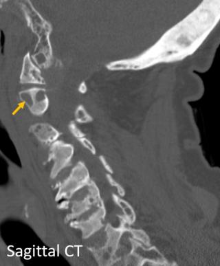
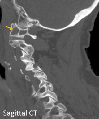
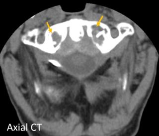
Renal osteodystrophy encompasses bone abnormalities related to chronic kidney disease, due to secondary hyperparathyroidism and osteomalacia. Imaging findings include osteosclerosis (diffuse or affecting only the vertebral endplates) and/or osteopenia, changes related to secondary hyperparathyroidism, such as brown tumors, and insufficiency fractures. This case reveals osteosclerosis of the occipital bone and vertebrae, with concomitant vertebral lytic lesions (brown tumors) affecting mainly C1 (B and C, arrows) and C2 (A, arrow), erosion of the vertebral endplates, and loss of height of several vertebral bodies. Secondary to the changes in the odontoid process, there is also thickening of the transverse ligament. Though gout can cause erosion of the odontoid process, it usually presents with juxta-articular soft-tissue lesions (tophi) with eccentric erosions of joint surfaces; likewise, diffuse osteosclerosis is not a typical feature.